
Main menu

  • Home
  • Content
    • Current
    • Ahead of print
    • Past Issues
    • JNM Supplement
    • SNMMI Annual Meeting Abstracts
    • Continuing Education
    • JNM Podcasts
  • Subscriptions
    • Subscribers
    • Institutional and Non-member
    • Rates
    • Journal Claims
    • Corporate & Special Sales
  • Authors
    • Submit to JNM
    • Information for Authors
    • Assignment of Copyright
    • AQARA requirements
  • Info
    • Reviewers
    • Permissions
    • Advertisers
  • About
    • About Us
    • Editorial Board
    • Contact Information
  • More
    • Alerts
    • Feedback
    • Help
    • SNMMI Journals
  • SNMMI
    • JNM
    • JNMT
    • SNMMI Journals
    • SNMMI

User menu

  • Subscribe
  • My alerts
  • Log in
  • My Cart

Search

  • Advanced search
Journal of Nuclear Medicine
  • SNMMI
    • JNM
    • JNMT
    • SNMMI Journals
    • SNMMI
  • Subscribe
  • My alerts
  • Log in
  • My Cart
Journal of Nuclear Medicine

Advanced Search

  • Home
  • Content
    • Current
    • Ahead of print
    • Past Issues
    • JNM Supplement
    • SNMMI Annual Meeting Abstracts
    • Continuing Education
    • JNM Podcasts
  • Subscriptions
    • Subscribers
    • Institutional and Non-member
    • Rates
    • Journal Claims
    • Corporate & Special Sales
  • Authors
    • Submit to JNM
    • Information for Authors
    • Assignment of Copyright
    • AQARA requirements
  • Info
    • Reviewers
    • Permissions
    • Advertisers
  • About
    • About Us
    • Editorial Board
    • Contact Information
  • More
    • Alerts
    • Feedback
    • Help
    • SNMMI Journals
  • View or Listen to JNM Podcast
  • Visit JNM on Facebook
  • Join JNM on LinkedIn
  • Follow JNM on Twitter
  • Subscribe to our RSS feeds
Meeting ReportPhysics, Instrumentation & Data Sciences - Instrumentation

Collimator fabrication techniques: a comparative study

Mark Ottensmeyer, Hamid Sabet, Lars Furenlid, Micaehla May and Matthew Kupinski
Journal of Nuclear Medicine June 2023, 64 (supplement 1) P1600;
Mark Ottensmeyer
1Massachusetts General Hospital
  • Find this author on Google Scholar
  • Find this author on PubMed
  • Search for this author on this site
Hamid Sabet
2Harvard Medical School & Massachusetts General Hos
  • Find this author on Google Scholar
  • Find this author on PubMed
  • Search for this author on this site
Lars Furenlid
3University of Arizona
  • Find this author on Google Scholar
  • Find this author on PubMed
  • Search for this author on this site
Micaehla May
4University of Arizona James C. Wyant College of Optical Science
  • Find this author on Google Scholar
  • Find this author on PubMed
  • Search for this author on this site
Matthew Kupinski
5J.C. Wyant College of Optical Sciences, University of Arizona
  • Find this author on Google Scholar
  • Find this author on PubMed
  • Search for this author on this site
  • Article
  • Figures & Data
  • Info & Metrics
Loading

Abstract

P1600

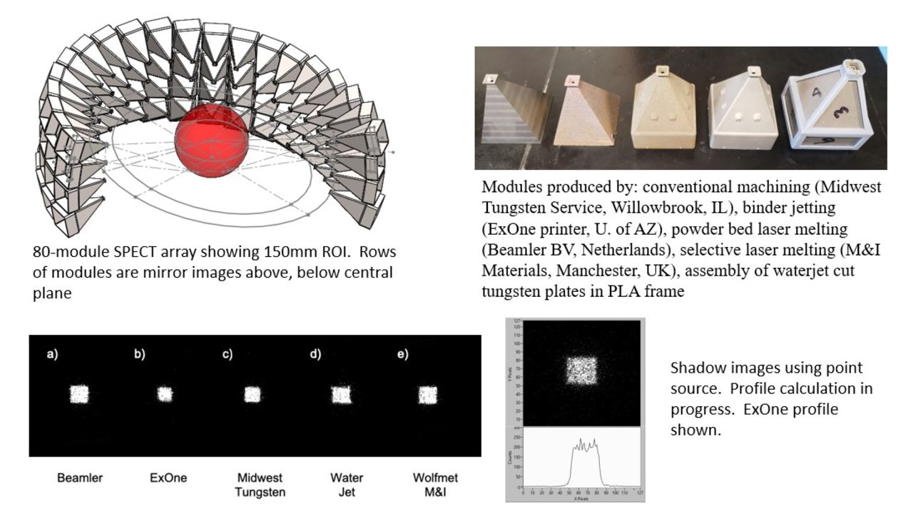
Introduction: Compared with conventional dual head gamma camera SPECT systems, SPECT scanners with larger numbers of detector/collimator modules to satisfy simultaneous spatial resolution and sensitivity requirements are becoming commonplace. Fabrication of modules for arrays that are symmetrical about the center of the region of interest (ROI) benefits from economies of scale through replication of the same module design throughout the array. When asymmetries are necessary however, as in our ongoing project which distributes pinhole camera modules on an ellipse centered on a patient’s thorax while focusing each module on an ROI centered on the heart, each module’s geometry is position dependent. Only symmetrical positioning in planes above and below the heart allows for replication of geometry between rows of modules, which individually are unique.

Tungsten is used for constructing pinhole collimator bodies of desired geometry having a density 1.7x that of lead. Its high hardness and melting temperature make fabrication by traditional methods difficult. Conventional machining is possible, however the non-circular pinhole geometry, sharp internal corners of the pyramidal bodies and creation of unique fixturing for each body argue for the use of other approaches.

We present an overview and geometric drivers of the 80-camera cardiac SPECT imager under development. Geometries closest to the center of the cardiac ROI were selected for fabrication of test units using conventional machining, three additive manufacturing methods (binder jetting, powder bed laser melting (PBLM), selective laser melting (SLM)), and assembly of water-jet-cut plates of pure tungsten fit into a 3D printed plastic support structure.

Methods: A constraint-driven geometric solution, implemented in parametric computer-aided design, generated the full set of 40 unique modules and mirror imaged counterparts above and below the plane of the heart. STEP-formatted geometry was exported for 3D printing. The same pinhole/collimator geometry defined the cut-plate design using two 0.040" thick layers and a 3D printed shell using PLA. The pinhole assembly was also cut from the same plate, yielding a finite-depth pinhole vs. the sharp-edged pinholes of other versions. The collimators are shown in the supporting figure.

140 keV shadow images of the pinholes were created using a 99mTc point source at 10" from the detector and the collimators resting on the detector surface.

Results: The machined collimator was fabricated using a tungsten/nickel/iron alloy (95/3.5/1.5%), yielding a density of 18.76 g/cm3. The binder-jet printed collimator used pure tungsten powder at 150 μm layer thickness with a lower density metal infiltrant , density = 14.60 g/cm3. PBLM used 100 μm layers of W/Ni/Fe powder (final stoichiometry 95% tungsten, 5% Ni/Fe binder), density = 18.20 g/cm3. SLM used a 100% tungsten powder, at a layer thickness of 30 μm, final density = 17.95 g/cm3, slightly less than the pure solid due to remaining porosity.

Conclusions: The fabrication methods were competitive with each other in terms of density (except binder-jetting). The shadow images were comparable in all cases. Water-jetting was the least expensive method and had potential for shortest lead-times given simpler material sourcing and machine set-up and availability of plastic 3D printing providers. The cut-plate pinhole was not sharp-edged, having image quality implications for upcoming tests. The external shell holding plates together increases the minimum overall array size and has a small negative impact on estimated resolution sensitivity. A collimator with cut-plate walls and printed pinhole is currently under design.

Figure
  • Download figure
  • Open in new tab
  • Download powerpoint
Previous
Back to top

In this issue

Journal of Nuclear Medicine
Vol. 64, Issue supplement 1
June 1, 2023
  • Table of Contents
  • Index by author
Article Alerts
Sign In to Email Alerts with your Email Address
Email Article

Thank you for your interest in spreading the word on Journal of Nuclear Medicine.

NOTE: We only request your email address so that the person you are recommending the page to knows that you wanted them to see it, and that it is not junk mail. We do not capture any email address.

Enter multiple addresses on separate lines or separate them with commas.
Collimator fabrication techniques: a comparative study
(Your Name) has sent you a message from Journal of Nuclear Medicine
(Your Name) thought you would like to see the Journal of Nuclear Medicine web site.
Citation Tools
Collimator fabrication techniques: a comparative study
Mark Ottensmeyer, Hamid Sabet, Lars Furenlid, Micaehla May, Matthew Kupinski
Journal of Nuclear Medicine Jun 2023, 64 (supplement 1) P1600;

Citation Manager Formats

  • BibTeX
  • Bookends
  • EasyBib
  • EndNote (tagged)
  • EndNote 8 (xml)
  • Medlars
  • Mendeley
  • Papers
  • RefWorks Tagged
  • Ref Manager
  • RIS
  • Zotero
Share
Collimator fabrication techniques: a comparative study
Mark Ottensmeyer, Hamid Sabet, Lars Furenlid, Micaehla May, Matthew Kupinski
Journal of Nuclear Medicine Jun 2023, 64 (supplement 1) P1600;
Twitter logo Facebook logo LinkedIn logo Mendeley logo
  • Tweet Widget
  • Facebook Like
  • Google Plus One
Bookmark this article

Jump to section

  • Article
  • Figures & Data
  • Info & Metrics

Related Articles

  • No related articles found.
  • Google Scholar

Cited By...

  • No citing articles found.
  • Google Scholar

More in this TOC Section

  • A multimodal approach to functional neuroimaging: Integrating eye-tracking and heart rate monitoring with ultra-high performance upright Prism-PET brain scanner
  • A Novel Approach to Cross Section Measurement: Prompt Gamma Isotope Yield Reconstruction with GammaSphere
  • Development of a portable and versatile PET System for Interactive Point-of-Care Imaging
Show more Physics, Instrumentation & Data Sciences - Instrumentation

Similar Articles

SNMMI

© 2025 SNMMI

Powered by HighWire