
Main menu

  • Home
  • Content
    • Current
    • Ahead of print
    • Past Issues
    • JNM Supplement
    • SNMMI Annual Meeting Abstracts
    • Continuing Education
    • JNM Podcasts
  • Subscriptions
    • Subscribers
    • Institutional and Non-member
    • Rates
    • Journal Claims
    • Corporate & Special Sales
  • Authors
    • Submit to JNM
    • Information for Authors
    • Assignment of Copyright
    • AQARA requirements
  • Info
    • Reviewers
    • Permissions
    • Advertisers
  • About
    • About Us
    • Editorial Board
    • Contact Information
  • More
    • Alerts
    • Feedback
    • Help
    • SNMMI Journals
  • SNMMI
    • JNM
    • JNMT
    • SNMMI Journals
    • SNMMI

User menu

  • Subscribe
  • My alerts
  • Log in
  • My Cart

Search

  • Advanced search
Journal of Nuclear Medicine
  • SNMMI
    • JNM
    • JNMT
    • SNMMI Journals
    • SNMMI
  • Subscribe
  • My alerts
  • Log in
  • My Cart
Journal of Nuclear Medicine

Advanced Search

  • Home
  • Content
    • Current
    • Ahead of print
    • Past Issues
    • JNM Supplement
    • SNMMI Annual Meeting Abstracts
    • Continuing Education
    • JNM Podcasts
  • Subscriptions
    • Subscribers
    • Institutional and Non-member
    • Rates
    • Journal Claims
    • Corporate & Special Sales
  • Authors
    • Submit to JNM
    • Information for Authors
    • Assignment of Copyright
    • AQARA requirements
  • Info
    • Reviewers
    • Permissions
    • Advertisers
  • About
    • About Us
    • Editorial Board
    • Contact Information
  • More
    • Alerts
    • Feedback
    • Help
    • SNMMI Journals
  • View or Listen to JNM Podcast
  • Visit JNM on Facebook
  • Join JNM on LinkedIn
  • Follow JNM on Twitter
  • Subscribe to our RSS feeds
Meeting ReportNeurosciences

Monitoring synaptic loss in a rat model of stroke: a PET imaging study with [18F]SynVesT-2, radiotracer for the synaptic vesicle protein 2A (SV2A)

Xueying Lyu, Takuya Toyonaga, Gang Deng, Yiyun Huang, Richard Carson, Jiangbing Zhou and Zhengxin Cai
Journal of Nuclear Medicine May 2020, 61 (supplement 1) 84;
Xueying Lyu
1Yale University New Haven CT United States
3Yale University New Haven CT United States
  • Find this author on Google Scholar
  • Find this author on PubMed
  • Search for this author on this site
Takuya Toyonaga
2PET center, Yale University New Haven CT United States
  • Find this author on Google Scholar
  • Find this author on PubMed
  • Search for this author on this site
Gang Deng
1Yale University New Haven CT United States
3Yale University New Haven CT United States
  • Find this author on Google Scholar
  • Find this author on PubMed
  • Search for this author on this site
Yiyun Huang
1Yale University New Haven CT United States
3Yale University New Haven CT United States
  • Find this author on Google Scholar
  • Find this author on PubMed
  • Search for this author on this site
Richard Carson
1Yale University New Haven CT United States
3Yale University New Haven CT United States
  • Find this author on Google Scholar
  • Find this author on PubMed
  • Search for this author on this site
Jiangbing Zhou
1Yale University New Haven CT United States
3Yale University New Haven CT United States
  • Find this author on Google Scholar
  • Find this author on PubMed
  • Search for this author on this site
Zhengxin Cai
1Yale University New Haven CT United States
3Yale University New Haven CT United States
  • Find this author on Google Scholar
  • Find this author on PubMed
  • Search for this author on this site
  • Article
  • Figures & Data
  • Info & Metrics
Loading

Abstract

84

Introduction: Stroke is a devastating disease with high mortality and morbidity rates. FDG PET has been used to probe brain tissue viability (penumbra) after acute ischemic stroke;1 while MR and CT have been used to image infarct through anatomical and perfusion changes.2 However, the transient hyperglycemia, a common phenomenon in acute stroke patients, could affect the FDG uptake and thereby confounding its interpretation.1 Because synapse is a crucial microstructure for brain functions and synaptic deficit is a hallmark of stroke, we, for the first time, tested in vivo PET imaging using our recently developed SV2A tracer [18F]SynVesT-2 to track the progression of synapse loss in a rat model of stroke.

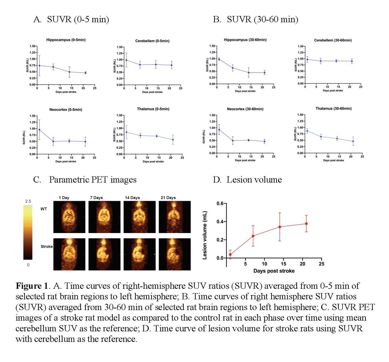
Methods: The stroke model was established through the middle cerebral artery occlusion (MCAO) procedure, with 4 h occlusion followed by reperfusion.3 A total of 6 stroke model rats underwent weekly PET scans for four weeks starting at 1-day post reperfusion. The SV2A PET ligand [18F]SynVesT-2 was synthesized via iodonium ylide precursor and was administered intravenously to the rats. Emission data were acquired from 0-60 min post-injection on the FOCUS 220 scanners. Images were reconstructed with attenuation and scatter correction. The Waxholm Space (WHS) rat brain template was manually registered to PET images to generate standardized uptake values (SUVs) for hippocampus, cerebellum, neocortex, and thalamus. The SUVs of ipsilateral brain regions averaged from either 30-60 min or 0-5 min were normalized with the SUVs of the contralateral regions to generate SUV ratio (SUVR). We chose the SUVR from later time window (30-60 min post tracer injection) for SV2A quantification, and early time window (0-5 min) as a surrogate of perfusion. The volume of lesion was assessed using SUVR normalized by cerebellum, after smoothing, masking, subtracting, thresholding, and binarizing. One-way repeated measure ANOVA and t-test were used in the statistical analysis.

Results: Using the contralateral brain regions as reference regions, the SUVR(30-60 min) of the infarct core regions (hippocampus, thalamus and neocortex) decreased over time (p < 0.01), most significantly from 1-day to 7-day post reperfusion (p < 0.006); but remained unchanged in cerebellum (p > 0.1). As a readout for blood flow, the SUVR(0-5 min)in the ischemic core regions was robustly reduced by ~50% at later time points; but highly variable at one-day post reperfusion, which might reflect a transient bilateral blood brain barrier opening after stroke (Figure 1A,B). Both the parametric SUVR(30-60 min) PET images (Figure 1C) and the infarct maps clearly identified the brain lesion regions and sizes. Based on the infarct lesion maps (Figure 1D), the infarct volume consistently increased over time (p < 0.01), most significantly from 1-day to 7-day post stroke (p = 0.0003; 0.037 ± 0.049 mL, 0.24 ± 0.11 mL, 0.34 ± 0.16 mL, and 0.38 ± 0.09 mL, at 1-day, 1-week, 2-week, and 3-week post reperfusion, respectively).

Conclusions: Using [18F]SynVesT-2 and PET imaging, we successfully detected synapse loss in rat MCAO model and tracked disease progression via lesion quantification. Synapse loss is mainly in hippocampus, thalamus, and neocortex, rather than cerebellum. Furthermore, most significant loss of synapses was observed to occur during the first week post reperfusion. Our results showed that the quantification of synaptic density by SV2A PET may provide an objective measure of disease progression and a primary endpoint to evaluate treatment efficacy of novel therapeutics for stroke in clinical trials. References:

  1. Bunevicius et al. Biomed Res Int 2013. 2013: 634598.

  2. Saver. Stroke 2011, 42: 2356-2364.

  3. Cai et al. Neurosci Lett 2015, 597: 127-31.

Figure
  • Download figure
  • Open in new tab
  • Download powerpoint

Previous
Back to top

In this issue

Journal of Nuclear Medicine
Vol. 61, Issue supplement 1
May 1, 2020
  • Table of Contents
  • Index by author
Article Alerts
Sign In to Email Alerts with your Email Address
Email Article

Thank you for your interest in spreading the word on Journal of Nuclear Medicine.

NOTE: We only request your email address so that the person you are recommending the page to knows that you wanted them to see it, and that it is not junk mail. We do not capture any email address.

Enter multiple addresses on separate lines or separate them with commas.
Monitoring synaptic loss in a rat model of stroke: a PET imaging study with [18F]SynVesT-2, radiotracer for the synaptic vesicle protein 2A (SV2A)
(Your Name) has sent you a message from Journal of Nuclear Medicine
(Your Name) thought you would like to see the Journal of Nuclear Medicine web site.
Citation Tools
Monitoring synaptic loss in a rat model of stroke: a PET imaging study with [18F]SynVesT-2, radiotracer for the synaptic vesicle protein 2A (SV2A)
Xueying Lyu, Takuya Toyonaga, Gang Deng, Yiyun Huang, Richard Carson, Jiangbing Zhou, Zhengxin Cai
Journal of Nuclear Medicine May 2020, 61 (supplement 1) 84;

Citation Manager Formats

  • BibTeX
  • Bookends
  • EasyBib
  • EndNote (tagged)
  • EndNote 8 (xml)
  • Medlars
  • Mendeley
  • Papers
  • RefWorks Tagged
  • Ref Manager
  • RIS
  • Zotero
Share
Monitoring synaptic loss in a rat model of stroke: a PET imaging study with [18F]SynVesT-2, radiotracer for the synaptic vesicle protein 2A (SV2A)
Xueying Lyu, Takuya Toyonaga, Gang Deng, Yiyun Huang, Richard Carson, Jiangbing Zhou, Zhengxin Cai
Journal of Nuclear Medicine May 2020, 61 (supplement 1) 84;
Twitter logo Facebook logo LinkedIn logo Mendeley logo
  • Tweet Widget
  • Facebook Like
  • Google Plus One
Bookmark this article

Jump to section

  • Article
  • Figures & Data
  • Info & Metrics

Related Articles

  • No related articles found.
  • Google Scholar

Cited By...

  • No citing articles found.
  • Google Scholar

More in this TOC Section

Neurosciences

  • PET CT Quantification to direct Alzheimer’s patients to anti-amyloid therapy
  • Multicenter trial study for usefulness of physical parameters toward the standardization of brain SPECT image: relation to visual analysis
  • Assessment of Brain Perfusion 123I-IMP SPECT imaging Using Low Energy High Resolution Collimator image reconstructed by Flash 3D
Show more Neurosciences

Basic Science - Synaptic activity and other mechanisms

  • NR2B
  • Single-cell analysis reveals the key role of enhanced endothelial glucose uptake in neurometabolic coupling
Show more Basic Science - Synaptic activity and other mechanisms

Similar Articles

SNMMI

© 2025 SNMMI

Powered by HighWire