
Main menu

  • Home
  • Content
    • Current
    • Ahead of print
    • Past Issues
    • JNM Supplement
    • SNMMI Annual Meeting Abstracts
    • Continuing Education
    • JNM Podcasts
  • Subscriptions
    • Subscribers
    • Institutional and Non-member
    • Rates
    • Journal Claims
    • Corporate & Special Sales
  • Authors
    • Submit to JNM
    • Information for Authors
    • Assignment of Copyright
    • AQARA requirements
  • Info
    • Reviewers
    • Permissions
    • Advertisers
  • About
    • About Us
    • Editorial Board
    • Contact Information
  • More
    • Alerts
    • Feedback
    • Help
    • SNMMI Journals
  • SNMMI
    • JNM
    • JNMT
    • SNMMI Journals
    • SNMMI

User menu

  • Subscribe
  • My alerts
  • Log in
  • My Cart

Search

  • Advanced search
Journal of Nuclear Medicine
  • SNMMI
    • JNM
    • JNMT
    • SNMMI Journals
    • SNMMI
  • Subscribe
  • My alerts
  • Log in
  • My Cart
Journal of Nuclear Medicine

Advanced Search

  • Home
  • Content
    • Current
    • Ahead of print
    • Past Issues
    • JNM Supplement
    • SNMMI Annual Meeting Abstracts
    • Continuing Education
    • JNM Podcasts
  • Subscriptions
    • Subscribers
    • Institutional and Non-member
    • Rates
    • Journal Claims
    • Corporate & Special Sales
  • Authors
    • Submit to JNM
    • Information for Authors
    • Assignment of Copyright
    • AQARA requirements
  • Info
    • Reviewers
    • Permissions
    • Advertisers
  • About
    • About Us
    • Editorial Board
    • Contact Information
  • More
    • Alerts
    • Feedback
    • Help
    • SNMMI Journals
  • View or Listen to JNM Podcast
  • Visit JNM on Facebook
  • Join JNM on LinkedIn
  • Follow JNM on Twitter
  • Subscribe to our RSS feeds
Meeting ReportPhysics, Instrumentation & Data Sciences

Single Subject Deep Learning-based Partial Volume Correction for PET using Simulated Data and Cycle Consistent Networks

Wei-jie Chen and Alan McMillan
Journal of Nuclear Medicine May 2020, 61 (supplement 1) 520;
Wei-jie Chen
1University of Wisconsin Madison WI United States
  • Find this author on Google Scholar
  • Find this author on PubMed
  • Search for this author on this site
Alan McMillan
1University of Wisconsin Madison WI United States
  • Find this author on Google Scholar
  • Find this author on PubMed
  • Search for this author on this site
  • Article
  • Figures & Data
  • Info & Metrics
Loading

Abstract

520

Introduction: Positron Emission Tomography (PET) can provide an unparalleled metabolic view of the human body, but spatial resolution is limited due to limitations of PET scanners (particularly clinical scanners). These resolution limits result in partial volume effects (PVE) where anatomic boundaries are blurred relative to the ground truth. PVE can be described when the object is less than twice of the Full Width at Half Maximum (FWHM) resolution in three dimensions of the imaging system. While partial volume correction (PVC) methods have been previously studied, these methods have traditionally been limited by scan time and necessary anatomic information. Magnetic Resonance Imaging (MRI), particularly enabled by simultaneous PET/MR, can serve as a high spatial resolution anatomical prior. In this work, we exploit deep learning (DL) techniques to develop PVC for PET images based on MRI priors alone. Traditional deep learning training is ground-truth-dependent. However, the ground truth of the underlying anatomy is often inaccessible. To overcome this barrier, transfer of a model trained on simulated data is used, which has easier access to ground truth.

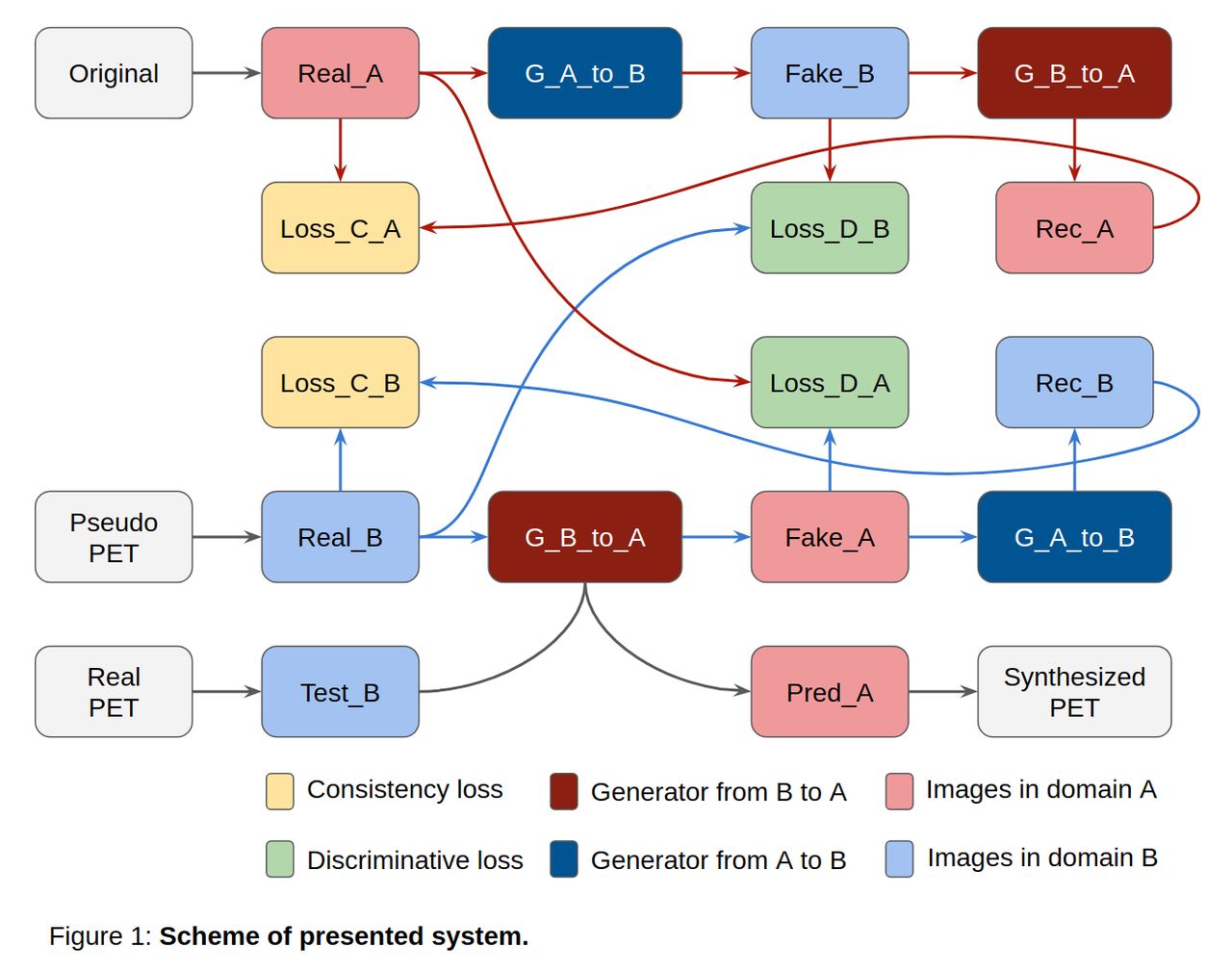
Methods: A cycle-consistent Generative Adversarial Network (cycle-GAN) is demonstrated in Figure 1.This system contains two image-to-image translators (generators): A to B and B to A, with corresponding discriminators to encourage indistinguishable outputs from real images. To regularize these generators, cycle-consistency loss is introduced for the insight that if we translate an image to another domain and back again, we should acquire the same reconstruction image as input. After training using Adam optimizer for around eight hours, we exploit the generator of translating Pseudo PET images (blurred high resolution images of similar objects) to Original (unblurred) images. Models trained for PVC on Pseudo PET images can then be applied to real PET images.Three training datasets from a single subject were collected (Figure 2), including fat-suppressed (water-only) breast MRI, T1-weighted brain MRI, whole body fat-suppressed MRI. A fourth, non-human dataset, (satellite cloud images) was also obtained. To generate Pseudo PET datasets, these images images were smoothed across a range of Gaussian FWHM kernels (3,5,7,7,9,11 mm) for a better interpretation of non-uniform blurring kernels. The contrast of the brain MRI dataset was inverted to make an image similar to a brain PET image.The Satellite-cloud-images dataset from were collected for a baseline of model isolated from anatomical images. All patient image data was obtained on a Signa PET/MR scanner (GE Healthcare, Waukesha, WI).

Results: As shown in Figure 3, the breast and brain PET images were evaluated over the four models. By observing the zoomed in (4x images), more accurate boundaries for regions with high intensity are seen. The model based on the Brain-T1(inverse) datasets shrinks the most partial volume from low metabolic regions (e.g., fat in breast, white matter in brain). Although the model trained on Satellite-cloud-images is able to deblur images, it introduces the most noise. Conclusion: PVC for PET images can be obtained based on a cycle-GAN trained on simulated PET images. This deep learning approach avoided the absence of ground truth of PET images and simulated images from the MR domain. Furthermore, the deep learning model was trained from datasets with only one subject provided, making this approach amenable to many applications in PET. Future work will explore improved simulation inputs for pseudo PET images and compare performance to existing PVC methods.

Figure
  • Download figure
  • Open in new tab
  • Download powerpoint
Figure
  • Download figure
  • Open in new tab
  • Download powerpoint
Figure
  • Download figure
  • Open in new tab
  • Download powerpoint
Previous
Back to top

In this issue

Journal of Nuclear Medicine
Vol. 61, Issue supplement 1
May 1, 2020
  • Table of Contents
  • Index by author
Article Alerts
Sign In to Email Alerts with your Email Address
Email Article

Thank you for your interest in spreading the word on Journal of Nuclear Medicine.

NOTE: We only request your email address so that the person you are recommending the page to knows that you wanted them to see it, and that it is not junk mail. We do not capture any email address.

Enter multiple addresses on separate lines or separate them with commas.
Single Subject Deep Learning-based Partial Volume Correction for PET using Simulated Data and Cycle Consistent Networks
(Your Name) has sent you a message from Journal of Nuclear Medicine
(Your Name) thought you would like to see the Journal of Nuclear Medicine web site.
Citation Tools
Single Subject Deep Learning-based Partial Volume Correction for PET using Simulated Data and Cycle Consistent Networks
Wei-jie Chen, Alan McMillan
Journal of Nuclear Medicine May 2020, 61 (supplement 1) 520;

Citation Manager Formats

  • BibTeX
  • Bookends
  • EasyBib
  • EndNote (tagged)
  • EndNote 8 (xml)
  • Medlars
  • Mendeley
  • Papers
  • RefWorks Tagged
  • Ref Manager
  • RIS
  • Zotero
Share
Single Subject Deep Learning-based Partial Volume Correction for PET using Simulated Data and Cycle Consistent Networks
Wei-jie Chen, Alan McMillan
Journal of Nuclear Medicine May 2020, 61 (supplement 1) 520;
Twitter logo Facebook logo LinkedIn logo Mendeley logo
  • Tweet Widget
  • Facebook Like
  • Google Plus One
Bookmark this article

Jump to section

  • Article
  • Figures & Data
  • Info & Metrics

Related Articles

  • No related articles found.
  • Google Scholar

Cited By...

  • No citing articles found.
  • Google Scholar

More in this TOC Section

Physics, Instrumentation & Data Sciences

  • Exploration of Multi-objective Optimization with Genetic Algorithms for PET Image Reconstruction
  • AI-based methods for nuclear-medicine imaging: Need for objective task-specific evaluation
  • Ultra-Fast Reconstruction of Short List-Mode PET Data Frames for Real-Time Visualization and Processing
Show more Physics, Instrumentation & Data Sciences

Artificial Intelligence for Image Enhancement/Analysis and Disease Assessment

  • A deep learning-based approach for lesion classification in 3D 18F-DCFPyL PSMA PET images of patients with prostate cancer
  • Machine learning based dual tracer FDG+68Ga-DOTATATE PET imaging for the diagnosis of neuroendocrine tumor
  • Quantitative Assessment of Deep Learning-enhanced Actual Ultra-low-dose Amyloid PET/MR Imaging
Show more Artificial Intelligence for Image Enhancement/Analysis and Disease Assessment

Similar Articles

SNMMI

© 2025 SNMMI

Powered by HighWire