
Main menu

  • Home
  • Content
    • Current
    • Ahead of print
    • Past Issues
    • JNM Supplement
    • SNMMI Annual Meeting Abstracts
    • Continuing Education
    • JNM Podcasts
  • Subscriptions
    • Subscribers
    • Institutional and Non-member
    • Rates
    • Journal Claims
    • Corporate & Special Sales
  • Authors
    • Submit to JNM
    • Information for Authors
    • Assignment of Copyright
    • AQARA requirements
  • Info
    • Reviewers
    • Permissions
    • Advertisers
  • About
    • About Us
    • Editorial Board
    • Contact Information
  • More
    • Alerts
    • Feedback
    • Help
    • SNMMI Journals
  • SNMMI
    • JNM
    • JNMT
    • SNMMI Journals
    • SNMMI

User menu

  • Subscribe
  • My alerts
  • Log in
  • My Cart

Search

  • Advanced search
Journal of Nuclear Medicine
  • SNMMI
    • JNM
    • JNMT
    • SNMMI Journals
    • SNMMI
  • Subscribe
  • My alerts
  • Log in
  • My Cart
Journal of Nuclear Medicine

Advanced Search

  • Home
  • Content
    • Current
    • Ahead of print
    • Past Issues
    • JNM Supplement
    • SNMMI Annual Meeting Abstracts
    • Continuing Education
    • JNM Podcasts
  • Subscriptions
    • Subscribers
    • Institutional and Non-member
    • Rates
    • Journal Claims
    • Corporate & Special Sales
  • Authors
    • Submit to JNM
    • Information for Authors
    • Assignment of Copyright
    • AQARA requirements
  • Info
    • Reviewers
    • Permissions
    • Advertisers
  • About
    • About Us
    • Editorial Board
    • Contact Information
  • More
    • Alerts
    • Feedback
    • Help
    • SNMMI Journals
  • View or Listen to JNM Podcast
  • Visit JNM on Facebook
  • Join JNM on LinkedIn
  • Follow JNM on Twitter
  • Subscribe to our RSS feeds
Meeting ReportGeneral Clinical Specialties

Evaluating the Relationship between Dynamic Na[18F]F-Uptake Parameters and MRI Knee Osteoarthritic Findings

Lauren Watkins, James MacKay, Bryan Haddock, Valentina Mazzoli, Scott Uhlrich, Garry Gold and Feliks Kogan
Journal of Nuclear Medicine May 2020, 61 (supplement 1) 182;
Lauren Watkins
1Stanford University Stanford CA United States
  • Find this author on Google Scholar
  • Find this author on PubMed
  • Search for this author on this site
James MacKay
2University of Cambridge Cambridge United Kingdom
  • Find this author on Google Scholar
  • Find this author on PubMed
  • Search for this author on this site
Bryan Haddock
3Rigshospitalet Copenhagen Denmark
  • Find this author on Google Scholar
  • Find this author on PubMed
  • Search for this author on this site
Valentina Mazzoli
1Stanford University Stanford CA United States
  • Find this author on Google Scholar
  • Find this author on PubMed
  • Search for this author on this site
Scott Uhlrich
1Stanford University Stanford CA United States
  • Find this author on Google Scholar
  • Find this author on PubMed
  • Search for this author on this site
Garry Gold
1Stanford University Stanford CA United States
  • Find this author on Google Scholar
  • Find this author on PubMed
  • Search for this author on this site
Feliks Kogan
1Stanford University Stanford CA United States
  • Find this author on Google Scholar
  • Find this author on PubMed
  • Search for this author on this site
  • Article
  • Figures & Data
  • Info & Metrics
Loading

Abstract

182

Objectives: Abnormal bone physiology is a potential mechanism for the progression of knee osteoarthritis (OA), a disease marked by degradation and loss of soft tissues like cartilage and the development of bone marrow lesions (BML) and osteophytes. Molecular information derived from kinetic modeling of Na[18F]F- ([18F]fluoride) uptake into subchondral bone enables quantitative analysis of bone metabolism that may be altered in the presence of OA features1,2,3. In this work, we evaluate the relationship between observed structural changes in bone and cartilage observed on MRI and quantitative subchondral bone metabolic parameters using hybrid PET/MRI.

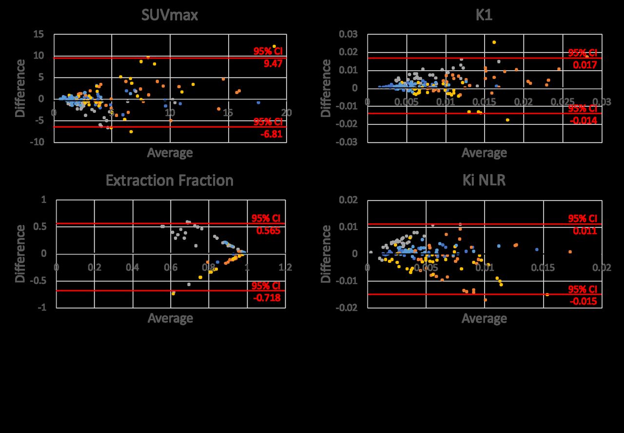
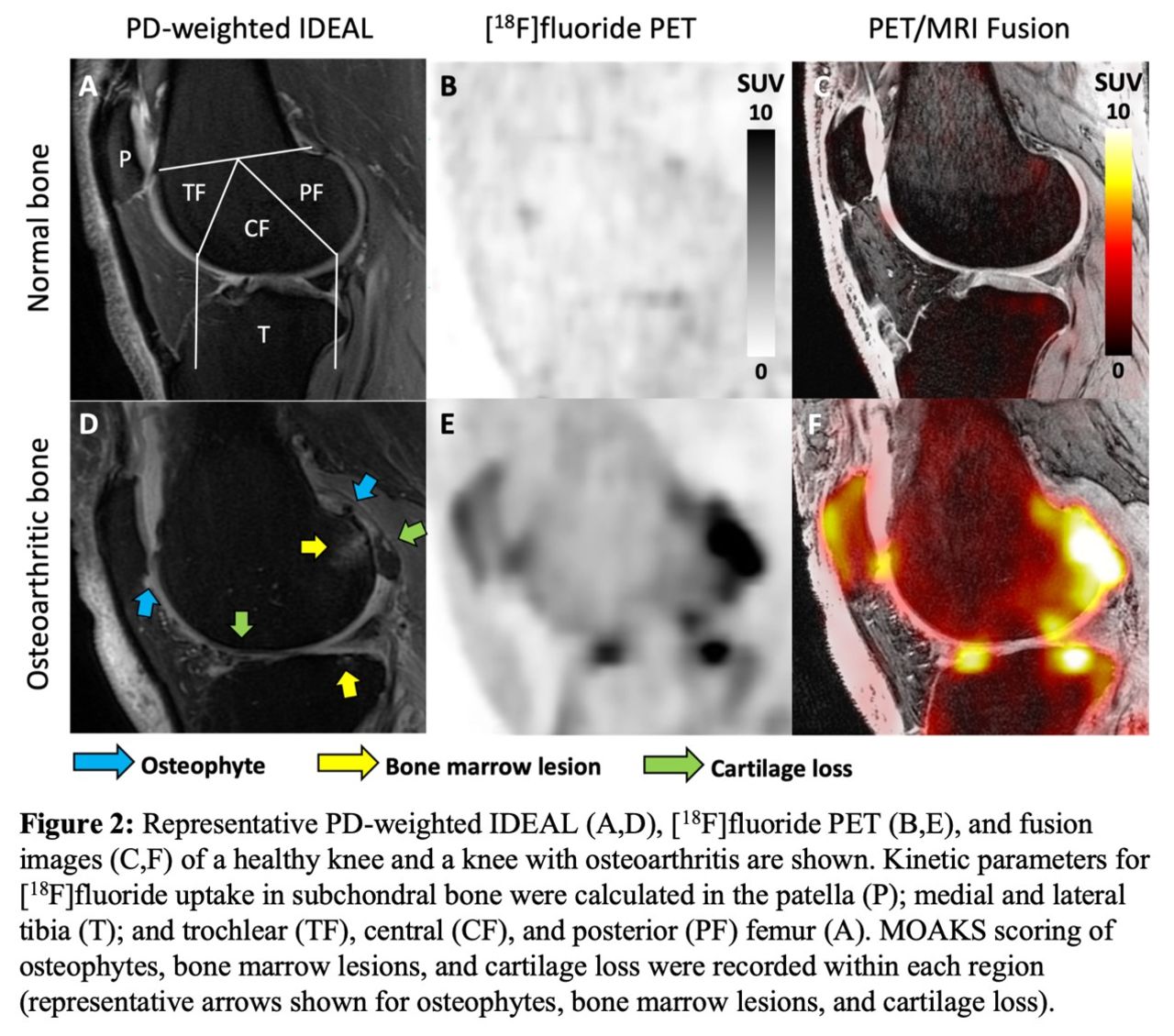
Methods: Both knees of twelve subjects with knee osteoarthritis (61.1 ± 8.8 years; BMI 27.1 ± 5.2; 10 female) were scanned using a 3T whole-body hybrid PET/MRI system under an approved IRB protocol. Upon injection of a 2.5 mCi dose of [18F]fluoride, dynamic PET and bilateral MRI data were obtained over 50 minutes. Five subjects were scanned twice, with at least 5 days between visits, to assess repeatability of the technique. MRI Osteoarthritis Knee Score (MOAKS)4 assessment of each knee was performed by a trained musculoskeletal radiologist using sagittal proton-density-weighted IDEAL and coronal T2-weighted fat-saturated images. Dynamic PET data were fit to a 2-tissue kinetic model to calculate the rates of bone perfusion (K1), tissue clearance (k2), and mineralization (k3), as well as tracer extraction fraction (k3/[k2+k3]) and total bone uptake rate (Ki = K1*extraction fraction). Kinetic fitting was performed for regions of interest representing the subchondral bone of the patella; medial and lateral tibia; and anterior, central, and posterior regions of the medial and lateral femur (Figure 2A).

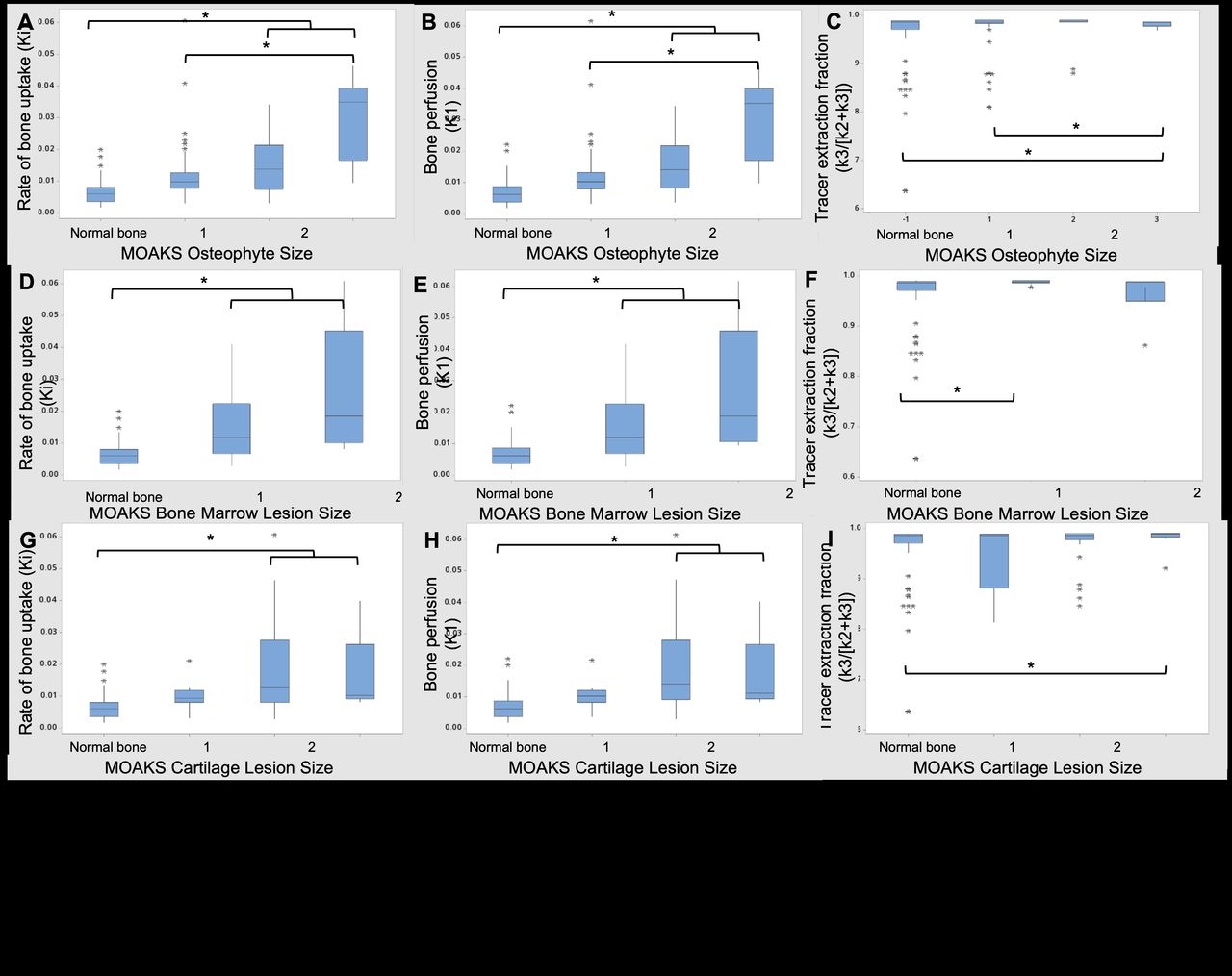
Results: Repeatability analysis showed minimal mean bias in SUVmax and kinetic parameters between visits (Figure 1). In the 24 knees studied, there were two knees without any MOAKS findings and a total of 81 bone regions with no MOAKS findings (“normal-appearing bone”). Within regions where there were one or more MOAKS findings, there were 54 regions with cartilage loss, 35 regions with BMLs, and 76 regions with osteophytes. Fifty-one of these regions had a single feature, while 26 had two features and 18 had all three features. There were no regions with size 3 bone marrow lesions. Representative PET/MRI fusion images are shown in Figure 2. Average and maximum SUV, bone perfusion, tissue clearance, extraction fraction, and total bone uptake rates of the PET tracer were significantly increased in regions with larger osteophytes [MOAKS 2/3] (p < 0.01, Figure 3 A-C); these and mineralization rates were significantly altered in BMLs (p < 0.01, Figure 3 D-F). Elevations in SUV and total bone uptake rates were driven by increased bone perfusion rates compared to other kinetic parameters. Additionally, subchondral bone uptake was significantly elevated in regions with adjacent cartilage lesions (Figure 3 G-I) compared to areas of normal-appearing bone and cartilage.

Conclusions: Kinetic parameters of the delivery and uptake of [18F]fluoride in bone regions with osteophytes, BML, and adjacent cartilage lesions were significantly different than in regions of normal bone. Kinetic parameters of [18F]fluoride uptake suggest that observed elevated tracer uptake in regions of subchondral bone with osteophytes and BMLs is driven by large increases in bone perfusion rates and smaller changes in bone extraction fraction rates. Additionally, cartilage lesion size was associated with altered metabolic parameters in adjacent subchondral bone, suggesting strong spatial relationships between subchondral bone metabolic abnormalities and changes in overlying cartilage. Kinetic parameters of [18F]fluoride uptake in subchondral bone are objective measures of bone metabolism with potential to provide functional information that complements assessments of structural abnormalities observed on MRI.

Figure
  • Download figure
  • Open in new tab
  • Download powerpoint
Figure
  • Download figure
  • Open in new tab
  • Download powerpoint
Figure
  • Download figure
  • Open in new tab
  • Download powerpoint
Previous
Back to top

In this issue

Journal of Nuclear Medicine
Vol. 61, Issue supplement 1
May 1, 2020
  • Table of Contents
  • Index by author
Article Alerts
Sign In to Email Alerts with your Email Address
Email Article

Thank you for your interest in spreading the word on Journal of Nuclear Medicine.

NOTE: We only request your email address so that the person you are recommending the page to knows that you wanted them to see it, and that it is not junk mail. We do not capture any email address.

Enter multiple addresses on separate lines or separate them with commas.
Evaluating the Relationship between Dynamic Na[18F]F-Uptake Parameters and MRI Knee Osteoarthritic Findings
(Your Name) has sent you a message from Journal of Nuclear Medicine
(Your Name) thought you would like to see the Journal of Nuclear Medicine web site.
Citation Tools
Evaluating the Relationship between Dynamic Na[18F]F-Uptake Parameters and MRI Knee Osteoarthritic Findings
Lauren Watkins, James MacKay, Bryan Haddock, Valentina Mazzoli, Scott Uhlrich, Garry Gold, Feliks Kogan
Journal of Nuclear Medicine May 2020, 61 (supplement 1) 182;

Citation Manager Formats

  • BibTeX
  • Bookends
  • EasyBib
  • EndNote (tagged)
  • EndNote 8 (xml)
  • Medlars
  • Mendeley
  • Papers
  • RefWorks Tagged
  • Ref Manager
  • RIS
  • Zotero
Share
Evaluating the Relationship between Dynamic Na[18F]F-Uptake Parameters and MRI Knee Osteoarthritic Findings
Lauren Watkins, James MacKay, Bryan Haddock, Valentina Mazzoli, Scott Uhlrich, Garry Gold, Feliks Kogan
Journal of Nuclear Medicine May 2020, 61 (supplement 1) 182;
Twitter logo Facebook logo LinkedIn logo Mendeley logo
  • Tweet Widget
  • Facebook Like
  • Google Plus One
Bookmark this article

Jump to section

  • Article
  • Figures & Data
  • Info & Metrics

Related Articles

  • No related articles found.
  • Google Scholar

Cited By...

  • No citing articles found.
  • Google Scholar

More in this TOC Section

General Clinical Specialties

  • Association between diffuse F18-FDG thyroid uptake and thyroid dysfunction
  • Bone Scintigraphy in Complex Regional Pain Syndrome Type I: A Pictorial Review
  • Genito-urinary uptake patterns and incidental lesions on a bone scan: Case based multimodality pictorial review.
Show more General Clinical Specialties

Musculoskeletal

  • Ultra-low-dose total-body 18F-FDG PET/CT in patients with autoimmune inflammatory arthritis: evaluation of image quality with shorter scan time
  • HUMAN STUDIES WITH 99mTc ZOLEDRONIC ACID (99mTc-ZA) FOR SKELETAL SCINTIGRAPHY A PRELIMINARY EXPERIENCE
  • PET/CT in localizing inflammation and microcalcification as potential causes of ongoing low back pain
Show more Musculoskeletal

Similar Articles

SNMMI

© 2025 SNMMI

Powered by HighWire