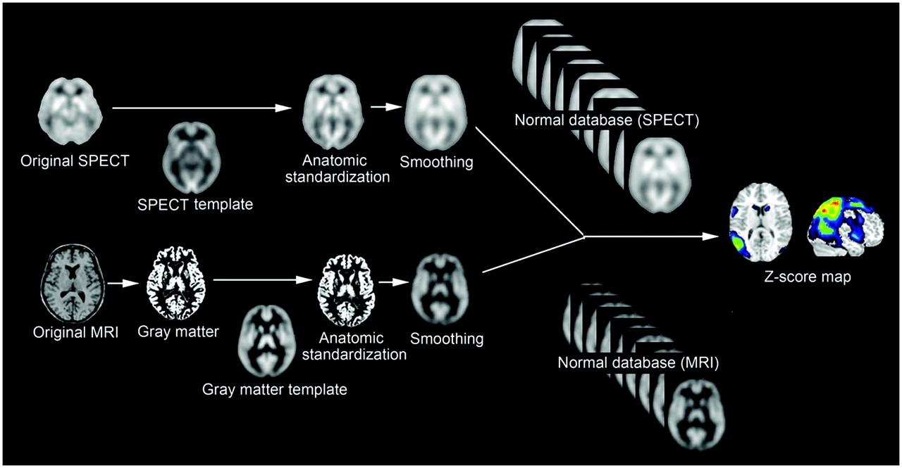
Main menu

  • Home
  • Content
    • Current
    • Ahead of print
    • Past Issues
    • JNM Supplement
    • SNMMI Annual Meeting Abstracts
    • Continuing Education
    • JNM Podcasts
  • Subscriptions
    • Subscribers
    • Institutional and Non-member
    • Rates
    • Journal Claims
    • Corporate & Special Sales
  • Authors
    • Submit to JNM
    • Information for Authors
    • Assignment of Copyright
    • AQARA requirements
  • Info
    • Reviewers
    • Permissions
    • Advertisers
  • About
    • About Us
    • Editorial Board
    • Contact Information
  • More
    • Alerts
    • Feedback
    • Help
    • SNMMI Journals
  • SNMMI
    • JNM
    • JNMT
    • SNMMI Journals
    • SNMMI

User menu

  • Subscribe
  • My alerts
  • Log in
  • My Cart

Search

  • Advanced search
Journal of Nuclear Medicine
  • SNMMI
    • JNM
    • JNMT
    • SNMMI Journals
    • SNMMI
  • Subscribe
  • My alerts
  • Log in
  • My Cart
Journal of Nuclear Medicine

Advanced Search

  • Home
  • Content
    • Current
    • Ahead of print
    • Past Issues
    • JNM Supplement
    • SNMMI Annual Meeting Abstracts
    • Continuing Education
    • JNM Podcasts
  • Subscriptions
    • Subscribers
    • Institutional and Non-member
    • Rates
    • Journal Claims
    • Corporate & Special Sales
  • Authors
    • Submit to JNM
    • Information for Authors
    • Assignment of Copyright
    • AQARA requirements
  • Info
    • Reviewers
    • Permissions
    • Advertisers
  • About
    • About Us
    • Editorial Board
    • Contact Information
  • More
    • Alerts
    • Feedback
    • Help
    • SNMMI Journals
  • View or Listen to JNM Podcast
  • Visit JNM on Facebook
  • Join JNM on LinkedIn
  • Follow JNM on Twitter
  • Subscribe to our RSS feeds
OtherCONTINUING EDUCATION

Role of Neuroimaging in Alzheimer's Disease, with Emphasis on Brain Perfusion SPECT

Hiroshi Matsuda
Journal of Nuclear Medicine August 2007, 48 (8) 1289-1300; DOI: https://doi.org/10.2967/jnumed.106.037218
Hiroshi Matsuda
  • Find this author on Google Scholar
  • Find this author on PubMed
  • Search for this author on this site
  • Article
  • Figures & Data
  • Info & Metrics
  • PDF
Loading
  1. FIGURE 1. 
    • Download figure
    • Open in new tab
    • Download powerpoint
    FIGURE 1. 

    Procedures for statistical image analysis of brain perfusion SPECT and MRI data. Anatomically standardized and then smoothed SPECT or gray matter image of subject is compared with each normal database by z score analysis. Obtained z score maps are displayed by overlay on anatomically standardized MRI template.

  2. FIGURE 2. 
    • Download figure
    • Open in new tab
    • Download powerpoint
    FIGURE 2. 

    Statistical image analysis for 78-y-old woman with amnestic MCI. (A) Brain perfusion SPECT data were analyzed with z score mapping system. Specific VOI for very early AD (bilateral posterior cingulate gyri and precunei and parietal association cortex) is demarcated by red lines on MRI template. Significantly decreased rCBF above z score of 2 was observed within this specific VOI (arrows). (B) Gray matter images extracted from MR images were analyzed with z score mapping system. Specific VOI for very early AD (bilateral medial temporal areas, including parahippocampal gyri and amygdaloid bodies) is demarcated by pink sections on MRI template. Significantly decreased gray matter concentration above z score of 2 was observed within this specific VOI (arrows).
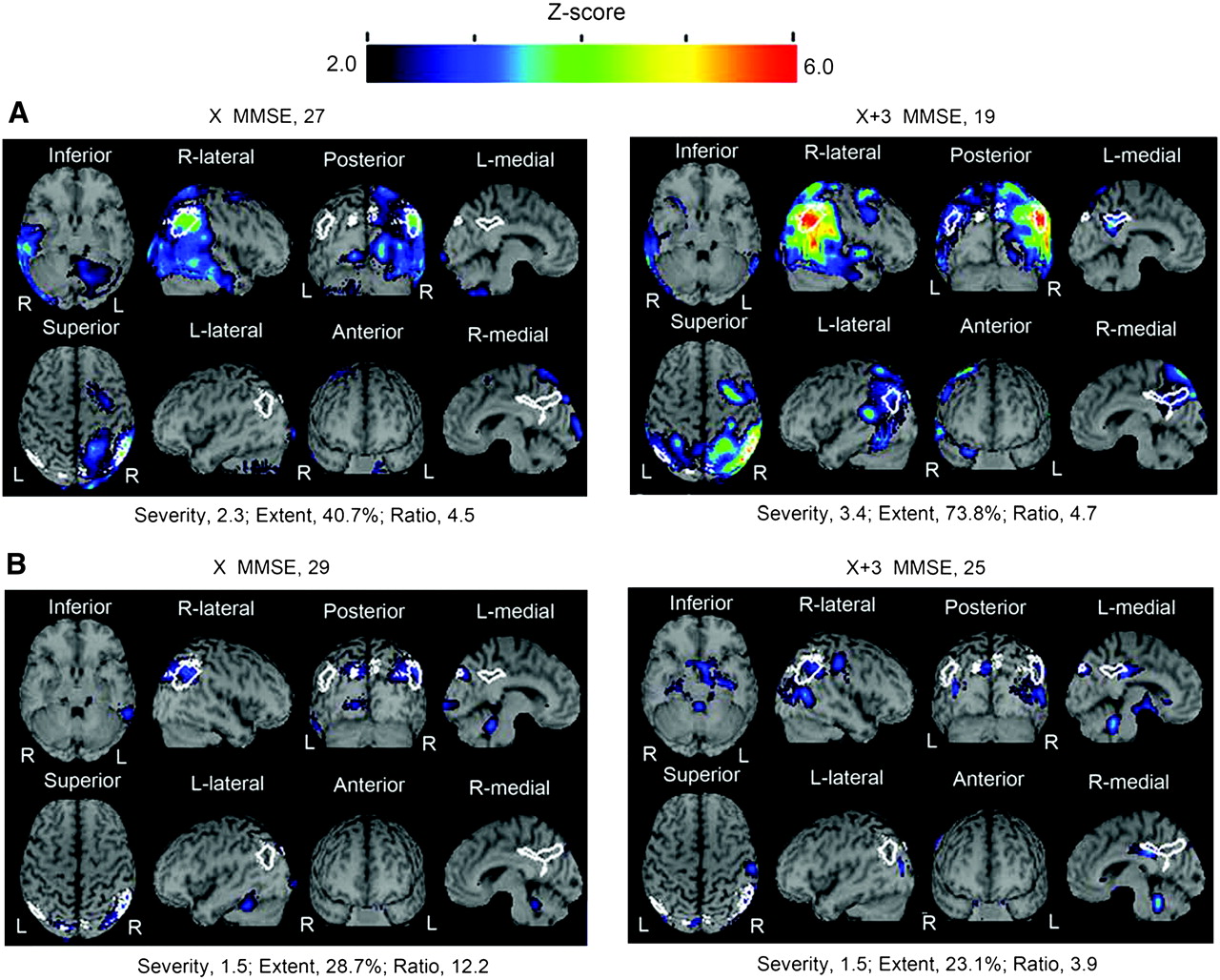
  3. FIGURE 3. 
    • Download figure
    • Open in new tab
    • Download powerpoint
    FIGURE 3. 

    Comparison of z score mapping results for brain perfusion SPECT for converters (from amnestic MCI to AD) and nonconverters over 3 y. (A) For 56-y-old woman who converted from MCI to AD, high values characterizing decreases in rCBF (severity and extent) were seen even at baseline. These values were markedly elevated 3 y later. (B) For 68-y-old man who did not convert from MCI to AD, low values for severity and extent were seen at baseline. These values were not elevated 3 y later. X MMSE = mean score on Mini-Mental State Examination (MMSE) at the initial study; X+3 MMSE = score on MMSE at 3 y.
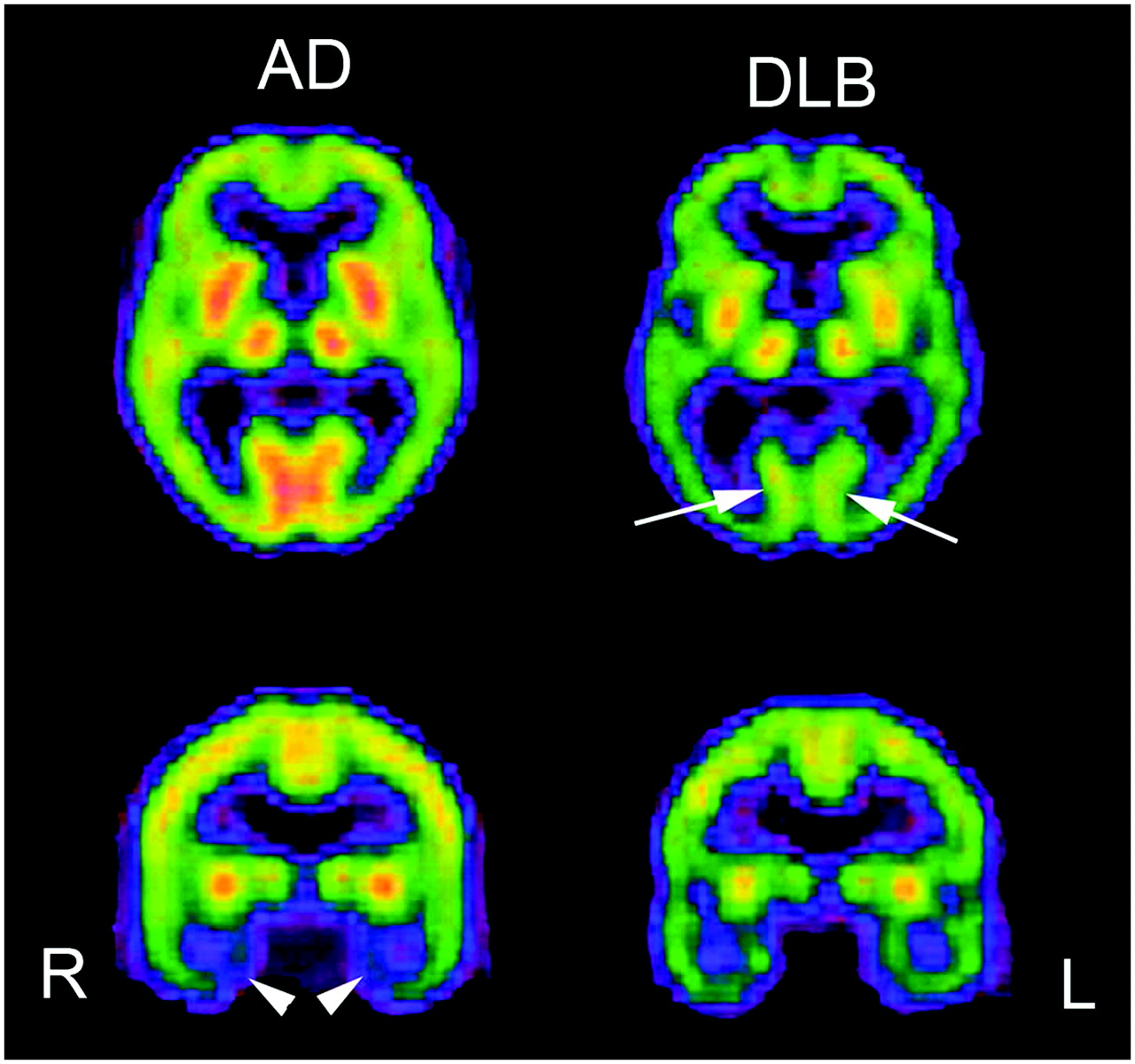
  4. FIGURE 4. 
    • Download figure
    • Open in new tab
    • Download powerpoint
    FIGURE 4. 

    Comparison of brain perfusion SPECT images for moderate AD and moderate DLB. DLB showed lower perfusion in occipital cortex than AD (arrows). In contrast, AD showed lower perfusion in medial temporal areas (arrowheads).
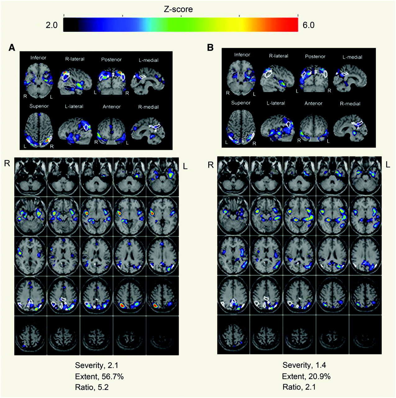
  5. FIGURE 5. 
    • Download figure
    • Open in new tab
    • Download powerpoint
    FIGURE 5. 

    Evaluation with z score mapping system of therapeutic effects of donepezil on brain perfusion SPECT for 68-y-old woman. (A) Baseline study 1 mo before donepezil showed significant decrease in rCBF within specific VOI for early AD. Mini-Mental State Examination (MMSE) score was 5 (out of 30). (B) At 3 mo after start of donepezil administration, all rCBF indices decreased from values at initial study, indicating amelioration of rCBF within specific VOI. MMSE score increased to 14 (out of 30).

PreviousNext
Back to top

In this issue

Journal of Nuclear Medicine: 48 (8)
Journal of Nuclear Medicine
Vol. 48, Issue 8
August 2007
  • Table of Contents
  • Table of Contents (PDF)
  • About the Cover
  • Index by author
Print
Download PDF
Article Alerts
Sign In to Email Alerts with your Email Address
Email Article

Thank you for your interest in spreading the word on Journal of Nuclear Medicine.

NOTE: We only request your email address so that the person you are recommending the page to knows that you wanted them to see it, and that it is not junk mail. We do not capture any email address.

Enter multiple addresses on separate lines or separate them with commas.
Role of Neuroimaging in Alzheimer's Disease, with Emphasis on Brain Perfusion SPECT
(Your Name) has sent you a message from Journal of Nuclear Medicine
(Your Name) thought you would like to see the Journal of Nuclear Medicine web site.
Citation Tools
Role of Neuroimaging in Alzheimer's Disease, with Emphasis on Brain Perfusion SPECT
Hiroshi Matsuda
Journal of Nuclear Medicine Aug 2007, 48 (8) 1289-1300; DOI: 10.2967/jnumed.106.037218

Citation Manager Formats

  • BibTeX
  • Bookends
  • EasyBib
  • EndNote (tagged)
  • EndNote 8 (xml)
  • Medlars
  • Mendeley
  • Papers
  • RefWorks Tagged
  • Ref Manager
  • RIS
  • Zotero
Share
Role of Neuroimaging in Alzheimer's Disease, with Emphasis on Brain Perfusion SPECT
Hiroshi Matsuda
Journal of Nuclear Medicine Aug 2007, 48 (8) 1289-1300; DOI: 10.2967/jnumed.106.037218
Twitter logo Facebook logo LinkedIn logo Mendeley logo
  • Tweet Widget
  • Facebook Like
  • Google Plus One
Bookmark this article

Jump to section

  • Article
    • Abstract
    • VOXEL-BASED SPECT AND PET ANALYSES
    • VOXEL-BASED MRI ANALYSIS
    • PARTIAL-VOLUME CORRECTION (PVC)
    • NORMAL AGING AND SEX DIFFERENCES
    • EARLY DIAGNOSIS OF AD BY NEUROIMAGING
    • PREDICTION OF CONVERSION FROM MCI TO AD
    • DIFFERENTIAL DIAGNOSIS OF AD AND OTHER TYPES OF DEMENTIA
    • EFFECTS OF CHOLINESTERASE INHIBITORS ON CEREBRAL BLOOD FLOW
    • CONCLUSION
    • Footnotes
    • References
  • Figures & Data
  • Info & Metrics
  • PDF

Related Articles

  • This Month in JNM
  • PubMed
  • Google Scholar

Cited By...

  • Cholinergic Modulation of Neurovascular Coupling and Neuroimaging Signals
  • Feasibility of 3-month melatonin supplementation for brain oxidative stress and sleep in mild cognitive impairment: protocol for a randomised, placebo-controlled study
  • Comparison of Early-Phase 11C-Deuterium-L-Deprenyl and 11C-Pittsburgh Compound B PET for Assessing Brain Perfusion in Alzheimer Disease
  • Imaging Approaches for Dementia
  • Brain perfusion patterns in familial frontotemporal lobar degeneration
  • Simultaneous Measurement of Arterial Transit Time, Arterial Blood Volume, and Cerebral Blood Flow Using Arterial Spin-Labeling in Patients with Alzheimer Disease
  • Comparison of 18F-FDG PET and Optimized Voxel-Based Morphometry for Detection of Alzheimer's Disease: Aging Effect on Diagnostic Performance
  • Google Scholar

More in this TOC Section

  • Approaches to Imaging Immune Activation Using PET
  • Large Language Models and Large Multimodal Models in Medical Imaging: A Primer for Physicians
  • Precision Oncology in Melanoma: Changing Practices
Show more Continuing Education

Similar Articles

SNMMI

© 2025 SNMMI

Powered by HighWire