


Abstract
In tinnitus, PET and other functional imaging modalities have shown functional changes not only in the auditory cortex but also in nonauditory regions such as the limbic, frontal, and parietal areas. Nonetheless, disparities in task dimension among studies, low statistical power due to small sample size, and the intrinsic uncertainty of a modality that measures activity indirectly limit the comprehensive understanding of the results from PET studies. These difficulties prompted us to undertake a metaanalysis of PET studies on tinnitus using a coordinate-based technique (activation-likelihood estimation) to retrieve the most consistent activation areas across different task dimensions and to compare the results with those from other imaging modalities. Methods: We performed 2 activation-likelihood estimation metaanalyses on data from 10 studies with 56 foci in which we examined the contrast between tinnitus individuals and controls and the difference in activation between sound stimuli and resting state in tinnitus individuals. Results: The studies show that the most consistently activated regions in tinnitus subjects, compared with controls, were the left primary and bilateral secondary auditory cortices, left middle and bilateral inferior temporal gyri, left parahippocampal area, left geniculum body, left precuneus, right anterior cingulate cortex, right claustrum, right middle and inferior frontal gyri, and right angular gyrus. The relatively activated area under sound stimuli, compared with resting state, in tinnitus subjects was the secondary auditory cortex. Our study reconfirms the findings of previous quantitative electroencephalography or magnetoencephalography studies because most of the 14 brain areas with significant activation found in our metaanalysis replicate these earlier data. Our results suggest that the areas described in the tinnitus network are solidly replicable regardless of the applied functional imaging technique. Conclusion: This study proves that PET is a useful modality for tinnitus research and solidifies human tinnitus research itself by confirming previously described brain areas involved in the generation and maintenance of tinnitus.
Tinnitus is an auditory phantom percept in the absence of any objective physical sound source. In most cases, this phantom perception resolves spontaneously within seconds or minutes. However, tinnitus persists in 5%–10% of the population in Western countries and interferes severely with the quality of life in 5%–26% of the tinnitus population (1). Moreover, the prevalence of chronic, subjective, and nonpulsatile tinnitus increases with age probably because of increased hearing loss associated with age, as tinnitus is often associated with sensorineural hearing loss (2).
Initially, it was proposed that tinnitus-related neural activity must originate from the cochlea. However, this belief has been challenged, and central mechanisms have been implicated in relation to, for example, the failure of tinnitus relief or the development of tinnitus after eighth cranial nerve sectioning (3). Recently, researchers have indeed found that experimentally induced hearing loss in animals increased spontaneous firing rates at several levels in the auditory brain stem and cortex (4). Moreover, auditory cortical map reorganization has been associated with increased auditory cortical activity demonstrating central changes in tinnitus (5).
The advent of brain-imaging tools provided neuroscientists with a window into the brain activity that orchestrates tinnitus. Specifically, magnetoencephalography detecting magnetic fields induced by synchronized neuronal currents, quantitative electroencephalography (qEEG) measuring electrical signals at the surface of the head that reflect activation of remote populations of neurons, functional MRI (fMRI) detecting blood oxygen level–dependent signals, and PET measuring regional cerebral blood flow (rCBF) or neurochemical changes have enabled researchers to study changes in neural activity in experimental placebo-controlled paradigms. It was demonstrated using these modalities that tinnitus is related to reorganization and hyperactivity of the auditory central nervous system with coactivation of nonauditory areas such as the insula and anterior and posterior cingulate cortices (ACC and PCC, respectively) (6). This finding has led to the concept that the unified tinnitus percept is the result of multiple dynamically adaptive overlapping networks, each representing another aspect of the tinnitus. A recent network theory proposed that sensory deafferentation causes neuroplastic changes resulting in increased activation of the auditory cortex, but awareness of tinnitus arises when this activity is connected to a larger coactivated perceptual network involving the ACC, PCC, precuneus, and parietal and frontal cortices and a salience network involving dorsal ACC and anterior insula (6). The theory also proposed that as a consequence of a constant learning process, the phantom percept becomes associated with distress by a distress network consisting of the ACC, anterior insula, and amygdala and becomes persistent by involvement of memory areas such as the parahippocampus, amygdala, and hippocampus (6).
Specific PET and fMRI studies in tinnitus, however, vary in task dimension (i.e., sound, somatosensory modulation, gaze change, lidocaine injection, and steady state) and are limited by statistical power and sensitivity. A metaanalysis permits the retrieval of the most consistent activation areas and comparison of results across different tasks. Metaanalyses are therefore essential for reviewing findings from different studies, comparing results in a standardized fashion, and summarizing statistical relations between study characteristics and findings. Hence, a metaanalysis of tinnitus studies using PET or fMRI (but actually no previous fMRI study met the inclusion criteria) was performed using a coordinate-based technique (activation-likelihood estimation [ALE]). Hereinafter, we report metaanalysis results of PET studies on tinnitus to elucidate significant neural correlates of tinnitus. Also, activated areas are compared with those from studies using qEEG or magnetoencephalography that record spontaneous brain activity, in contrast to the indirect brain activity measure used by PET.
MATERIALS AND METHODS
Search Criteria
According to the guidelines of PRISMA (Preferred Reporting Items for Systematic reviews and Meta-Analyses) (7), we conducted Pubmed (http://www.ncbi.nlm.nih.gov/pubmed/) searches to identify all PET and fMRI studies on tinnitus. Keywords used in these searches were “PET” and “tinnitus”, and “fMRI” and “tinnitus”, with activated limits (article types other than review, human species, and English language).
To be included in the current analysis, the studies needed to be published in a peer-reviewed journal and based on a data-driven whole-brain approach. That is, studies based on selected regions of interest (ROIs) were excluded. In addition, the results had to be reported in standard stereotactic spaces such as Montreal Neurologic Institute (MNI) or Talairach and Tournoux and driven by categoric contrasts rather than correlation analyses. Furthermore, studies with a t value of 3 or greater or a z score of 2.33 or greater were included to ensure comparable specificity.
Metaanalysis Algorithm
The software GingerALE (http://brainmap.org/ale/index.html) desktop application (version 2.1.1) was used for the transformation of all reported coordinates into stereotactic standard Talairach and Tournoux space (8). Coordinates reported in MNI space were converted to Talairach coordinates. The method we used for our metaanalysis is a variation of ALE published originally by Turkeltaub et al. (9) and improved by Eickhoff et al. (10). The improvements include the adaptive estimation of the width of the gaussian kernel for each included experiment and the possibility of performing a random-effects analysis.
For each experiment, every reported maximum was modeled by a 3-dimensional gaussian probability distribution centered at the given coordinate. The width of the gaussian probability distribution was determined individually for each experiment on the basis of empiric estimates of between-subject variability, taking into account the number of subjects in each experiment (10). For each voxel, ALE was calculated from the union of the gaussian probability distributions associated with the different foci. In a random-effects analysis, ALE values were combined across experiments and tested against a null hypothesis of random distribution of ALE values (10), thereby identifying those regions in which empiric ALE values were higher than could be expected by chance. A threshold of P < 0.05 was applied to the resulting ALE map (corrected for multiple comparisons by false-discovery rate). Statistically significant voxels represent the convergence of the investigated effect across the several studies. ALE results were overlaid onto an optimized individual anatomic T1 template (http://www.brainmap.org/ale/Colin1.1.nii), and cluster centers were anatomically located by using MRIcron software (http://www.sph.sc.edu/comd/rorden/mricron/). All peak coordinates and their designated locations both as brain regions and as Brodmann areas (BAs) were reconfirmed using Talairach and Tournoux’s atlas (8).
RESULTS
PET and fMRI Studies
Of the 29 retrieved studies, 19 coordinate-based PET studies on tinnitus were initially identified. After the results were limited by the criteria described, 10 studies were considered eligible. Initially, we attempted to perform a separate metaanalysis on fMRIs, but all 6 fMRI studies were ROI-based and thus excluded. In total, 157 tinnitus subjects and 23 controls were included (Table 1). The literature search, selection, and compilation of coordinates for the contrast were performed independently by 2 investigators. For PET studies, a total of 56 peak coordinates were reported for changes in brain metabolism (Table 1).
Studies Included in ALE Metaanalyses
Significant ALE Clusters for Tinnitus Subjects
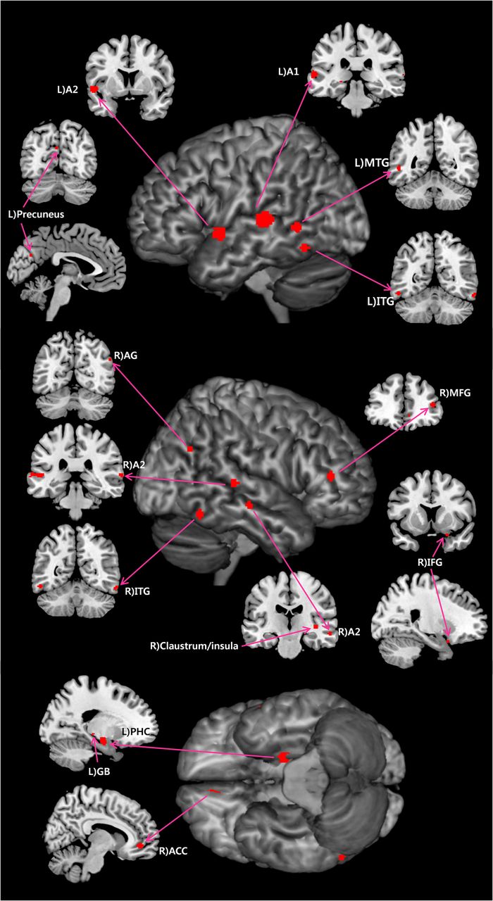
The ALE metaanalysis revealed 14 significant clusters. As illustrated in Figure 1 and Table 2, we found a significant increase in rCBF in tinnitus subjects in the left primary and bilateral secondary auditory cortices (A1 and A2, respectively; BAs 42/21/22), the left middle and bilateral inferior temporal gyri (MTG and ITG, respectively; BAs 37/22/20), the left parahippocampal area (PHC; BAs 27/34), the left geniculum body (GB), the left precuneus (BAs 31/7), the right ACC (BA 32), the right claustrum, the right middle and inferior frontal gyri (MFG and IFG, respectively; BAs 10/47), and the right angular gyrus (AG; BA 39).

Patterns of regional increases in metabolism in tinnitus subjects. A1 = primary auditory cortex; A2 = secondary auditory cortex; ACC = anterior cingulate cortex; AG = angular gyrus; GB = geniculate body; IFG = inferior frontal gyrus; ITG = inferior temporal gyrus; L) = left; MFG = middle frontal gyrus; MTG = middle temporal gyrus; PHC = parahippocampal area; R) = right.
Overview of ALE Metaanalysis Results in Tinnitus Patients
A second analysis by reversing the contrast showed no significant effect for decreased rCBF in tinnitus subjects.
Significant ALE Clusters for Contrast Between Sound Stimuli and Resting State in Subjects
A second ALE metaanalysis looking at the specific contrasts (sound stimuli minus resting state) demonstrated 2 clusters. As shown in Table 3 and Figure 2, we found a significant increase in rCBF during sound stimuli in the left A2 (BA 22) in comparison to resting state. The contrast “resting state minus sound stimuli” revealed no significant activations.
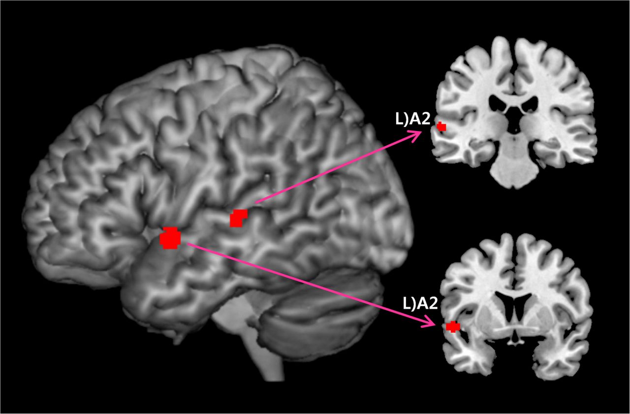
Patterns of regional increases in metabolism during sound stimuli vs. resting state in tinnitus subjects. Two areas in left secondary auditory cortex were significantly activated.
Overview of ALE Metaanalysis Results for Contrast (Sound Stimuli Minus Resting State) in Tinnitus Patients
DISCUSSION
The brain regions that have consistently increased rCBF in tinnitus participants in comparison to controls, irrespective of the applied stimulus, were determined using an ALE metaanalysis. Almost all brain regions found in this study have already been retrieved in previous qEEG and magnetoencephalography studies, reinforcing the utility of PET in tinnitus research. Of 14 significant clusters found in the current study, 12 areas (except the ITG and GB) have been mentioned in previous qEEG studies (11,12). Moreover, although described in more general terms such as ACC or frontal lobe or temporal cortices probably due to poor spatial resolution, many of the similar areas have been described in previous magnetoencephalography studies (13,14). Hereinafter, we discuss the brain regions found in this metaanalytic study and compare these with previous studies using other imaging methods.
Increased rCBF in the left A1 in the absence of an external stimulus has been elucidated in a metaanalysis of schizophrenia patients with auditory hallucinations (15). In the same context, researchers have long been paying attention to the changes in the A1 in individuals with a simpler form of auditory phantom perception, namely tinnitus. Our results demonstrating increased rCBF of the A1 are in line with previous qEEG or magnetoencephalography studies. A previous magnetoencephalography study has demonstrated that cortical map plasticity in the A1 is associated with tinnitus, and the degree of the reorganization is positively correlated with tinnitus loudness (14). Moreover, a qEEG study on unilateral tinnitus subjects has shown a strong positive correlation between resting state γ-band activity in the contralateral A1 and subjective tinnitus intensity (16). Increased rCBF of the A2 (BAs 21/22) in tinnitus subjects in the current study also replicated previous fMRI or qEEG study results. In an fMRI study, significant signal change has been found bilaterally in the A2 (17). Also in a recent qEEG study, unilateral tinnitus individuals have demonstrated an increased γ-band activity in the bilateral A2 (18). One discrepancy that should be addressed is that in our study only the left A1 (BA 42) was more active than normal, but magnetoencephalography and fMRI studies have suggested that the A1 contralateral to the tinnitus side or even bilateral A1 might be the location of neural generator (19). From a clinical point of view, further research to reach a definite conclusion is needed because this discrepancy among studies also affects the side for repeated transcranial magnetic stimulation. Indeed, one group preferred to stimulate the left auditory cortex irrespective of the tinnitus side while others preferred to stimulate contralaterally to the tinnitus side (19).
The PHC (BAs 27/34) has been hypothesized to play a central role in memory recollection and relaying information (6). In a human electrophysiologic study, cells in the PHC responded to a novel stimulus with an increase in firing, but the response to a repetition of the stimulus decreased dramatically, suggesting a rapid habituation (20). A dysfunction in this habituation mechanism has been posited as an explanation for auditory phantom percepts such as auditory hallucinations (21). Likewise, tinnitus has been suggested as a result of the constant sending of stored auditory information from the hippocampus to the auditory association areas by persistent parahippocampal activity (6). The increased rCBF in the left PHC is also in line with previous qEEG studies. In tinnitus subjects with higher distress, increased α2 (10.5–12.5 Hz)-band activity has been elucidated in a qEEG study (11). In this regard, we may interpret our results as reconfirmation of the role of the PHC in tinnitus, presumably for constant perception and distress.
Although the ALE metaanalysis designated that the peak coordinate (−3, −69, 27) is in BA 31, the coordinate is located in BA 7 in the Talairach’s atlas and thus marked as BAs 31/7 (Table 2). The precuneus is a highly integrative structure supposed to be involved in visuospatial imagery, episodic memory, self-consciousness, and attention shifting (22). A qEEG study has shown decreased precuneal α1 (8–10 Hz)-band activity in tinnitus subjects with higher distress and decreased α1- and α2-band activity in noncoping tinnitus subjects (11). Together with our results, the precuneal hyperactivity may be explained as a failure of inhibiting conscious awareness of the tinnitus. The discrepancy between decreased α-band activity in the study by Vanneste et al. (11) and increased rCBF in the current study might be related to the fact that the α2 decreases in relation to increasing distress and that there is no clear-cut correlation between α-activity and rCBF on PET at a cortical level.
The MTG (BAs 37/22) has been suggested to be involved in cognitive processes including language, semantic memory, and multimodal sensory integration (23). A qEEG study has demonstrated higher α2-band activity in the MTG in tinnitus subjects with higher distress (11). Although the role of the MTG is not supported by sufficient data, the results of the qEEG study and our study may advocate a yet-undetermined but still important role of the left MTG. The ITG (BA 20) is known to be involved in visual perception, memory consolidation, and multimodal sensory integration (24). Although there are few qEEG or magnetoencephalography studies mentioning the ITG, improvement of tinnitus after a single session of transcranial magnetic stimulation of the ITG designates a role of this area in the pathophysiology of tinnitus (25). In this regard, the bilateral rCBF increase of the ITG in our study may be attributed to a mechanism of maladaptive memory consolidation.
The peak at Talairach (−22.88, −25.03, −4.08) corresponds to the left GB rather than the medial geniculate body (MGB). Only 1 of 10 included studies indicated that the thalamic activity correlated with tinnitus (the study by Lockwood et al. (40); Table 1), and in particular, 2 of 3 thalamic coordinates presented in that study were posterior thalamus between MGB. Considering that the LGB, the primary relay center for visual information, has never correlated with tinnitus in the literature, we speculate that our result may indicate the MGB rather than the LGB because of the possible spatial resolution problems with PET. Previous research on the MGB in murine tinnitus models has already elucidated changes in the spontaneous firing rates at a single-cell level, cell density reductions in all subdivisions by immunohistochemical staining, and significant damage and compensatory plastic changes using diffusion tensor imaging (26). Because of the anatomic subcortical location, the MGB has never been mentioned in tinnitus studies using qEEG or magnetoencephalography. However, a previous ROI-based fMRI study has indicated significant blood oxygen level–dependent signal changes in bilateral MGB (17). The increased rCBF in the GB in the current study may be in line with these previous reports, but still further studies using PET with improved spatial resolution are needed to locate more accurate thalamic regions.
The peak ALE value at Talairach (8.09, 44.17, −5) (Table 2) in BA 32 corresponds to the right pregenual ACC (pgACC). The pgACC has been implicated as the affective subdivision of the ACC. This area is an important component of a network for mood regulation (27). The average increase of rCBF in the pgACC corresponds to previous qEEG studies. In a qEEG study, modulation of the pgACC has been proved to suppress tinnitus intensity and tinnitus-related distress (28). Therefore, increased rCBF of the right pgACC may be ascribed to its presumptive role in tinnitus perception and tinnitus-related distress.
The claustrum, a structure mainly located in the insular cortex, has been suggested to orchestrate mixed information from various cortical regions, and the claustrum–insula region has been reported to integrate conceptually related sounds and pictures (29). Few articles regarding tinnitus have mentioned the claustrum alone, but the insula has been suggested to be an important area responsible both for a salience network and for a distress network for tinnitus (6). Our finding of the right claustal rCBF increase is in line with previous results using qEEG. Increased insular α2-band activity in noncoping patients has been reported in a qEEG study (11). Another qEEG study has revealed that tinnitus questionnaire scores are correlated with the neural activity in the bilateral anterior insula (30). Taken together, the claustrum–insula region was reconfirmed as a crucial area for tinnitus by the current study.
The MFG (BA 10), also known as the frontopolar cortex, forms the apex of the executive system underlying decision making (31). The activation of the MFG by acoustic stimulation, measured by an ROI study using fMRI, was more prominent in tinnitus subjects than in controls (32). A previous study using qEEG has shown that increased δ-activity could be revealed in the right lateral frontopolar cortex for individuals with pure-tone tinnitus in comparison to those with narrow-band-noise tinnitus, and this difference was attributed to pitch-specific memory retrieval in cases of pure-tone tinnitus (12). When the findings of qEEG studies are considered in combination with the results of other previous functional imaging studies, an rCBF increase in the right MFG may be considered reflective of the pitch of the tinnitus.
The IFG (BA 47), or the ventrolateral prefrontal cortex (VLPFC), implements cognitive reappraisal, strategic operations at encoding, and retrieval (33). Previous studies have revealed that the VLPFC plays a functional role in nonspatial auditory cognition and congruity (34). A previous qEEG study has also mentioned that bilateral tinnitus subjects showed decreased δ-activity in the VLPFC in comparison to unilateral subjects and increased bilateral β1 in comparison to healthy controls (35). Tinnitus has been suggested to be related to temporal incongruity (6). In case of deafferentation, the loss of frequency-specific auditory input in comparison to what used to arrive as auditory input in the same context can result in a temporal incongruity, which might be reflected by the VLPFC activation. Thus, the rCBF increase in the VLPFC in our study, together with previous qEEG study results, may indicate a role in cognitive reappraisal of tinnitus or may be the result of temporal incongruity.
The AG (BA 39) has been reported to be associated with recollection-related activity, auditory stimulus processing, sound location monitoring, and multisensory integration (36). In this regard, some investigators have proposed that the AG is a part of the tinnitus perception network (6). A previous qEEG study has demonstrated increased β3- and γ-band activity in the AG for subjects with unilateral tinnitus in comparison to bilateral tinnitus. In this study, increased synchronized β3-activity in the AG was shown for both left- and right-sided tinnitus in comparison to bilateral tinnitus (35). In accordance with previous qEEG studies, increased rCBF in the right AG may be ascribed to the role of the AG as a spatial localizer of tinnitus or as a multisensory integrator.
The brain regions of increased rCBF found in the current study could be fitted to the aforementioned network theory (6). The A1 and A2 may be attributable to sensory deafferentation; the ACC, precuneus, and frontal cortices to the perceptual network; the ACC and insula to the salience network; the ACC and anterior insula to the distress network; and the PHC to memory consolidation. In this way, the network theory can also be replicated with the regions found in the current study. However, because no functional connectivity analysis has been performed it cannot be stated how these areas interact at a network level.
Of various functional imaging techniques used for tinnitus research, qEEG and magnetoencephalography are partly advantageous over PET or fMRI in that the former two have much higher temporal resolution than latter two. In addition, inasmuch as PET and fMRI measure brain activity indirectly, qEEG and magnetoencephalography are advantageous in that these modalities measure direct neural synchrony. Moreover, especially for fMRI, inherent scanner noise (≤130 dB) (37) is problematic when performing tinnitus research, whereas qEEG and magnetoencephalography are nearly noise-free. In addition, fMRI studies are mainly ROI-based and can thus be biased by a priori hypotheses. This bias might be related to the scanner noise making it more difficult to obtain statistical results when performing a whole-brain analysis because of a decreased signal-to-noise ratio. Furthermore, fMRI, with the exception of resting-state fMRI, is always task-related and does not measure spontaneous activity, whereas PET, qEEG, and magnetoencephalography can easily measure resting-state activity. This ability is particularly important in tinnitus research, because tinnitus is specifically acknowledged by resting-state hyperactivity.
In contrast, PET and fMRI are advantageous over qEEG or magnetoencephalography with regard to spatial resolution. Although PET does not have as much spatial resolution as fMRI, the spatial resolution of both (5–10 mm) is good enough to differentiate A1 from A2. Furthermore, in contrast to magnetoencephalography and electroencephalography, PET, like fMRI, is not restricted to the study of cortical structures (38). Additionally, PET retains considerable advantages over fMRI for tinnitus research. First, PET is more appropriate for assessing the IFG (because of susceptibility artifact in fMRI) (37). Furthermore, PET is better suited for tinnitus subjects with cochlear or other implants and for claustrophobic subjects. Finally, PET is much quieter than fMRI; thus, PET is better tolerated by patients with hyperacusis and it is easier to mask the scanner noise by applying sound-attenuating insert earphones or headphones. In these regards, PET may be useful for tinnitus research despite the drawbacks.
Considering that tinnitus percept has been proposed to be a result of overlapping networks, functional connectivity studies aiming to determine multiple brain areas with similar temporal activity profiles are necessary. Although not as suitable as qEEG or magnetoencephalography because of limitations in temporal resolution, functional connectivity studies using PET data have been performed in other fields of cognitive neuroscience (39). Therefore, functional connectivity analysis should be performed in PET studies on tinnitus.
To the best of our knowledge, this is the first study that used a coordinate-based metaanalytic approach to systematically determine consistency across PET studies on tinnitus. In fact, there are considerable disparities among PET studies with regard to the character of the study subjects, stimulation paradigm, imaging modality, and analysis methods. These disparities are major obstacles to finding unequivocal neural correlates that are related to the generation and maintenance of tinnitus. However, from the current study, it is clear that most of the important brain regions for tinnitus generation and maintenance were also identifiable by PET studies.
The ALE metaanalysis has a greater level of spatial accuracy than the previously used more global characterization methods. In particular, compared with previous methods, ALE shows greater involvement of the ACC, one of the most crucial parts of the brain in subjects with tinnitus. The principal strength of this quantitative metaanalysis of imaging studies is that it is based on multiple peer-reviewed studies—in our case with a total of 165 tinnitus participants and 23 controls. Thus, the results from the present tinnitus-related brain activation maps are more robust than those of any individual imaging study on tinnitus.
A limitation of the ALE metaanalysis is that it includes only reported local maxima and does not take into account the level of statistical significance and the cluster size. However, it is unlikely that the variation in statistical thresholds has otherwise significantly biased the obtained results because false-positives from a single study are averaged out when multiple studies are combined. Moreover, although we included all the studies available on PubMed, the disparities among individual PET studies, such as missing control groups in some studies, restricted diverse comparisons of the studies with multiple contrasts. To obtain more reliable metaanalytic results, further PET studies with sufficient controls and tinnitus subjects should be performed.
CONCLUSION
Our metaanalysis on PET studies confirms the findings of previous qEEG and magnetoencephalography studies in tinnitus because most of the 14 brain areas with a significant rCBF increase are localized in areas described in previous reports using qEEG or magnetoencephalography. This confirmation suggests that the areas described in the tinnitus network are solidly replicable regardless of the applied functional imaging technique. This study therefore solidifies human tinnitus research by confirming previously described brain areas involved in the generation and maintenance of tinnitus.
DISCLOSURE STATEMENT
The costs of publication of this article were defrayed in part by the payment of page charges. Therefore, and solely to indicate this fact, this article is hereby marked “advertisement” in accordance with 18 USC section 1734.
Acknowledgments
This work was supported by the Research Foundation Flanders (FWO); the Tinnitus Research Initiative; TOP project, University Antwerp; and the Korean Science and Engineering Foundation (KOSEF) grant funded by the Korean government (MOST) (no. 2012-0030102). No other potential conflict of interest relevant to this article was reported.
Footnotes
Published online Aug. 23, 2012.
- © 2012 by the Society of Nuclear Medicine and Molecular Imaging, Inc.
REFERENCES
- Received for publication January 10, 2012.
- Accepted for publication April 30, 2012.


