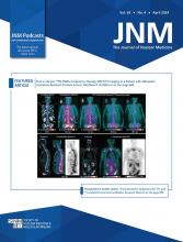
Visual Abstract
Abstract
Prostate Imaging Reporting and Data System (PI-RADS) category 3 lesions remain a diagnostic challenge for detecting clinically significant prostate cancer (csPCa). This article evaluates the added value of 68Ga-labeled prostate-specific membrane antigen-11 (68Ga-PSMA) PET/MRI in classifying PI-RADS 3 lesions to avoid unnecessary biopsies. Methods: Sixty biopsy-naïve men with PI-RADS 3 lesions on multiparametric MRI were prospectively enrolled between February 2020 and October 2022. In all, 56 participants underwent 68Ga-PSMA PET/MRI and prostate systematic biopsy. 68Ga-PSMA PET/MRI was independently evaluated and reported by the 5-level PRIMARY score developed within the PRIMARY trial. Receiver-operating-characteristic curve analysis was used to estimate the diagnostic performance. Results: csPCa was detected in 8 of 56 patients (14.3%). The proportion of patients with csPCa and a PRIMARY score of 1, 2, 3, 4, and 5 was 0% (0/12), 0% (0/13), 6.3% (1/16), 38.5% (5/13), and 100% (2/2), respectively. The estimated area under the curve of the PRIMARY score was 0.91 (95% CI, 0.817–0.999). For a PRIMARY score of 4–5 versus a PRIMARY score of 1–3, the sensitivity, specificity, positive predictive value, and negative predictive value were 87.5%, 83.3%, 46.7%, and 97.5%, respectively. With a PRIMARY score of at least 4 to make a biopsy decision in men with PI-RADS 3 lesions, 40 of 48 patients (83.3%) could avoid unnecessary biopsies, at the expense of missing 1 of 8 (12.5%) csPCa cases. Conclusion: 68Ga-PSMA PET/MRI has great potential to classify patients with PI-RADS 3 lesions and help avoid unnecessary biopsies.
Prostate multiparametric MRI (mpMRI) is currently recommended before prostate biopsy because of its promising value in the detection and localization of clinically significant prostate cancer (csPCa) (1,2). Over the last decade, mpMRI has been increasingly used as a biopsy-triage tool and has been used in guiding targeted biopsy (2–4). The Prostate Imaging Reporting and Data System (PI-RADS) is a 5-point scale based on imaging features and is designed to standardize radiology reporting (5). PI-RADS category 3, which presents an equivocal suggestion of csPCa, remains a significant diagnostic challenge. Although biopsy is recommended under the current guidelines, less than 20% of PI-RADS 3 lesions contain csPCa (6,7). PI-RADS 3 lesions present a dilemma to both urologists and patients because immediate biopsy could be unnecessary; however, a monitoring strategy could lead to some missed diagnoses of csPCa. Hence, specifically identifying csPCa among PI-RADS 3 patients will have significant implications for clinical diagnosis.
68Ga-labeled prostate-specific membrane antigen (68Ga-PSMA) PET/CT has exhibited increasing value in assessing the presence of primary intraprostatic tumors (8,9). Studies indicate that 68Ga-PSMA PET/CT has some superiority over mpMRI in detecting primary tumors (10–12). Our previous study found that the combination of mpMRI and 68Ga-PSMA PET/CT improved the diagnostic accuracy of csPCa compared with mpMRI alone, with significant improvement in accurate diagnosis in PI-RADS 3 lesions (13). Recently, 68Ga-PSMA PET/CT showed some value in stratifying PI-RADS 3 lesions in a retrospective study (14). However, to the best of our knowledge, the value of 68Ga-PSMA PET/MRI, which provides anatomic localization and radiologic information simultaneously, in classifying PI-RADS 3 lesions has never been investigated.
To assess whether 68Ga-PSMA PET/MRI can classify patients with PI-RADS 3 lesions on mpMRI and avoid unnecessary biopsies, this study prospectively included men with PI-RADS 3 lesions and assigned the patients for 68Ga-PSMA PET/MRI scanning before prostate biopsy was performed. The 5-level PRIMARY system developed within the PRIMARY trial was applied to report 68Ga-PSMA PET/MRI findings. Using the pathologic results of the biopsies as the reference, we analyzed the diagnostic utility of 68Ga-PSMA PET/MRI to verify our hypothesis.
MATERIALS AND METHODS
Patients
Between February 2020 and October 2022, consecutive men were assessed for eligibility in the study. Inclusion criteria included biopsy-naïve men with solitary or multiple PI-RADS 3 lesions on mpMRI within 3 mo. Patients with concurrent PI-RADS 4–5 lesions were excluded. This prospective study was approved by the Ethics Committee of Nanjing Drum Tower Hospital (approval 2020-173-02) and registered on Clinicaltrials.gov (NCT 04573179). Written informed consent from the patients was obtained.
mpMRI Imaging Acquisition and Analysis
All candidates underwent pelvic mpMRI using an Achieva 3.0-T TX scanner (Philips) with a 16-channel phased-array coil as described previously (15). Image sequences of the prostate included T1-weighted images, T2-weighted images, and diffusion-weighted images at b values of 50, 800, and 1500, respectively, and dynamic-contrast enhanced images after gadolinium injection. The apparent diffusion coefficient was generated using Philips WorkStation software. For participant selection, images with full technical quality were reevaluated independently by 2 dual-trained board-certified radiologists who had over 10 y of experience with a subspecialty in urologic radiology. The images were then scored according to PI-RADS version 2. Concordant conclusions regarding the location of mpMRI findings and whether they were benign or malignant (i.e., PI-RADS 3 vs. PI-RADS 4–5) were required of the 2 readers.
68Ga-PSMA PET/MRI Acquisition
68Ga-PSMA-11 was synthesized as described previously (16) and intravenously injected at a median activity of 142 MBq (range, 111–185 MBq). Patients were required to void their bladders the moment before the scan. After a median of 60 min (range, 50–80 min), simultaneous PET/MRI was performed on a uPMR 790 PET/MRI system (United Imaging Healthcare).
The PET/MRI acquisition consisted of 2 parts: a whole-body PET/MRI following the workflow of the protocol reported previously (17) and a dedicated MRI examination of the pelvis for prostate access (18). This required 5 bed positions with 6 min per bed position with free breathing and 4 min per bed position during breath-hold. MR-based attenuation correction started simultaneously with the PET scan, thus ensuring optimal temporal and regional correspondence between MRI and PET data. Whole-body PET/MRI acquisition was performed from the thigh to the brain, including 3-dimensional gradient-echo T1-weighted imaging sequences, transverse fast spin-echo T2-weighted image sequences with fat suppression, diffusion-weighted images, cranial 3-dimensional T1-weighted imaging sequences, and cranial 3-dimensional T2 fluid-attenuated inversion recovery sequences. Dedicated PET/MRI acquisition in the pelvis with a 12-channel phased-array coil was subsequently started, and sagittal and transverse high-resolution T2-weighted images, transverse and coronal T2-weighted images with fat suppression, transverse high-resolution T1-weighted images, and diffusion-weighted image sequences (4,452/78-ms repetition time/echo time; 3 mm thick with an intersection gap of 10 mm; 340 × 220 field of view; 600 × 800 matrix; 0/800/1,500 s/mm2 b-factor) were acquired. Image reconstruction was performed with time of flight and an iterative ordered-subset expectation maximization algorithm.
68Ga-PSMA PET/MRI Analysis
All 68Ga-PSMA PET/MR images were independently reviewed by 2 experienced nuclear medicine specialists masked to the previous mpMRI and clinical outcomes. The 5-level PRIMARY system, which is based on a combination of 68Ga-PSMA pattern information and intensity, was used to report prostate 68Ga-PSMA PET/MRI findings according to a previous study (19). In contrast, the prostate zones were delineated on MRI, which provided more accurate zonal anatomy than did the CT images.
In brief, lesions of concern were identified on PET (higher uptake than background) and analyzed on the fused PET/MR images. Readers interrogated the PET/MR images for specific patterns: diffuse transition zone (TZ) activity (pattern A), symmetric central zone activity (pattern B), focal TZ activity (pattern C), and focal peripheral zone activity (pattern D) (Fig. 1). All patterns were documented, and the most clinically significant pattern was used in cases of multiple patterns. Uptake (SUVmax) was measured in each prostate quadrant, with the single highest value used for analysis. Afterward, a PRIMARY score using a combination of pattern information and SUVmax was assigned to each participant. Descriptions and examples of different PRIMARY scores are shown in Figure 1.
68Ga-PSMA PET/MRI examples of PRIMARY scores. T2WI = T2-weighted image; PZ = peripheral zone; CZ = central zone.
Prostate Biopsy and Histopathology Examination
All patients underwent transperineal 12-core systematic biopsy and mpMRI/ultrasound fusion-guided targeted biopsy (20). Lesions of concern from mpMRI and 68Ga-PSMA PET were both targeted if all treating physicians agreed on the abnormalities on the basis of the key images. All biopsies were performed by the same urologist who had over 5 y of experience with prostate biopsy. Tumor grade was assigned according to the 2014 grade group guidelines of the International Society of Urological Pathology (21). csPCa was defined as an International Society of Urological Pathology grade group of greater than 1, whereas clinically insignificant PCa (International Society of Urological Pathology group 1) and benign conditions were defined as non-csPCa (22).
Statistical Analysis
In addition to basic descriptive statistics, receiver-operating-characteristic curve analysis was performed to evaluate the diagnostic performance of the PRIMARY score, and a 95% CI was calculated as proposed by Obuchowski (23). The optimal cutoff value was chosen using the Youden method. The area under the curve (AUC) was compared using the DeLong test. Statistical analysis was performed using IBM SPSS statistics software, version 26.0. A P value of less than 0.05 was considered to indicate a significant difference.
RESULTS
Participant Characteristics
Of 80 potential candidates, 56 men were qualified for analysis (Fig. 2). 68Ga-PSMA PET/MRI was performed with a median interval of 2.4 wk (interquartile range, 0.7–6.0 wk) from mpMRI scanning and with a median interval of 1 wk (interquartile range, 0.5–1.0 wk) from biopsy. Baseline characteristics are shown in Table 1. Twelve participants (16.2%) had no pattern on 68Ga-PSMA PET/MRI, 15 (20.3%) exhibited pattern A, 3 (4.1%) exhibited pattern B, 25 (33.8%) had pattern C, and 19 (25.7%) had pattern D. Among the 56 participants, csPCa was detected in 8 patients (14.3%) by biopsy, whereas insignificant PCa was detected in 6 (10.7%) patients. Of the patients with csPCa, 2 of 8 cases (25%) were missed by systematic biopsy, whereas no csPCa was missed by targeted biopsy.
Flowchart showing final eligible participants.
Baseline Features of Included Cases (n = 56)
Diagnostic Performance of 68Ga-PSMA PET/MRI
The rates of detection of csPCa by the PRIMARY score is shown in Table 2. The proportion of men with csPCa and a PRIMARY score of 1, 2, 3, 4, or 5 was 0% (0/12), 0% (0/13), 6.3% (1/16), 38.5% (5/13), and 100% (2/2), respectively. The estimated AUC of the PRIMARY score was 0.91 (95% CI, 0.817–0.999) (Fig. 3). The Youden-selected threshold was 4. For a PRIMARY score of 4–5 versus a PRIMARY score of 1–3, the sensitivity, specificity, positive predictive value, and negative predictive value were 87.5%, 83.3%, 46.7%, and 97.5%, respectively (Table 3).
Results of Tumor Detection with PRIMARY Scores
Receiver-operating-characteristic curve for PRIMARY score.
Performance Characteristics of PRIMARY Score at Cutoff Points of 3 and 4 for Patients with PI-RADS 3 Lesions
When a PRIMARY score of at least 4 was used to make a biopsy decision in men with PI-RADS 3 lesions, 40 of 48 (83.3%) participants could avoid unnecessary biopsies, including 4 of 6 (66.7%) men with insignificant cancer and 36 of 42 (85.7%) men with benign conditions, at the expense of missing 12.5% (1/8) of csPCa cases.
DISCUSSION
Diagnosis of PI-RADS category 3 lesions remains a significant clinical challenge. In this study, 56 biopsy-naïve men with PI-RADS 3 lesions on mpMRI were prospectively included, and 68Ga-PSMA PET/MR scanning was performed before prostate biopsy. 68Ga-PSMA PET/MRI was independently reviewed, and a PRIMARY score was assigned to each participant. As a result, 14.3% of the patients harbored csPCa, in line with the 13%–21% reported in previous articles (24,25). The estimated AUC of the PRIMARY score in predicting csPCa was 0.91. With a threshold of a PRIMARY score of 4, sensitivity and specificity for csPCa were both high (87.5% and 83.3%, respectively). Adding a PRIMARY score of at least 4 into the biopsy decision could avoid 83.3% (40/48) unnecessary biopsies, at the expense of missing 12.5% (1/8) of csPCa cases. Further validation is warranted before clinical implementation. To the best of our knowledge, this is the first prospective study to show the value of 68Ga-PSMA PET/MRI in classifying PI-RADS 3 lesions and to avoid unnecessary biopsy.
Several studies have assessed the added value of traditional clinical parameters (e.g., age (26), previous biopsy status (26), prostate-specific antigen density (26,27)) and imaging parameters (e.g., prostate volume (28,29), lesion location (30), apparent diffusion coefficient value (28,29) on mpMRI) in classifying PI-RADS 3 lesions. Despite the variety of parameters presented, prostate-specific antigen density is most commonly used to assist with biopsy decision for PI-RADS 3 lesions, with AUC ranging from 0.62 to 0.67 (26,31) and being 0.72 in our cohort (Supplemental Table 1; supplemental materials are available at http://jnm.snmjournals.org). In a recently retrospective high-volume multicenter study, prostate-specific antigen density with a cutoff of 0.15 was used to predict csPCa in patients with PI-RADS 3 lesions, and 58.4% of biopsies would have been avoided, at the cost of missing 6.5% of csPCa cases (26). Several prediction models have been developed combining clinical and imaging parameters for the same purpose. Recently, a risk model based on clinical and MRI-derived parameters was developed in a multiinstitutional study, with an AUC of 0.78; the most desirable outcome of this model was avoiding 25% unnecessary biopsies at the expense of missing only 5% of csPCa cases (30). Nevertheless, the added values of the aforementioned parameters or models seem to be limited, varied in cohorts, and lacking validation in prospective settings.
As is well known, mpMRI shows high sensitivity (0.91; 95% CI, 0.83–0.95) but low specificity (0.37; 95% CI, 0.29–0.46) for csPCa detection (32). 68Ga-PSMA PET may bridge the gap because of its superb specificity in identifying 84.6%–95.0% of csPCa cases (10,12). The improved specificity in detecting csPCa was reported when mpMRI was combined with 68Ga-PSMA PET (11,33). The recently introduced PRIMARY score may further improve the specificity because the classification relies mainly on pattern rather than intensity. The 2 low-risk patterns including the diffuse TZ activity and symmetric central zone activity, both occurring centrally in the prostate, are shown to be potential causes of false positives (34,35). It is reported that increasing SUVmax did not raise the probability of csPCa in these 2 patterns (19).
The present study performed 68Ga-PSMA PET/MRI, and the 5-level PRIMARY score was applied. Using the MRI phase to demarcate prostatic zones, improved tumor location was offered and better specificity of 68Ga-PSMA patterns could be expected indisputably. In our cohort, no csPCa was found in low-risk patterns including no pattern, pattern A, and pattern B. The good performance could be attributed to the combination of the improved tumor localization offered by MRI and the improved specificity offered by PET imaging and 68Ga-PSMA patterns. In addition, the top score with an SUVmax greater than 12 remains at 100% specificity, as reported in the PRIMARY study (36). In all, the AUC of the PRIMARY score was estimated to be 0.91, which significantly outperformed the clinical prostate-specific antigen density parameter (Supplemental Table 1).
In this study, a PRIMARY score of 3 was rated to be negative as determined by the Youden index, which was different from that of the PRIMARY study. For one reason, the presence of csPCa in the TZ is not common and is even rarer among PI-RADS 3 patients. As shown in our cohort, only 12.5% (1/8) of csPCa cases were detected from the TZ. For another reason, compared with a threshold of 3 and a cutoff of 4, specificity was improved significantly from 52.08% to 83.33%, while preserving a high level of sensitivity (decreasing from 100% to 87.5%). As a result, as much as 83.3% (40/48) of PI-RADS category 3 patients could avoid unnecessary biopsies.
Our study still has some limitations. First, this was a single-center study with a relatively small sample size. Although all patients were enrolled and tested prospectively, more large-volume studies are needed to confirm the conclusions of the current study. Second, the subjectivity in scoring the PI-RADS system may impose difficulty in applying our findings to other medical centers. To compensate for this and to make sure the PI-RADS 3 participants in this study adhered to the PI-RADS version 2.0 standardization, participants with PI-RADS 3 lesions were reevaluated independently by 2 senior radiologists, and concordant conclusions were required for inclusion. Third, our work supported the benefits of performing 68Ga-PSMA PET/MRI for the mpMRI PI-RADS 3 population, but further work is required to determine whether this potential reduction in biopsy by adding an imaging modality is cost-effective.
CONCLUSION
68Ga-PSMA PET/MRI showed a promising performance in classifying patients with PI-RADS 3 lesions on mpMRI. Using a PRIMARY score of 4–5 versus a PRIMARY score of 1–3, the sensitivity and specificity for csPCa were 87.5% and 83.3%, respectively. Unnecessary biopsies could be avoided in 83.3% of cases with the sacrifice of missing 12.5% (1/8) of csPCa cases, which needs to be further investigated in larger-cohort studies.
DISCLOSURE
This study was supported by grants from the Sino-German Mobility Programme (M-0670), the National Natural Science Foundation of China (82172639), and the Natural Science Foundation of Jiangsu Province (BE2020622). No other potential conflict of interest relevant to this article was reported.
KEY POINTS
QUESTION: Could 68Ga-PSMA PET/MRI classify patients with PI-RADS 3 lesions on mpMRI and help with biopsy decisions?
PERTINENT FINDINGS: A PRIMARY score derived from 68Ga-PSMA PET/MRI exhibited high diagnostic accuracy among PI-RADS 3 patients, with an AUC of 0.91. Using the threshold of 4, the sensitivity (87.5%) and specificity (83.3%) for csPCa were both high. As much as 83.3% of unnecessary biopsies could be avoided but at the sacrifice of missing 12.5% of csPCa cases.
IMPLICATIONS FOR PATIENT CARE: PI-RADS 3 patients could be referred for 68Ga-PSMA PET/MRI before prostate biopsy.
Footnotes
↵* Contributed equally to this work.
Published online Mar. 14, 2024.
- © 2024 by the Society of Nuclear Medicine and Molecular Imaging.
REFERENCES
- Received for publication September 24, 2023.
- Revision received January 23, 2024.


