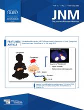
Visual Abstract
Abstract
Our purpose was to evaluate the association of a new biochemical recurrence (BCR) risk stratification system with PSMA-targeted PET/CT findings. Methods: Two prospective studies that included patients with BCR were pooled. Findings on PSMA PET were catalogued. Patients were characterized according to the European Association of Urology BCR risk categories. Univariable and multivariable analyses were performed by logistic regression. Results: In total, 145 patients were included (45 low-risk and 100 high-risk). High-risk BCR patients had a higher positive rate than low-risk patients (82.0% vs. 48.9%; P < 0.001) and reached independent predictor status for positive PSMA PET/CT scan results on multivariable logistic regression (odds ratio, 6.73; 95% CI, 2.41–18.76; P < 0.001). The area under the curve using the combination of BCR risk group and prostate-specific antigen was higher than that using prostate-specific antigen alone (0.834 vs. 0.759, P = 0.015). Conclusion: The European Association of Urology BCR risk groups define the candidates who can most benefit from a PSMA PET/CT scan when BCR occurs.
Prostate cancer is the second most common cancer type and the fifth leading cause of cancer death in men worldwide (1). In patients who receive either radical prostatectomy (RP) or radiotherapy to treat their primary tumors, approximately 30% will develop biochemical recurrence (BCR) (2). Since, by definition, prostate cancer at this stage is invisible on conventional imaging, it is of importance to stratify BCR patients into different risk groups in order to give intensive treatment to patients with aggressive disease phenotypes.
The European Association of Urology (EAU) BCR risk stratification system was proposed by the EAU prostate cancer guideline update, which defines low-risk BCR after RP as patients with a prostate-specific antigen (PSA) doubling time of more than 12 mo and a Gleason score of less than 8; high-risk BCR after RP is defined as patients with a PSA doubling time of no more than 12 mo or a Gleason score of at least 8 (3). Validation of this risk stratification system in 1,125 patients demonstrated that the 5-y metastatic progression-free and prostate cancer–specific mortality-free survival rates were significantly higher among patients with low-risk BCR. Multivariable analysis confirmed the EAU risk stratification as an independent predictor of metastatic progression and prostate cancer–specific mortality (4).
With the recent advances in prostate-specific membrane antigen (PSMA) PET/CT, our current definition of BCR may soon be obsolete. We may need to begin rephrasing our clinical questions in the context of PSMA positivity. We previously reported that more than 60% of post-RP BCR patients had positive findings on PSMA PET/CT, and according to a metaanalysis, the positive predictive value of PSMA PET/CT was 0.99 based on a histopathologic gold standard (5–7).
The aim of the current study was to compare the detection rates and the localization of PSMA-avid lesions in low-risk versus high-risk BCR patients after RP and to evaluate the association of this new risk stratification system with PSMA PET/CT findings.
MATERIALS AND METHODS
Patients
We pooled cohorts of patients with BCR from 2 prospective studies at tertiary referral centers (Johns Hopkins Hospital and Renji Hospital). The inclusion criteria of the patients in each cohort, as well as technical details of the PSMA PET/CT scan (e.g., scanner, scan protocol, and scan interpretation) have been previously reported (5,6). Risk stratification was performed as proposed by Van den Broeck et al. (3).
Pelvis-confined disease was defined by uptake of the radiotracer in the prostate bed, pelvic soft tissue, or pelvic lymph nodes. PSA doubling time was calculated as previously described (6), using the 3 most recent PSA values before PSMA PET/CT. If the slope of the linear regression was 0 (elevated but constant PSA) or negative (decreasing PSA after initial increase), the PSA doubling time was set as at least 12 mo.
Statistical Analysis
Logistic regression models were conducted for univariable and multivariable analyses, calculating odds ratios with 95% CIs to estimate the associations between BCR risk stratification and outcomes, adjusting for potential confounders. The predictive value of BCR risk stratification was assessed using the receiver-operating-characteristic curve and the area under the curve. Statistical testing was based on 2-sided tests at the 5% level of significance. SAS software (version 9.4; SAS Institute) was used.
RESULTS
Patients
In total, 145 patients were enrolled; 94 were scanned with 18F-DCFPyL PET/CT (Johns Hopkins Hospital), and 51 were scanned with 68Ga-PSMA-11 PET/CT (Renji Hospital). Low-risk BCR was present in 45 patients, and high-risk BCR in 100. Table 1 summarizes the clinical and pathologic characteristics of these patients.
Demographics and Clinical Data for Study Cohort
Imaging Findings
Of the 145 patients, 104 (71.7%) had at least one PSMA-positive lesion on the PSMA PET/CT scan. High-risk BCR patients had a significantly higher positive rate than the low-risk BCR group (82.0% vs. 48.9%; P < 0.001; Fig. 1A). On multivariable logistic regression analyses adjusted for age, PSA at the time of the scan, disease-free time, pathologic tumor stage (pT stage), and cohort (Johns Hopkins Hospital or Renji Hospital), the BCR risk group was an independent predictor for a positive PSMA PET/CT result (odds ratio, 6.73; 95% CI, 2.41–18.76; P < 0.001; Table 2). The median number of PSMA-positive lesions is 0 (interquartile range, 0–1) for low-risk BCR and 1 (interquartile range, 1–3) for high-risk BCR. The multivariable linear regression model was used to estimate the associations between BCR risk group and lesion number. The model parameter β is 0.85, with statistical significance (P = 0.037).
(A and B) Percentage of positive PSMA PET/CT scans among all patients (A) and among PSA subgroups (B). (C) Prevalence of pelvis-confined disease in each risk group. (D) Area under curve for detection of prostate cancer stratified by BCR risk group, PSA, and combination of BCR risk group and PSA. Each receiver-operating-characteristic multivariable analysis model also includes age, disease-free time, and pT stage. LN = lymph node; PB = prostate bed; ROC = receiver operating characteristic.
Univariable and Multivariable Logistic Regression Models Stratified According to EAU BCR Risk Groups Predicting Positive Findings, Pelvis-Confined Disease, and Disease Location on PSMA PET/CT Imaging
In PSA subgroups, the positive rates of patients with low-risk BCR remained the same (40%) in groups with a PSA of less than 0.5 ng/mL and with a PSA of 0.5–1.0 ng/mL, whereas higher positive rates were observed with increasing PSA values in patients with high-risk BCR. Nearly 95% of patients with a PSA of more than 1.0 ng/mL in the high-risk group had detectable disease on PSMA PET/CT, whereas the positive rate was 66.7% for low-risk patients in the same PSA subgroup (Fig. 1B).
Of the 104 scan-positive patients, 56 (53.8%) had pelvis-confined disease. The BCR risk group was not associated with pelvis-confined disease (Table 2; Fig. 1C). Receiver-operating-characteristic curves were generated to demonstrate the ability of the BCR risk group and PSA to predict positive PSMA PET/CT results. The areas under the curve using the BCR risk group or PSA alone were comparable (0.761 vs. 0.759, P = 0.96; Fig. 1D), whereas the area under the curve using the combination of BCR risk group and PSA was higher than PSA alone (0.834 vs. 0.759, P = 0.015; Fig. 1D).
Of the 145 total patients, 68 (46.9%) had recurrence or metastasis in lymph nodes, 28 (19.3%) had bone metastasis, and 31 (21.4%) had prostate bed recurrence. On multivariable logistic regression analyses, the BCR risk group was independently associated with lymph node involvement on PSMA PET/CT in all patients, including those with negative scan results (odds ratio, 2.38; 95% CI, 1.04–5.49; P = 0.041; Table 2). However, in 104 patients with positive scan results, the BCR group was not associated with the location of PSMA-avid lesions (Fig. 2; Table 2).
Percentage of all patients with lymph node involvement (A), bone metastasis (B), and prostate bed recurrence (C) (n = 145), and percentage of PSMA scan–positive patients with lymph node involvement (D), bone metastasis (E), and prostate bed recurrence (F) (n = 104).
DISCUSSION
We demonstrated that patients with EAU high-risk BCR were more likely to have PSMA PET/CT–detectable disease, suggesting that tumor volume and distribution may help to explain the worse prognosis of those patients. Notably, even patients with low-risk BCR had relatively high detection rates on PSMA PET/CT, and the rates of extrapelvic disease on positive scans was similar between high- and low-risk groups, suggesting that patients across the BCR spectrum may be good candidates for PSMA PET/CT imaging.
Previously, PSA has been reported as the strongest predictor of a positive PSMA PET/CT result (8). In this study, the added value of the EAU BCR risk groups has been demonstrated in a diverse population. It further stratifies the patients in each PSA subgroup, defining the patients who are most likely to have a positive PSMA PET/CT result. Use of EAU risk groups can serve as a simple and clinically applicable nomogram for predicting whether patients will have a positive scan result. The survival benefits from salvage pelvic radiation or focal treatment of oligometastases in different BCR risk groups in the context of PSMA PET/CT should be further explored.
The EAU BCR risk groups are associated with meaningful oncologic outcomes such as metastatic progression-free and prostate cancer–specific mortality-free survival rates (4), suggesting that PSMA-targeted PET imaging will yield imaging biomarkers. Imaging specialists, urologists, and oncologists working with PSMA imaging should focus on the design of prospective trials that can discover and validate the prognostic significance of findings.
The limitations of this work include the relatively small number of cases, post hoc evaluation of prospectively acquired data, use of more than one PSMA-targeted radiotracer, and lack of central review or a specific read paradigm. Future work is needed to confirm these findings in multicenter, larger prospective cohorts.
CONCLUSION
The EAU BCR risk groups define the candidates who can most benefit from a PSMA PET/CT scan when BCR occurs.
DISCLOSURE
Martin Pomper is a coinventor on a U.S. patent covering 18F-DCFPyL and as such is entitled to a portion of any licensing fees and royalties generated by this technology. This arrangement has been reviewed and approved by the Johns Hopkins University in accordance with its conflict-of-interest policies. Michael Gorin has served as a consultant to Progenics Pharmaceuticals, the licensee of 18F-DCFPyL. Steven Rowe is a consultant to Progenics Pharmaceuticals. Kenneth Pienta, Martin Pomper, Michael Gorin, and Steven Rowe have received research funding from Progenics Pharmaceuticals. Funding was received from Progenics Pharmaceuticals, the Prostate Cancer Foundation Young Investigator Award, the National Institutes of Health (grants CA134675, CA183031, CA184228, and EB024495), Program of Shanghai Subject Chief Scientist (19XD1402300), and Program for Outstanding Medical Academic Leader (2019LJ11). No other potential conflict of interest relevant to this article was reported.
KEY POINTS
QUESTION: Are the EAU BCR risk groups associated with findings on PSMA PET?
PERTINENT FINDINGS: In men with BCR after RP, the EAU high-risk group is more likely to have visible sites of recurrent disease on PSMA PET. However, low-risk and high-risk men have the same likelihood of having non–pelvis-confined disease.
IMPLICATIONS FOR PATIENT CARE: Risk stratification using the EAU BCR risk groups can help select men who are most likely to benefit from imaging with PSMA PET.
ACKNOWLEDGMENTS
We thank Meghan Pienta and Morgan D. Kuczler for their kind help in data collection.
Footnotes
Published online May 28, 2021.
- © 2022 by the Society of Nuclear Medicine and Molecular Imaging.
REFERENCES
- Received for publication April 7, 2021.
- Revision received May 20, 2021.


