


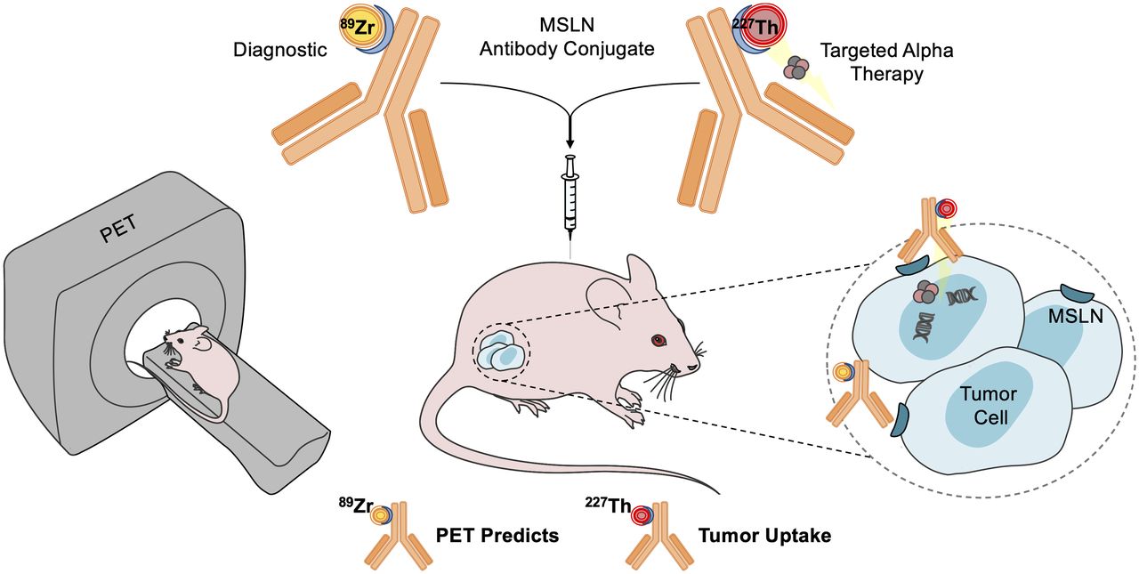
Visual Abstract
Abstract
The mesothelin (MSLN)-targeted 227Th conjugate is a novel α-therapy developed to treat MSLN-overexpressing cancers. We radiolabeled the same antibody–chelator conjugate with 89Zr to evaluate whether PET imaging with 89Zr-MSLN matches 227Th-MSLN tumor uptake, biodistribution, and antitumor activity. Methods: Serial PET imaging with protein doses of 4, 20, or 40 μg of 89Zr-MSLN and 89Zr-control was performed up to 168 h after tracer injection in human tumor–bearing nude mice with high (HT29-MSLN) and low (BxPc3) MSLN expression. 89Zr-MSLN and 227Th-MSLN ex vivo tumor uptake and biodistribution were compared at 6 time points in HT29-MSLN and in medium–MSLN-expressing (OVCAR-3) tumor–bearing mice. 89Zr-MSLN PET imaging was performed before 227Th-MSLN treatment in HT29-MSLN and BxPc3 tumor–bearing mice. Results: 89Zr-MSLN PET imaging showed an SUVmean of 2.2 ± 0.5 in HT29-MSLN tumors. Ex vivo tumor uptake was 10.6% ± 2.4% injected dose per gram at 168 h. 89Zr-MSLN tumor uptake was higher than uptake of 89Zr-control (P = 0.0043). 89Zr-MSLN and 227Th-MSLN showed comparable tumor uptake and biodistribution in OVCAR-3 and HT29-MSLN tumor–bearing mice. Pretreatment SUVmean was 2.2 ± 0.2 in HT29-MSLN tumors, which decreased in volume on 227Th-MSLN treatment. BxPc3 tumors showed an SUVmean of 1.2 ± 0.3 and remained similar in size after 227Th-MSLN treatment. Conclusion: 89Zr-MSLN PET imaging reflected MSLN expression and matched 227Th-MSLN tumor uptake and biodistribution. Our data support the clinical exploration of 89Zr-MSLN PET imaging together with 227Th-MSLN therapy, both using the same antibody–chelator conjugate.
Despite anticancer therapy advancements, several unmet medical needs remain. For instance, patients with mesothelioma and high-grade serous ovarian cancer would benefit from novel treatment options (1,2).
Recently, targeted α-therapy has emerged as a potential cancer treatment option. α-particle–emitting radionuclides targeted to the tumor enable potent antitumor activity while limiting toxicity to healthy tissues because of their high linear energy transfer and short range in tissue (3,4). Currently, 223Ra-dichloride for metastatic castration-resistant prostate cancer is the only approved targeted α-therapy (5,6). Unlike 223Ra, its progenitor 227Th forms a stable complex with an N-methyl-3-hydroxypyridine-2-one (3,2-HOPO) chelator conjugated to tumor-associated antigen–targeting antibodies (7–9). Targeted 227Th conjugates showed efficacy in mice, including those targeting mesothelin (MSLN), prostate-specific membrane antigen, CD33, and CD70 (10–14). Tumor-associated antigen binding of targeted 227Th conjugates enables local tumor cell killing via double-strand DNA breaks caused by 227Th decay (9).
MSLN is a glycosyl-phosphatidylinositol cell membrane–anchored protein involved in cell–cell adhesion and metastatic spread (15–17). MSLN expression by healthy tissues is limited to the peritoneum, pleura, and pericardium. However, it is overexpressed by several human cancers, such as mesothelioma and ovarian cancer (18). Therefore, MSLN is attractive for targeted cancer therapy, such as antibody–drug conjugates, chimeric antigen receptor T-cells, and targeted radionuclide therapy, currently tested in preclinical and clinical studies (19–22).
The MSLN-targeted 227Th conjugate comprises 3,2-HOPO covalently attached to fully human anti-MSLN monoclonal antibody anetumab and stably complexed with the α-particle emitter 227Th (13). The conjugate is reactive only to human MSLN. Understanding 227Th-MSLN tumor uptake and biodistribution may be valuable to guide clinical development. PET can noninvasively visualize the biodistribution of monoclonal antibodies, also targeting MSLN (23–25). We developed a PET tracer complexing the 3,2-HOPO–MSLN conjugate with 89Zr. By using the same antibody–chelator conjugate, we aim to avoid chelator-driven differences in pharmacokinetic properties. In mice bearing human MSLN–overexpressing tumors, we evaluated whether 89Zr-MSLN PET was able to specifically visualize MSLN, whether this imaging could predict 227Th-MSLN tumor uptake and biodistribution, and whether 89Zr-MSLN tumor uptake matches 227Th-MSLN antitumor activity.
MATERIALS AND METHODS
Radiolabeling and Quality Control of 227Th-MSLN, 89Zr-MSLN, and 89Zr-Control
The radionuclides 227Th and 89Zr were coupled to fully human IgG1 anti-MSLN monoclonal antibody and an IgG1-isotype control with 3,2-HOPO. This chelator is an octadentate with 4 bidentate 3,2-HOPO metal-complexation units and a carboxylic arm for monoclonal antibody conjugation, via amide coupling (8). Bayer AG provided conjugates 3,2-HOPO–MSLN and 3,2-HOPO–control with chelator-to-antibody ratios of 0.5. 227Th radiolabeling of 3,2-HOPO–MSLN, resulting in 227Th-MSLN, and quality control were performed as described previously (13). For PET studies, 3,2-HOPO–MSLN and 3,2-HOPO–control were radiolabeled with 89Zr-oxalate (PerkinElmer) in 0.5 M 2-[4-(2-hydroxyethyl)piperazin-1-yl]ethanesulfonic acid, pH 6.7, for 1–2 h at 37°C. 89Zr-MSLN tended to form radioactive dimers. For the 4-μg dose, the radioactive dimer formation was 10% at a 0.1 mg/mL antibody concentration, with protein desalting purification in 10 mM histidine and 130 mM glycine at pH 7.4 in water. The 20- and 40-μg dose preparation required higher antibody concentrations of 0.2 and 0.4 mg/mL during radiolabeling, resulting in 30% and 60% radioactive dimers, respectively. The effect of 60% and 10% radioactive dimer content on 89Zr-MSLN biodistribution was compared at the 4-μg dose. To induce 60% dimers at this dose, an additional radiolabeling was performed at a 0.4 mg/mL concentration of antibody with purification via Vivaspin (Sartorius Stedim Biotech) centrifugation in 0.9% NaCl. For 89Zr-MSLN quality control, size-exclusion ultra-performance liquid chromatography was used with a TSK-Gel SW column G3000SWXL (5 μm, 7.8 mm; Joint Analytic Systems), elution buffer phosphate-buffered saline (140.0 mM NaCl, 9.0 mM Na2HPO4, 1.3 mM NaH2PO4), and a 0.7 mL/min flow rate (absorbance detection, 280 nm; radioactivity detection). Radiochemical purity was assessed by trichloroacetic acid precipitation assay (26). To determine the immunoreactive fraction, a 10-fold molar excess of recombinant MSLN extracellular domain (catalog no. 3265-MS-050; R&D Systems) was added to 89Zr-MSLN and assessed by radioactivity chromatogram overlay peak intersection of bound 89Zr-MSLN versus unbound 89Zr-MSLN.
Cell Lines
BxPc3 human pancreatic and OVCAR3 human ovarian cancer cells were obtained from American Type Culture Collection, and HT29-MSLN transfected human colon cancer cells were obtained from Bayer AG, generated at Natural and Medical Sciences Institute (respectively, 4,200, 37,877, and 242,413 MSLN molecules per cell) (13). All cell lines were Mycoplasma-negative. The genetic origin of the cell lines was authenticated by BaseClear using short-tandem-repeat profiling. BxPc3 and OVCAR-3 cells were cultured in Dulbecco modified Eagle medium/Ham F12, and HT29-MSLN cells were cultured in RPMI 1640 medium and hygromycin B, 600 μg/mL. All cells were cultured in 10% fetal calf serum and 1% penicillin/streptomycin and were incubated at 37°C, with 5% CO2 in a humidified incubator.
Animal Studies
Animal experiments conformed with animal welfare laws in The Netherlands and Norway. Female nude mice, 4–10 wk old and weighing 25–35 g, received 200 μg of irrelevant IgG2A (Sigma-Aldrich) within 24 h before 89Zr-MSLN, 89Zr-control, or 227Th-MSLN injection to limit unspecific uptake in liver and spleen (27). A 20-μg dose was used as the standard dose, in line with published data (13). To investigate 89Zr-MSLN dose effect, 4 and 20 μg of 89Zr-MSLN were applied, equaling 0.14 mg/kg and 0.75 mg/kg in a previous publication (13) with an additional dose of 40 μg. Only a 20-μg nonspecific 89Zr-control was used, as we were not expecting a dose effect. Tumor volumes were measured with calipers and calculated with the formula [long side × short side2]/2, expressed as cubic millimeters. Mice with similar tumor sizes were balanced between groups. Ex vivo tissue uptake between 89Zr-MSLN and 227Th-MSLN was compared in female BALB/c nude-Foxn1nu mice (Janvier Labs). In this experiment, inclusion of 25–30 mm3 tumor sizes was accepted given the challenging tumor growth of the OVCAR-3 model. For PET experiments, Female NMRI-Foxn1nu mice (Taconic Biosciences) were used, enabling direct comparison with published data (13). To reliably quantify PET data, the inclusion criterion for the imaging studies was a tumor size of more than 150 mm3. Therefore, the OVCAR-3 tumor model was excluded for PET imaging.
To investigate 89Zr-MSLN tumor and healthy tissue uptake, dose effect, and radioactive dimers, NMRI-Foxn1nu mice were inoculated with 1.0 × 10⁶ HT29-MSLN cells 14 d or 2.5 × 10⁶ BxPc3 cells 21 d before the start of PET studies. HT29-MSLN tumor–bearing mice received 20 μg of 89Zr-MSLN or 89Zr-control (3–4 MBq, n = 6). BxPc3 tumor–bearing mice received 4, 20, or 40 μg of 89Zr-MSLN or 20 μg of 89Zr-control (1–5 MBq, n = 6). Given radioactive dimer formation, we did not exceed 40 μg. Only 20- and 40-μg dose groups could undergo PET imaging 24, 72, and 168 h after injection. Ex vivo biodistribution was performed for all groups at 168 h after injection. The effect of radioactive dimers on tissue uptake was tested at 4 μg of 89Zr-MSLN with 10% versus 60% radioactive dimers in BxPc3 tumor–bearing mice. Female BALB/c nude-Foxn1nu mice were inoculated with 1.0 × 10⁶ HT29-MSLN cells 5 d or 5.0 × 10⁶ OVCAR-3 cells 28 d before comparison of ex vivo tissue uptake of 89Zr-MSLN versus 227Th-MSLN. Mice received 20 μg of 89Zr-MSLN (0.20 MBq) or 20 μg of 227Th-MSLN (0.015 MBq) and were killed 0.5, 2, 6, 24, 72, and 168 h after injection (n = 4–5). To study whether 89Zr-MSLN PET tumor uptake coincided with 227Th-MSLN antitumor activity, BxPc3 and HT29-MSLN tumor–bearing NMRI-Foxn1nu mice underwent 89Zr-MSLN PET imaging 168 h after injection (4 MBq, 20 μg) and received a 0.75 mg/kg (500 kBq/kg) dose of 227Th-MSLN 5 d after imaging (n = 7–8, no treatment: n = 2). In this time frame, no changes in MSLN tumor expression were to be expected. Tumor sizes were measured until 21 d after treatment.
The mice were imaged with a Focus 220 PET scanner (CTI Siemens). PET data were reconstructed and corrected for decay, random coincidences, scatter, and attenuation. Tumor and heart uptake was quantified with PMOD software, version 4.004, as SUVmean. Ex vivo blood and tissues were weighed and radioactivity measured in a Wizard γ-counter (PerkinElmer) or a germanium detector (Ortec). Ex vivo uptake was expressed as percentage injected dose per gram (%ID/g).
Ex Vivo Analysis of Plasma and Tumor
Tracer integrity of 89Zr-MSLN in the plasma of mice killed at 168 h after injection was studied by sodium dodecyl sulfate polyacrylamide gel electrophoresis. Mini-Protean TGX precast protein gels, 4%–15% (BioRad), were loaded with 80 μg of plasma protein. A control sample including intact tracer and free 89Zr was generated by storing 89Zr-MSLN at room temperature for a week. Gels ran for 30–45 min at 100 V. Formalin-fixed tumor tissues were paraffin-embedded and sliced into 4-μm sections. Gels and tumor sections were exposed overnight to a multipurpose phosphor plate (PerkinElmer) at −20°C and captured with Cyclone phosphor imager (PerkinElmer).
MSLN immunohistochemistry was executed on the autoradiography tumor sections, as described earlier (28). A Ventana Discovery automated stainer was used. After washing, the primary anti-MSLN antibody (clone SP74; Spring Biosciences) at 0.25 mg/mL was detected with horseradish peroxidase–labeled antimouse polymer (Dako) and 3,3′-diaminobenzidine solution. Sections were fixed at 4°C for 5 min, air-dried, and washed with double-distilled H2O before incubation with Dako blocking solution (10 min, room temperature). After washing, primary antibody was detected with horseradish peroxidase–labeled antimouse polymer (Dako) and 3,3′-diaminobenzidine solution. Hematoxylin–eosin staining was performed on adjacent tumor sections. Digital scans were acquired by a Hamamatsu NanoZoomer 2.0-HT multislide scanner and analyzed with NanoZoomer Digital Pathology viewer software.
Statistical Analysis
Similarity between 2 groups was analyzed using a Mann–Whitney U test. When there were multiple groups or time points, a Bonferroni multiple-comparison correction was applied. All data are presented with SD. All statistical tests were performed in GraphPad Prism 8, and P values of less than 0.05 were considered significant.
RESULTS
Quality Control 89Zr-MSLN
89Zr-MSLN was produced with a radiolabeling efficiency of 64% ± 10%, a radiochemical purity of 98% ± 1%, and 4% ± 1% antibody dimers and 15% ± 2% radiolabeled dimers (n = 6) (Supplemental Fig. 1A; supplemental materials are available at http://jnm.snmjournals.org). The immunoreactive fraction was 0.8 (Supplemental Fig. 1B). We observed by radioactive detection that 89Zr-MSLN tended to dimerize, not observed at 280 nm. Favorable and unfavorable conditions are shown in Supplemental Table 1. The radiolabeling conditions and quality control results of the experiments are shown in Supplemental Figures 1C and 1D and Supplemental Table 2.
Tumor Uptake and Biodistribution of 89Zr-MSLN
PET evaluation of 89Zr-MSLN in HT29-MSLN tumor–bearing mice showed 1.8-fold higher tumor uptake and tumor-to-blood ratio for 89Zr-MSLN than for 89Zr-control (SUVmean, 2.2 ± 0.5 vs. 1.2 ± 0.2 168 h after injection, P = 0.0043; Figs. 1A and 1B). Ex vivo biodistribution confirmed the PET data, showing 2.5-fold higher tumor uptake of 89Zr-MSLN than of 89Zr-control at 168 h (10.6% ± 2.4% vs. 4.2% ± 0.7 %ID/g, P = 0.0043), whereas uptake in all other tissues was similar (Fig. 2). No low-molecular-weight species of 89Zr-MSLN or free 89Zr were present in blood 168 h after injection (Supplemental Fig. 1E). Autoradiography showed MSLN-specific 89Zr-MSLN tumor uptake compared with 89Zr-control (Fig. 3; Supplemental Figs. 2A and 2B).

In vivo tumor uptake and biodistribution in 89Zr-MSLN HT29-MSLN tumor–bearing mice (6 per group). (A) Representative coronal PET images at 24, 72, and 168 h after injection of 20 μg of 89Zr-MSLN and 20 μg of 89Zr-control (3–4 MBq). Uptake is presented as SUV. (B) PET quantification of 89Zr-MSLN and 89Zr-control uptake in tumor and blood at 24, 72, and 168 h after injection. 89Zr-MSLN and 89Zr-control uptake is shown as SUVmean ± SD. Tumor-to-blood ratio is indicated at 168 h. B = bone; C = circulation; T = tumor; TBR = tumor-to-blood ratio.

Ex vivo tumor uptake and biodistribution of 89Zr-MSLN HT29-MSLN tumor–bearing mice (6 per group). Shown is ex vivo tumor and healthy tissue uptake of 20 μg of 89Zr-MSLN and 20 μg of 89Zr-control at 168 h after injection. Data are %ID/g, as mean ± SD; 89Zr-MSLN and 89Zr-control batches each contained 30% radioactive dimers. *P < 0.05. **P < 0.01.

Intratumoral 89Zr-MSLN distribution. Shown is MSLN immunohistochemistry, autoradiography, and hematoxylin and eosin staining of HT29-MSLN and formalin-fixed, paraffin-embedded tumor sections that received 89Zr-MSLN (A) or 89Zr-control (B). MSLN immunohistochemistry and autoradiography were performed on same tumor section, and hematoxylin and eosin staining were performed on adjacent tumor section. Radioactivity in A and B is simultaneously scaled, shown from high to low 89Zr signal intensity. Representative data are shown (n = 3–5; rest are shown in Supplemental Fig. 2). H&E = hematoxylin and eosin.
Dose Effect of 89Zr-MSLN on Tumor Uptake and Biodistribution
In vivo, uptake and tumor-to-blood ratios were lower in 89Zr-MSLN BxPc3 tumors than in HT29-MSLN tumors. 89Zr-MSLN BxPc3 tumor uptake and tumor-to-blood ratios were similar between 20 μg and 40 μg (SUVmean, 1.6 ± 0.2 vs. 1.9 ± 0.3) and higher than in 20-μg 89Zr-control (SUVmean, 1.1 ± 0.2; Figs. 4A and 4B). Ex vivo, 89Zr-MSLN tumor and liver uptake were higher at 40 μg than at 4 μg 168 h after injection. In addition, mice that received 4 μg of 89Zr-MSLN with 60% radioactive dimers showed higher tumor (10.0% ± 2.2% vs. 6.1% ± 1.7 %ID/g) and liver (8.8% ± 1.4% vs. 4.9% ± 1.2 %ID/g) uptake compared with mice that received 4 μg of 89Zr-MSLN with 10% radioactive dimers. The excretion rate was not affected by radioactive dimers. Bone uptake was mainly in cortical bone and not in bone marrow (Supplemental Figs. 3A–3D).

Dose effect of 89Zr-MSLN on tumor uptake and biodistribution in BxPc3 tumor–bearing mice (5–6 per group). (A) Representative coronal PET images of 40 μg and 20 μg of 89Zr-MSLN and 20 μg of 89Zr-control at 24, 72, and 168 h after injection. Uptake is presented as SUV. (B) Quantification of tumor and blood at 24, 72, and 168 h after injection, shown as SUVmean ± SD. Tumor-to-blood ratio is indicated at 168 h. B = bone; C = circulation; T = tumor; TBR = tumor-to-blood ratio.
89Zr-MSLN Versus 227Th-MSLN Tumor Uptake and Biodistribution
Ex vivo OVCAR3 and HT29-MSLN tumor uptake of 89Zr-MSLN and 227Th-MSLN was comparable except at 168 h after injection, when 89Zr-MSLN HT29-MSLN tumor uptake was lower (33.1% ± 9.0% vs. 89.8% ± 26.3 %ID/g, P = 0.016; Fig. 5A). Tumor-to-blood ratios were similar for 89Zr-MSLN and 227Th-MSLN in both models (Fig. 5B). 89Zr-MSLN liver uptake was higher than 227Th-MSLN uptake up to 24 h in HT29-MSLN tumor–bearing mice but not at 72 and 168 h, the clinically relevant time points. 89Zr-MSLN femur uptake was higher than 227Th-MSLN uptake from 24 to 168 h in both models—for example, 12.3% ± 1.3 %ID/g versus 4.9% ± 0.6 %ID/g 168 h after injection in the HT29-MSLN tumor–bearing mice, resulting in lower blood and kidney levels at 72 h and 168 h (Supplemental Figs. 4A and 4B).

Tumor uptake of 89Zr-MSLN compared with 227Th-MSLN. (A and B) HT29-MSLN tumor uptake and OVCAR3 tumor uptake (A) and respective tumor-to-blood ratios (B) of 20 μg of 89Zr-MSLN (0.20 MBq) vs. 20 μg of 227Th-MSLN (0.015 MBq) total antibody dose at 0.5, 2, 6, 24, 72, and 168 h. Data are median %ID/g and interquartile range, including single data points. *P < 0.05 with Bonferroni adjustment.
89Zr-MSLN PET Before 227Th-MSLN Treatment
89Zr-MSLN PET imaging before 227Th-MSLN treatment revealed 1.8-fold higher tumor SUVmean in HT29-MSLN than in BxPc3 tumors (2.2 ± 0.2 vs. 1.2 ± 0.3, P = 0.0003; Figs. 6A and 6B). Because of the 18.7-d half-life of 227Th, treatment effect is not observed in the first 9 d. From day 9 until day 21 after 227Th-MSLN administration, HT29-MSLN tumors decreased 0.7 ± 0.1-fold in volume (from 432.4 ± 131.2 mm3 to 317.4 ± 130.1 mm3). Tumors of untreated mice grew individually 1.3-fold and 1.7-fold. In the same time frame, BxPc3 tumors did not grow after 227Th-MSLN administration (1.0 ± 0.3-fold, 310.2 ± 166.5 mm3 at day 9 and 288.3 ± 112.3 mm3 at day 21) whereas tumors of untreated animals individually grew 1.4-fold (Figs. 7A and 7B). Absolute tumor growth is shown in Supplemental Figure 5. BxPc3 tumors of untreated- versus 227Th-MSLN–treated animals were slightly larger at day 0. Therefore, tumor sizes were normalized to the size at day 0 (29). 227Th-MSLN treatment increased DNA double-strand breaks compared with tumors of untreated mice, confirming the molecular mode of action of 227Th-MSLN (Supplemental Figs. 6A and 6B).

89Zr-MSLN PET before 227Th-MSLN treatment. (A) Representative coronal PET images of HT29-MSLN and BxPc3 tumor–bearing mice 168 h after 20 μg of 89Zr-MSLN. Tumor uptake is presented as SUV. (B) Quantification of 89Zr-MSLN in HT29-MSLN and BxPc3 tumors at 168 h after injection (n = 7–8 per group). 89Zr-MSLN uptake is shown as SUVmean ± SD, including single data points. ***P < 0.001. B = bone; T = tumor.

Tumor growth after 227Th-MSLN treatment. (A and B) Tumor growth after 227Th-MSLN treatment (0.75 mg/kg, 500 kBq/kg) in HT29-MSLN tumor–bearing mice (n = 8) (A) and in BxPc3 tumor–bearing mice (n = 7) and per model untreated mice (n = 2), normalized to day 0. Absolute tumor sizes are shown in Supplemental Figure 5.
DISCUSSION
This study showed that 89Zr-MSLN PET imaging reflects 227Th-MSLN tumor uptake and biodistribution in mice bearing tumors overexpressing human MSLN. We showed the dual use of an antibody–chelator conjugate, 3,2-HOPO–MSLN, radiolabeled with 89Zr for PET imaging and with 227Th for targeted α-therapy as a theranostic. Even though some studies have shown direct molecular imaging of 227Th, this remains a challenge due to the low abundance of measurable photons in the decay chain of 227Th (30). Studies on patients and mice showed the theranostic potential of 89Zr PET for β-particle–emitting therapeutic radionuclides, such as 177Lu and 90Y (31,32). Therefore, we hypothesized that 89Zr might serve as a PET surrogate radioisotope for the α-particle emitter 227Th as well. Estimating 227Th-MSLN whole-body distribution with 89Zr-MSLN PET before treatment may be of value to guide clinical development. In addition, this study encourages 89Zr-MSLN PET exploration to select patients and predict 227Th-MSLN efficacy. Moreover, PET imaging with 89Zr may be amenable to other targeted α-therapies.
89Zr-MSLN tumor uptake might correlate with response to 227Th-MSLN. However, we did not use the isogenic cell systems required to exclude differences in sensitivity to 227Th-MSLN. We did not perform tumor biopsies to assess whether changes in MSLN expression occurred in the 5-d time frame between PET scan and start of treatment. A change is unlikely, as tumor growth was relatively consistent. We observed a trend in antitumor activity of 227Th-MSLN comparable to the earlier in vivo study (13). A firm conclusion on predicting 227Th-MSLN antitumor activity is precluded given the low number of animals in the control groups.
Variability in 89Zr-MSLN and 227Th-MSLN tumor uptake may have been a result of small tumors in the ex vivo biodistribution comparison. Higher 89Zr-MSLN bone uptake might be explained by dissociated 89Zr from 3,2-HOPO, tending to accumulate in the growing bone in young mice, not seen in humans (33,34). Ex vivo blood samples at 168 h showed intact 89Zr-MSLN, indicating that the tracer available in the circulation for tissue uptake is intact. This finding suggests that free 89Zr clears from the blood immediately into cortical bone. Although desferrioxamine-based chelators are commonly used for complexation with 89Zr, HOPO-based chelators are a proven alternative (35,36). To avoid chelator-driven discrepancies in pharmacokinetics between 89Zr-MSLN and 227Th-MSLN (37,38), we developed the PET tracer using the same 3,2-HOPO–MSLN conjugate with the additional advantage of having the intermediate product clinical grade right at hand. We showed in mice that 89Zr-MSLN uptake can predict tumor targeting of 227Th-MSLN. In patients, 89Zr-MSLN PET imaging might detect MSLN-positive lesions in mesothelioma, ovarian cancer, and pancreatic cancer and clarify whether the antibody can reach these lesions. Yet, calculating an exact 227Th radiation dose per organ is limited by the observed 89Zr-MSLN bone uptake. For potential dosimetry purposes, we should aim to improve the in vivo stability of HOPO-based 89Zr chelators.
Higher liver uptake of 89Zr-MSLN with a high radioactive dimer content indicates faster clearance of dimers and aggregates than of monomers (39). Higher tumor uptake could be explained by increased retention at the target-binding site due to an avidity effect (40) combined with an enhanced permeability and retention effect (41). Higher tumor uptake at 40 μg than at 4 μg of total antibody dose may, therefore, most likely be a dimer effect instead of a dose effect. Dimerization appears specific for 89Zr-3,2-HOPO because the control antibody revealed similar radioactive dimer content, not observed in combination with 227Th to the same extent (13). In a clinical setting, the specification should be set at less than 15%. This is feasible with the optimized radiolabeling procedure that we describe.
CONCLUSION
Our study revealed the potential of 89Zr-MSLN PET to predict 227Th-MSLN tumor uptake and biodistribution. Furthermore, it addressed the potential of 89Zr-MSLN PET as a tool to estimate 227Th-MSLN antitumor activity. Our data support clinical investigation of 89Zr-MSLN PET imaging in combination with 227Th-MSLN therapy.
DISCLOSURE
This study was supported by a research grant from Bayer to Elisabeth de Vries. Payments were made available to her institution (University Medical Center Groningen). Bayer developed and owns the intellectual property rights of 3,2-HOPO–MSLN and 227Th-MSLN. Elisabeth de Vries reports institutional financial support for a consulting and advisory role from Sanofi, Daiichi Sankyo, NSABP, Crescendo Biologics, and Cyprus Cancer Research Institute and institutional financial support for clinical trials or contracted research from Amgen, Genentech, Roche, AstraZeneca, Synthon, Regeneron, Chugai Pharma, CytomX Therapeutics, Servier, Nordic Nanovector, G1 Therapeutics, and Radius Health, all outside the submitted work. No other potential conflict of interest relevant to this article was reported.
KEY POINTS
QUESTION: Can 89Zr-MSLN PET imaging predict 227Th-MSLN behavior?
PERTINENT FINDINGS: 89Zr-MSLN PET imaging shows tumor uptake and biodistribution similar to that of 227Th-MSLN in MSLN-expressing tumor–bearing nude mice.
IMPLICATIONS FOR PATIENT CARE: These data support the theranostic potential of 89Zr-MSLN PET imaging to guide 227Th-MSLN therapy in patients.
ACKNOWLEDGMENTS
We thank Manuela Brand for performing the immunohistochemistry and immunofluorescent staining (Bayer AG) and Linda Pot-de Jong for assisting with the animal experiments (Department of Medical Oncology, University Medical Center Groningen).
Footnotes
Published online Apr. 14, 2022.
- © 2022 by the Society of Nuclear Medicine and Molecular Imaging.
REFERENCES
- Received for publication August 19, 2021.
- Revision received March 23, 2022.


