Abstract
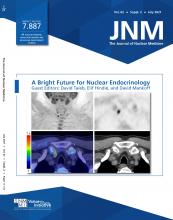
After exclusion of exogenous iodine overload, radioiodine uptake (RAIU) testing with 123I or 131I enables the accurate evaluation and quantification of iodine uptake and kinetics within thyroid cells. In addition, scintigraphic evaluation with 123I or 99mTc-pertechnetate (99mTc04-) provides the topographic distribution of thyroid cell activity and allows the detection and localization of ectopic thyroid tissue. Destructive thyrotoxicosis is characterized by abolished or reduced uptake whereas productive thyrotoxicosis (i.e., hyperthyroidism “sensu strictu”) is characterized by high RAIU with scintigraphically diffuse (i.e., Graves disease and diffuse thyroid autonomy) or focal (i.e., autonomously functioning thyroid nodules [AFTN]) overactivity. Accordingly, RAIU or thyroid scintigraphy are widely used to differentiate different causes of thyrotoxicosis. In addition, several radiopharmaceuticals are also available to help in differentiating benign from malignant thyroid nodules and inform clinical decision making. In fact, AFTNs can be safely excluded from fine-needle aspiration biopsy while either 99mTc-methoxyisobutylisonitrile (MIBI) and 18F-FDG may complement the work-up of cytologically indeterminate cold nodules and contribute to reducing the need for diagnostic lobectomies/thyroidectomies. Finally, RAIU studies are also useful for calculating the administered therapeutic activity of 131I to treat hyperthyroidism and euthyroid multinodular goiter. All considered, thyroid molecular imaging allows us to characterize molecular/functional aspects of different thyroid diseases, even before clinical symptoms become manifest and remains integral to properly managing such conditions. Our present paper summarizes basic concepts, clinical applications, and potential developments of thyroid molecular imaging in patients affected by thyrotoxicosis and thyroid nodules.
Radioiodine uptake (RAIU) testing and thyroid scintigraphy (TS) remained the only methods to evaluate both functional and morphologic aspects of the thyroid gland and its diseases for decades. Subsequently, with the development of immunometric assays, multiparametric thyroid ultrasound (MPUS), and fine-needle aspiration (FNA), the use of TS and RAIU tests has decreased. However, TS remains the only method able to differentiate “productive” from “destructive” thyrotoxicosis and detect thyroid functional autonomy (TFA) (1,2). Although the spatial resolution of TS is approximately 5–7 mm compared with 1 mm for MPUS, smaller sites of hyperfunctioning thyroid tissue may be detectable provided the target–to–background count rate ratio is adequately high (i.e., > 2.5 times) as it is expected in thyroid autonomy. Such foci are rendered in the image larger than actual size (i.e., partial-volume effect) and this should be considered; however, functional information remain clinically relevant. Additionally, different tracers able to evaluate the proliferation rate of cold thyroid nodules are available to support the management of cytologically indeterminate cold nodules and limit unnecessary lobectomies/thyroidectomies (3,4). The aim of our paper is to summarize molecular imaging methods available for thyroid evaluation and provide practical suggestions for their optimized use in clinical practice.
THYROID MOLECULAR IMAGING: BASIC CONCEPTS
Normal thyroid follicular cells trap stable iodine by the sodium (i.e., natrium) iodide symporter, a laterobasal transmembrane glycoprotein. Then, iodine is transported at the apical membrane into the follicular lumen and incorporated into selected tyrosyl residues of thyroglobulin to form thyroxine (T4) and triiodothyronine (T3) (5,6). Under physiologic conditions the thyroid-stimulating-hormone (TSH), leading in first approximation an inverse log-linear relationship TSH/FT4 (7) and a positive linear relationship TSH/RAIU, respectively (8), mainly regulates the process. The thyroid gland mainly produces T4, which accounts for 85%–90% of thyroid hormones, whereas the bioactive T3 largely derives from peripheral conversion of T4 under the action of liver deiodinases. More than 99% of T4 and T3 molecules are tightly bound to the carrier proteins, thyroid-binding globulin, transthyretin, and albumin, and only a small percentage circulates as free hormones (9,10).
Tracing Thyroid Function
123I is an ideal thyroid radiopharmaceutical because of its low radiation burden and optimal imaging quality compared with 131I, which is strongly discouraged for routine diagnostic use because of its much higher radiation burden to the thyroid. 124I is a positron-emitting isotope that allows high-quality imaging of the thyroid: its use, however, is restricted to clinical trials involving individuals with differentiated thyroid cancer (1). Different tracers, such as 99mTc-pertechnetate (99mTc04-) and 18F-tetrafluoroborate, are also trapped by the sodium iodide symporter, but wash out completely from thyroid cells in about 30 min and are not organified. In most cases, 99mTc04- imaging provides the clinical information needed (1,11) and, being inexpensive and readily available from on-site generators, is widely used in clinical practice. On the other hand, 123I allows a precise quantification of both iodine trapping and organification and supports individual dosimetric therapy planning (3,11,12).
Tracing Thyroid Growth and Proliferation
99mTc-MIBI is a lipophilic cation that crosses the cell membrane and penetrates reversibly into the cytoplasm and then irreversibly into the mitochondria (13). Transmembrane glucose transporters and hexokinases mediate the cellular uptake and retention of 18F-FDG, respectively. Notably, higher 99mTc-MIBI and 18F-FDG uptake are expected in thyroid cancers due to the higher electrical gradient of mitochondrial membrane and insulin-independent glucose consumption of cancer cells compared with either normal cells and those forming benign thyroid nodules (13,14).
Diagnostic Procedures
Extensive information on radiopharmaceutical activities, instrumentations, imaging protocols, interpretation criteria, and reporting can be found in recently published joint EANM practice guideline/SNMMI procedure standard for RAIU and TS (1).
THYROTOXICOSIS
Thyrotoxicosis refers to the clinical syndrome of excess circulating thyroid hormones, irrespective of the source, whereas hyperthyroidism is characterized by increased thyroid hormone synthesis and secretion from the thyroid gland (15). The clinical presentation of thyrotoxicosis ranges from asymptomatic to thyroid storm, depending by its intensity and duration. Accordingly, besides clinical examination serum TSH measurement is integral to confirm (TSH < 0.1 mUI/L) or exclude (TSH > 0.4 mUI/L) hyperthyroidism, with values between 0.1 and 0.4 mUI/L considered as a gray area requiring serial testing for surveillance. Serum concentrations of free T4 and free T3 differentiate subclinical from overt hyperthyroidism and assess the severity of the latter (16–18). The most common cause of hyperthyroidism is Graves disease (GD), followed by TFA. The latter may present as unifocal or multifocal autonomously functioning thyroid nodules (AFTNs) or disseminated gland overactivity (19,20). TFA is rare (<10%) in countries with adequate iodine supply but its prevalence significantly increases (up to 20% and more) in countries with current or previous iodine deficiency (21). Other causes of thyrotoxicosis include destructive thyroiditis, iodine-induced and drug-induced thyroid dysfunction, and factitious ingestion of excess thyroid hormones. Then, an accurate differential diagnosis is required to properly treat patients (22,23). Depending on the available resources, TSH receptor antibody (TRAb) measurement (24), TS or RAIU (25,26), and MPUS with color flow Doppler evaluation can be used (27). A recent study on 124 consecutive patients with newly diagnosed and untreated hyperthyroidism compared 2 TRAb assays, TS and MPUS, respectively (28). Thyroid MPUS was less accurate than both TRAb and TS, with the exception of a vascular thyroid inferno pattern, which provides a high positive predictive value (PPV) for GD. TS was the most reliable tool for differential diagnosis, and easily delineated different GD and TFA variants, including Marine-Lenhardt syndrome and compensated autonomy (2,19,29–31). However, depending on locally available facilities, TRAb assays can be adopted as a first-line diagnostic option limiting the use of TS to TRAb-negative patients. Notably, exogenous iodine overload should be always investigated as it may lead to a sustained increase in hormone synthesis and, eventually, thyrotoxicosis with inhibition of TSH and decreased uptake due to an expanded iodine pool mimicking the early phase of destructive thyroiditis on TS. Finally, conventional TS is unsuitable to differentiate type 1 from type 2 amiodarone-induced hyperthyroidism (AIH) because iodine trapping and organification are reduced by exogenous iodine overload. Using 99mTc-MIBI TS, which demonstrates preserved MIBI uptake in type I AIH and decreased MIBI uptake in type 2 AIH, may be useful in such cases for determining clinical management (32,33). Etiology, pathophysiology, and differential diagnosis of thyrotoxicosis are summarized in Table 1.
Thyrotoxicosis: Etiology, Pathophysiology, and Relevant Points for Differential Diagnosis
THYROID NODULES
Thyroid nodules are commonly detected in clinical practice, and thus it is necessary to decide which ones carry a significant risk of malignancy and require further workup with FNA (34). MPUS provides an accurate assessment of morphologic features, which have been recently used to produce a standardized risk assessment for thyroid malignancy under the different Thyroid Imaging And Data Reporting Systems (TI-RADS) to mitigate the high interoperator variability (35,36). Different TI-RADS systems combine, with some differences, ultrasound features such as shape, margins, echogenicity, composition, and microcalcifications in hierarchical risk categories and, overall, demonstrated a satisfactory performance (37). However, almost all information on the reliability of different TI-RADS are based on studies that included papillary thyroid carcinoma, the cancer most prevalent among all thyroid malignancies. Notably, however, the accuracy of such systems is significantly reduced in other, more aggressive, thyroid cancers such as follicular histotypes (38). Additionally, FNA is inappropriately performed in a large proportion of AFTNs, based on TI-RADS classifications (27% to 90%, depending on different TI-RADS) (39,40). AFTNs have a 96%–99% negative predictive value (NPV) for malignancy but indeterminate cytologic features are frequently reported: accordingly FNA procedures are discouraged in such cases (41). Even if TS is essential to detect AFTNs it is neither recommended nor, in some countries, reimbursed in patients with thyroid nodules but normal TSH. Although this is reasonable in countries with adequate iodine supply, the TSH levels may remain normal in the presence of AFTNs when iodine supply is reduced as, especially early, the low synthesis rate of thyroid hormones is insufficient to suppress the TSH secretion (42,43) (Fig. 1). Accordingly, up to 50% of AFTNs show normal TSH values in Europe (2), leading to different recommendations on a national or regional basis (1,41–47). In this case, the integration of various information (i.e., regional iodine intake, local TSH reference range, and size and MPUS features of the nodule) may better inform the decision to perform TS or not, instead of just applying fixed TSH thresholds (Fig. 2). Conversely, nonautonomous nodules are managed based on TI-RADS to select high-risk ones for FNA and cytopathology assessment (41,42). Thyroid cytopathology is currently reported using the standardized Bethesda system (48), which is highly accurate in detecting or excluding malignancy in most nodules. Unfortunately, up to 25% of nodules are reported as follicular lesion of undetermined significance or atypia of undetermined significance (Bethesda III) and follicular neoplasm (Bethesda IV). As the attached risk of malignancy ranges from 10% to 50%, diagnostic lobectomies/thyroidectomies are frequently performed but most indeterminate nodules are ultimately found to be benign on definitive pathology (49). Currently, both molecular testing on FNA material and molecular imaging with 99mTc-MIBI and 18F-FDG should be considered before diagnostic surgery (50) (Fig. 3). Overall, molecular imaging accurately rules out whereas FNA mutational panels are more useful to rule in malignancy. 99mTc-MIBI– or 18F-FDG–negative nodules carry a risk of malignancy of only 0%–5% and can be safely ruled out from surgery (high NPV). Unfortunately, only 35%–60% of 99mTc-MIBI– or 18F-FDG–positive nodules are cancers (low PPV). On the other hand, BRAF mutation analysis is 100% predictive of papillary thyroid carcinoma (high PPV), but most cancers are BRAF-negative (very low NPV) (51–53). More recently, different gene expression classifier tests have become available, demonstrating different performance in terms of PPV and MPV (54,55). Overall, mutation mapping and gene expression analysis on FNA samples and molecular imaging can be used to refine the diagnosis in patients with indeterminate thyroid nodules. However, all methods largely depend on local cancer prevalence and pretest probabilities, and additionally high costs limit their use in many countries. Accordingly, no definitive guidelines exist, and a locally adapted multimodality stepwise approach, ideally combining one rule-in and one rule-out test, likely offers the most accurate diagnosis (50).
Sonographically suspicious left thyroid nodule in patient with normal TSH level (i.e., 1.24 mUI/L). FNA biopsy demonstrated follicular neoplasm (Bethesda IV cytology). MPUS shows slightly irregular hypoechoic nodule (A) with increased vascularization (B). 123I TS depicted AFTN (C) excluding malignancy.
Diagnostic work-up of thyroid nodules. Local TSH threshold is mainly based on local iodine supply: in general, levels below lower limit of reference range (i.e., <0.3–0.4 mUI/L) are adopted in countries with adequate iodine supply whereas higher values are suggested in countries with previous or current iodine deficiency (up to 2.5–3.0 mUI/L) (1,44–48).
Cytologically indeterminate thyroid nodules at FNA. TS demonstrates photopenic defect on 99mTc-pertechnetate scan consistent with hypofunctioning nodule within right thyroid lobe (A) with uptake and retention of 99mTc-sestaMIBI (B). Histology: papillary thyroid cancer, follicular variant.
PREDICTIVE MOLECULAR IMAGING AND THERANOSTICS
In addition to overt thyrotoxicosis even latent forms of hyperthyroidism are responsible for an increase in cerebrovascular and cardiovascular morbidity and mortality and overall mortality (56). Recently, the risk of heart failure events was also shown to correlate with low but unsuppressed TSH values (0.1–0.44 mU/L) (57,58). In such a context, TS is the most reliable tool to differentiate true TSH-independent thyroid overactivity from nonspecific TSH fluctuations (59). In such cases, performing TS after administration of 25–60 μg T3 daily over 5–10 d allowed for the discrimination of TSH-responsive from autonomously functioning tissue (Fig. 4). Particularly, a quantified 123I uptake at 24 h greater than 2% or technetium thyroid uptake (TcTU) at 15 min greater than 1%–1.8% under suppression are both consistent with significant thyroid autonomy (60). Alternatively, 123I uptake (mpU) can be also estimated integrating actual 123I uptake and TSH levels at baseline (61). Interestingly, the TcTU at baseline or after suppression reliably predicts an evolution toward overt hyperthyroidism in patients with compensated autonomy and identifies patients at higher risk to decompensate after exogenous iodine exposure (62). In addition, TcTU provides estimates of functional volume, especially useful in planning multifocal or disseminated autonomy treatment with 131I (63). Accordingly Dunkelmann et al. obtained a cure rate of 91.5% and a very low rate (1%) of hypothyroidism in 641 patients with multifocal and disseminated autonomy integrating the dosimetric Marinelli’s formula with TcTU-adapted target doses (i.e., 150 Gy for TcTU < 3% to 250 Gy for TcTU > 12%), respectively (64). Furthermore, functional volumes and activity quantification based on SPECT/CT measurements is now possible, allowing real-time quantification of radiation-absorbed dose in target volumes of interest based on empiric data on 131I biokinetics in tissue (65). Therefore, simplified SPECT/CT-based dosimetric protocols and the development of dedicated software should further improve patient-centered approaches to radioiodine thyroid theranostics (66). Furthermore, although current radiomic predictors are less reproducible than hoped for, integrating radiomics information with pathology, biochemical, and molecular features will make possible a refined diagnosis of thyroid nodules using artificial intelligence (67,68).
Chronically fluctuating low TSH values in woman with no biologic diagnosis for autoimmune thyroid disease (i.e., negative TRAb and TPOAb) and unremarkable MPUS (not shown). (A) Baseline TS: normal 123I thyroid uptake (13.1%) and homogeneous tracer distribution. (B) Suppressed TS: not suppressed 123I thyroid uptake (9.2%) and well-contrasted thyroid image after administration of liothyronine (50 μg/d) for 5 d demonstrate diffuse thyroid autonomy.
CONCLUSION
For decades, nuclear medicine methods have allowed molecular/functional characterization and tailored radioiodine therapy of different thyroid diseases. Even as accurate nonradioisotopic methods emerged over time, the selective and optimized use of TS and RAIU remains integral in differentiating the causes of hyperthyrodism and properly informs clinical and therapeutic decisions. Furthermore, among patients with thyroid nodules TS remains the only method able to detect AFTNs while aspecific tracers such as 99mTc-MIBI and 18F-FDG may contribute to refining the clinical management of cytologically indeterminate cold nodules, avoiding inappropriate invasive procedures.
DISCLOSURE
No potential conflict of interest relevant to this article was reported.
KEY POINTS
QUESTION: What is the current role of molecular imaging in evaluating thyrotoxicosis and thyroid nodules?
PERTINENT FINDINGS: Thyroid scintigraphy with 123I or 99mTc-pertechnetate allows a real-time differentiation of different forms of thyrotoxicosis, the exclusion of autonomously functioning thyroid nodules from inappropriate biopsy, and the detection of ectopic thyroid tissue. The use of either 99mTc-methoxyisobutylisonitrile (MIBI) and 18F-FDG may complement the work-up of cytologically indeterminate cold nodules and contribute to reducing the need for diagnostic lobectomies/thyroidectomies.
IMPLICATIONS FOR PATIENT CARE: Thyroid molecular imaging characterizes molecular/functional aspects of different thyroid diseases, even before clinical symptoms become manifest and remains integral to properly managing such conditions.
- © 2021 by the Society of Nuclear Medicine and Molecular Imaging.
REFERENCES
- Received for publication July 1, 2020.
- Accepted for publication September 21, 2020.


