


Visual Abstract
Abstract
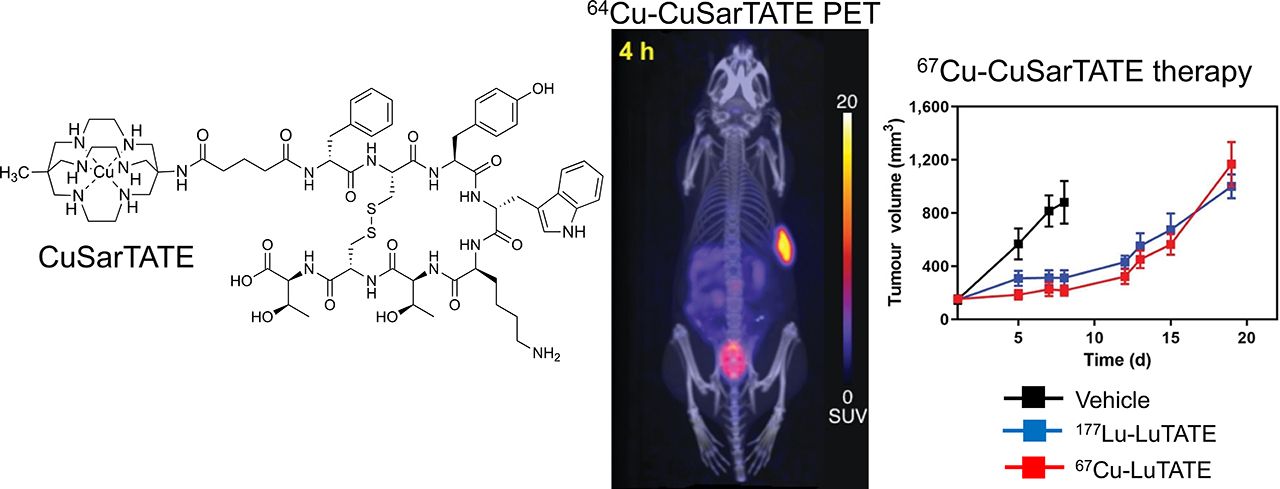
Peptide receptor radionuclide therapy (PRRT) using radiolabeled octreotate is an effective treatment for somatostatin receptor 2–expressing neuroendocrine tumors. The diagnostic and therapeutic potential of 64Cu and 67Cu, respectively, offers the possibility of using a single somatostatin receptor–targeted peptide conjugate as a theranostic agent. A sarcophagine cage amine ligand, MeCOSar (5-(8-methyl-3,6,10,13,16,19-hexaaza-bicyclo[6.6.6]icosan-1-ylamino)-5-oxopentanoic acid), conjugated to (Tyr3)-octreotate, called 64Cu-CuSarTATE, was demonstrated to be an imaging agent and potential prospective dosimetry tool in 10 patients with neuroendocrine tumors. This study aimed to explore the antitumor efficacy of 67Cu-CuSarTATE in a preclinical model of neuroendocrine tumors and compare it with the standard PRRT agent, 177Lu-LuDOTA-Tyr3-octreotate (177Lu-LuTATE). Methods: The antitumor efficacy of various doses of 67Cu-CuSarTATE in AR42J (rat pancreatic exocrine) tumor–bearing mice was compared with 177Lu-LuTATE. Results: Seven days after a single administration of 67Cu-CuSarTATE (5 MBq), tumor growth was inhibited by 75% compared with vehicle control. Administration of 177Lu-LuTATE (5 MBq) inhibited tumor growth by 89%. Survival was extended from 12 d in the control group to 21 d after treatment with both 67Cu-CuSarTATE and 177Lu-LuTATE. In a second study, the efficacy of fractionated delivery of PRRT was assessed, comparing the efficacy of 30 MBq of 67Cu-CuSarTATE or 177Lu-LuTATE, either as a single intravenous injection or as two 15-MBq fractions 2 wk apart. Treatment of tumors with 2 fractions significantly improved survival over delivery as a single fraction (67Cu-CuSarTATE: 47 vs. 36 d [P = 0.036]; 177Lu-LuTATE: 46 vs. 29 d [P = 0.040]). Conclusion: This study demonstrates that 67Cu-CuSarTATE is well tolerated in BALB/c nude mice and highly efficacious against AR42J tumors in vivo. Administration of 67Cu-CuSarTATE and 177Lu-LuTATE divided into 2 fractions over 2 wk was more efficacious than administration of a single fraction. The antitumor activity of 67Cu-CuSarTATE in the AR42J tumor model demonstrated the suitability of this novel agent for clinical assessment in the treatment of somatostatin receptor 2–expressing neuroendocrine tumors.
Overexpression of the somatostatin subtype 2a receptor on certain types of neuroendocrine tumors, as well as neuroblastoma, pheochromocytoma/paraganglioma, Merkel cell carcinoma, and meningioma, leads to this receptor being a valid target for diagnostic imaging and peptide receptor radionuclide therapy (PRRT). Diagnostic PET imaging with the 68Ga(III) (t1/2 = 68 min) complex of DOTATATE, in which the macrocycle DOTA is conjugated to Tyr3-octreotate, an 8-amino-acid peptide analog of somatostatin, has emerged as a valuable tool to identify patients suitable for PRRT with the β−-emitting lutetium complex 177Lu-LuDOTA-Tyr3-octreotate (177Lu-LuTATE) (1–5). The safety and efficacy of PRRT relies on selectively delivering the highest possible dose of radiation to the tumor while sparing organs from radiation toxicity, particularly the kidney, which is a critical target organ for radionuclide therapy. Accurate prospective dosimetry would allow prescription of an administered activity that maximizes therapeutic efficacy within the tolerance of organs such as the kidney. The use of a short-lived radionuclide (68Ga, t1/2 = 68 min) to predict dosimetry for subsequent therapy with the long-lived radionuclide 177Lu (half-life [t1/2] = 6.65 d, β− = 100%, Eβ− (mean) = 134 keV) introduces limitations in modeling dosimetry. Furthermore, the use of 2 different chemical elements (gallium and lutetium) with different chemistries can lead to inconsistent tissue biodistribution, as it is likely that peptide–metal complex assemblies prepared with different metal ions do not have the same binding and internalization interactions and altered excretory pathways (6–8). Furthermore, the use of the same element for both imaging and therapy would represent an important advance for radionuclide therapy, particularly if the t1/2 of the diagnostic agent is sufficient to evaluate clearance kinetics from critical target organs. There are 2 copper radionuclides, 64Cu and 67Cu, that offer a matched theranostic pair. Positron-emitting 64Cu (t1/2 = 12.7 h, β+ = 17.4%, Eβ+ (mean) = 278 keV) can be used as a companion diagnostic agent to plan use of PRRT using the β−-emitting 67Cu (t1/2 = 61.9 h, β− = 100%, Eβ− (mean) = 141 keV) (9–11). The β−-emissions of 67Cu have a mean range of 0.2 mm and are appropriate for the treatment of small tumors down to 5 mm in diameter and disseminated metastatic disease (12–14). 67Cu has a higher fraction of γ-emission than 177Lu, but the mean energies of their relevant peaks are similar (67Cu: 185 keV, 49%, and 93 keV, 16%; 177Lu: 208 keV, 11%, and 113 keV, 6%) (15), making it suitable for posttreatment whole-body scintigraphy for calculation of percentage retention of administered dose on planar or quantitative SPECT dosimetry.
Pioneering studies that identified the therapeutic potential of 67Cu used derivatives of the tetraazamacrocycles, such as cyclam, to coordinate the copper(II) radionuclide (16). In one example, a monofunctionalized cyclam derivative, 4-[(l,4,8,11-tetraazacyclotetradec-l-yl)methyl]benzoic acid, was conjugated to an antibody against anticarcinoembryonic antigen (AB35) and the conjugate radiolabeled with 67Cu. Evaluation of this conjugate in a mouse LoVo tumor model demonstrated high tumor uptake (15% ± 3% injected activity [%IA]/g after 24 h and 32 ± 7 %IA/g after 96 h) (17). This work was extended to a comparison of the biodistribution of 67Cu-labeled AB35 with 125I-labeled AB35 in 6 patients with primary colorectal cancer. The 67Cu conjugate had higher tumor uptake than the radioiodinated antibody but also high nonspecific liver and bowel uptake (18).
The therapeutic efficacy of both 64Cu- and 67Cu-labeled mouse antihuman colorectal cancer monoclonal antibody (called 1A3) was investigated in a GW39 human colon carcinoma carried in hamsters. Both the 64Cu- and the 67Cu-labeled antibodies were able to cause complete remission in small tumors, but to account for the different physical decay and radioactive half-lives of 64Cu and 67Cu, 5 times more 64Cu agent was administered (9). In another demonstration of the therapeutic potential of 67Cu, a monoclonal antibody (Lym-1) that preferentially targets malignant tissue was functionalized with a derivative of the tetraazamacrocycle 1,4,7,11-tetraazacyclotetradecane-N,N′,N″,N″-tetraacetic acid (TETA) to permit radiolabeling with 67Cu. An evaluation of this conjugate in 11 lymphoma patients resulted in tumor regressions despite the fact that patients received only imaging doses (126–477 MBq) (19,20). The potential for PRRT with a 64Cu-labeled somatostatin-targeting complex, 64Cu-CuTETA-TATE, has been demonstrated previously (21). Relatively high doses (555 MBq) were required to reduce tumor burden in CA20948 tumor–bearing rats (21).
Recent improvements in developing copper chelators and in the methods of linear accelerator–based production of 67Cu have reinvigorated interest in this radionuclide (22). The use of copper radionuclides in radiopharmaceuticals is best achieved using chelators that form complexes with copper(II) that are stable in vivo. Hexamine cage ligands of the bicyclo[6.6.6]icosane type, given the trivial name of sarcophagines, form complexes with copper(II) that are more stable in vivo than copper(II) complexes of DOTA (23–28). For example, incubation of 67Cu-[Cu(sar)]2+ in blood plasma for 174 h revealed that less than 2% of the copper(II) dissociated from the complex (25).
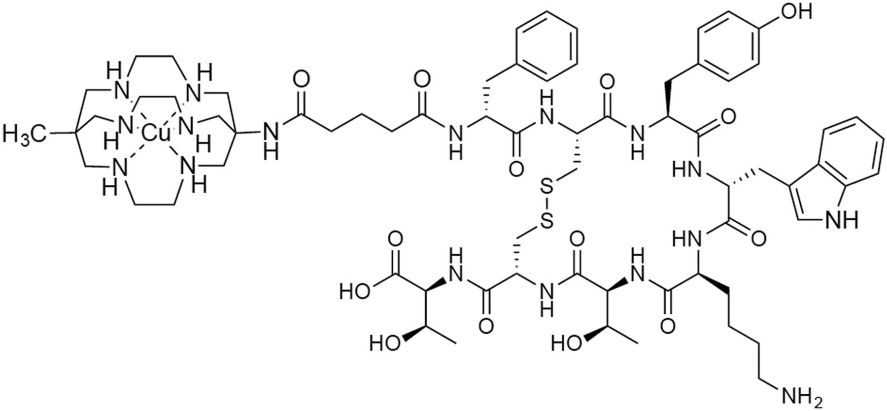
Preclinical evaluation of a sarcophagine ligand functionalized with Tyr3-octreotate, 64Cu-CuMeCOSar-Tyr3-octreotate (64Cu-CuSarTATE, Fig. 1), in a murine xenograft model revealed high uptake of 64Cu-CuSarTATE in somatostatin receptor–expressing tumors at 2 h after injection (63.0 ± 15.0 %IA/g) and remained high 24 h after injection (105 ± 27 %IA/g) (29). This agent was subsequently evaluated in 10 patients with neuroendocrine neoplasia and displayed high and late retention in tumor tissue, suggesting the agent is suitable for diagnostic purposes and for prospective dosimetry for peptide receptor radionuclide therapy (PRRT) with 67Cu-CuSarTATE (30).

Chemical structure of 64/67Cu-CuSarTATE.
In this work, we confirmed that 64Cu-CuSarTATE binds to somatostatin subtype 2a receptor–positive tumors in an AR42J (rat pancreatic exocrine tumor) xenograft model using PET imaging and then compared the therapeutic efficacy of 67Cu-CuSarTATE with that of 177Lu-LuTATE, which has been proven to be effective in multiple studies (31).
MATERIALS AND METHODS
Materials
SarTATE was prepared by Auspep (Tullamarine) using a modified version of a method reported previously (29). 64Cu-CuCl2 (700 MBq in 0.05 M HCl was provided by the Molecular Imaging and Therapy Research Unit of the South Australian Health and Medical Research Institute. 67Cu-CuCl2 was provided by Idaho Accelerator Center of Idaho State University as a solution (≤4 GBq) in 0.05–0.1 M HCl. 177Lu-LuTATE was prepared by the in-house radiopharmacy of Peter MacCallum Cancer Centre following standard protocols (32).
Preparation of 64Cu-CuSarTATE
64Cu-CuSarTATE was prepared on the iPhase MultiSyn synthesizer unit using a modified version of a previously described method (29,30). Briefly, 64Cu-CuCl2 (700 MBq in 0.05 M HCl [300 μL]) was added to SarTATE (20 μg, 13.7 nmol) in a solution of 10% ethanol in 0.1 M ammonium acetate (5 mL) containing gentisic acid, sodium salt (38 mg). The reaction mixture was incubated for 25 min at room temperature and then passed through a Strata X 33-μm polymeric reverse-phase cartridge (Phenomenex, Inc.). Cartridge-retained 64Cu-CuSarTATE was rinsed with saline for injection and then eluted with ethanol followed by saline for injection into a vial. The contents of this vial were filtered through a 0.22-μm filter. 64Cu-CuSarTATE was recovered in 60%–80% radiochemical yield with more than 95% radiochemical purity.
Preparation of 67Cu-CuSarTate
Briefly, 67Cu-CuCl2 (∼4 GBq, 0.05–0.1 M HCl) was added to SarTATE (60 μg, 41.2 nmol) in a solution of 10% ethanol in 0.1 M ammonium acetate (5 mL) containing gentisic acid, sodium salt (38 mg). The reaction mixture was incubated for 30 min at room temperature and then passed through a Strata-X 33-μm polymeric reverse-phase cartridge (30 mg). Cartridge-retained 67Cu-CuSarTATE was rinsed with saline for injection before elution with ethanol into a vial containing saline for injection. The contents of this vial were passed through a 0.22-mm filter to give 67Cu-CuSarTATE in 60%–80% radiochemical yield with more than 95% radiochemical purity.
PET Imaging and Biodistribution Studies
All in vivo studies were performed with the approval of the Peter MacCallum Cancer Centre Animal Experimentation Ethics Committee and in accordance with the Australian code for the care and use of animals for scientific purposes, eighth edition (2013). AR42J cells were obtained from the American Type Culture Collection. Cells (3 × 10⁶) in 50% Matrigel (Corning) in phosphate-buffered saline were implanted subcutaneously into the right flank of 6- to 7-wk-old female BALB/c nude mice (Australian Bioresources). Once tumors reached a volume of 100–150 mm3, the mice were injected intravenously with 3 MBq (0.24 nmol) of 64Cu-CuSarTATE. At 1 and 4 h after injection, the mice were anesthetized with 2% isoflurane in oxygen and placed into a G8 PET/CT scanner (Perkin Elmer). A CT image was then acquired over 1 min, followed by a 10-min static PET image. The PET images were reconstructed using the on-board maximum-likelihood expectation maximization algorithm and then analyzed using the VivoQuant (Invicro) software package. On completion of the 4 h of imaging, the mice were euthanized. Organs (blood, lungs, heart, liver, kidneys, muscle, spleen, and tumor) were excised and weighed, and the amount of radioactivity present in each organ was quantified using a Capintec (Captus 4000e) γ-counter. Data are presented as %IA/g tissue.
In Vivo Therapy Studies
In the therapy experiments, BALB/c nude female mice with subcutaneous AR42J xenografts (100–150 mm3) were intravenously injected with vehicle (saline), 5 MBq (0.07 nmol) of 177Lu-LuTATE, or 5 MBq (0.37 nmol) of 67Cu-CuSarTATE in a final volume of 100 μL (n = 7 for each group), or they were intravenously injected with vehicle (saline), 30 MBq (0.4 nmol) of 177Lu-LuTATE, or 30 MBq (2.2 nmol) of 67Cu-CuSarTATE either as a single administration or as a fractionated dose 14 d apart (15 MBq × 2) (n = 8 for each group). The mice were monitored twice weekly for tumor growth using calipers and evaluation of general health. They were euthanized when tumor volume exceeded 1,200 mm3. Tumor volume was calculated as length (mm) × width (mm) × height (π/6).
Data Analysis
Percentage tumor growth inhibition was calculated as 100 × (1 − ΔT/ΔC), where ΔC and ΔT were determined by subtracting the mean tumor volume (in the vehicle control and treated groups, respectively) on day 1 of treatment from the mean tumor volume on the last day all mice remained in the study. Statistical analysis was performed using GraphPad Prism, version 8.0. An ANOVA was performed, followed by a Dunnett post hoc test to compare tumor growth in the treated groups with that in the vehicle control group. Survival curves were analyzed using the Mantel–Cox log-rank test, where survival was defined as time for a tumor to reach a volume of at least 1,200 mm3.
RESULTS
Both 64Cu-CuSarTATE and 67Cu-CuSarTATE could be prepared in ammonium acetate buffer at room temperature in 30 min to give the complexes in 60%–80% radiochemical yield with more than 95% radiochemical purity.
64Cu-CuSarTATE Has High Tumor Uptake in Somatostatin Receptor 2–Positive AR42J Xenograft Model
PET images were acquired after administration of 64Cu-CuSarTATE (3 MBq, 0.24 nmol) via tail-vein injection to AR42J tumor–bearing female BALB/c nude mice. PET/CT images at 1 and 4 h after injection revealed very high tumor uptake and low background (Fig. 2). The tumors were clearly identified at 1 h after injection (SUVmax, 16.3 ± 1.7 at 1 h after injection), with excellent tumor to background ratios (66 ± 7 at 1 h after injection). Clearance over 4 h led to a further increase in the tumor-to-background ratios (76 ± 7; P = 0.027 vs. 1 h). The high tumor uptake was confirmed with an ex vivo biodistribution analysis in which the animals were euthanized after imaging at 4 h after injection and the amount of radioactivity in the tumor and organs was quantified (Fig. 2B). The tumor uptake 4 h after injection was 61.8 ± 2.4 %IA/g. Kidney uptake was 16.0 ± 0.7 %IA/g, and lung uptake was 12.2 ± 0.8 %IA/g.

(A) Representative maximum-intensity-projection PET/CT images of AR42J tumor–bearing female BALB/c nude mice after injection of 64Cu-CuSarTATE (3 MBq, 0.24 nmol of peptide) at 1 and 4 h after injection. (B) Ex vivo biodistribution expressed as %IA/g (mean ± SEM, n = 3) was performed after imaging at 4 h after injection.
67Cu-CuSarTATE Is Highly Efficacious Against AR42J Tumors In Vivo
Mice were inoculated with AR42J cells, and once the tumors reached a volume of approximately 150 mm3, the mice were randomized to receive saline, 177Lu-LuTATE, or 67Cu-CuSarTATE via intravenous injection. The treatments were well tolerated, causing only a transient reduction in animal body weight that did not exceed 5% of that at baseline (Fig. 3A). Seven days after a single administration of 67Cu-CuSarTATE (5 MBq, 0.37 nmol), tumor growth was inhibited by 75% (P = 0.0001 vs. control) compared with vehicle control (Fig. 3B). Administration of 177Lu-LuTATE (5 MBq) inhibited tumor growth by 89% (P ≤ 0.0004 vs. control) (Fig. 3B). Survival, defined as the time to a tumor volume of more than 1,200 mm3, was extended from 12 d in the control group to 21 d after treatment with both 67Cu-CuSarTATE and 177Lu-LuTATE (Fig. 3C).

Inhibition of AR42J tumor growth by 67Cu-CuSarTATE. AR42J tumor–bearing mice were treated with vehicle, 5 MBq of 177Lu-LuTATE, or 5 MBq of 67Cu-CuSarTATE on day 1 (vehicle and 177Lu-LuTATE) or day 2 (67Cu-CuSarTATE). (A and B) Percentage body weight change from baseline (A) and tumor volumes (B) were determined every 3–4 d. Data are shown until first mouse from group was removed from study. Data are expressed as mean ± SEM; n = 7 mice per group. (C) Kaplan–Meier survival analysis was performed on data in B, with survival defined as time to tumor volume ≥ 1,200 mm3.
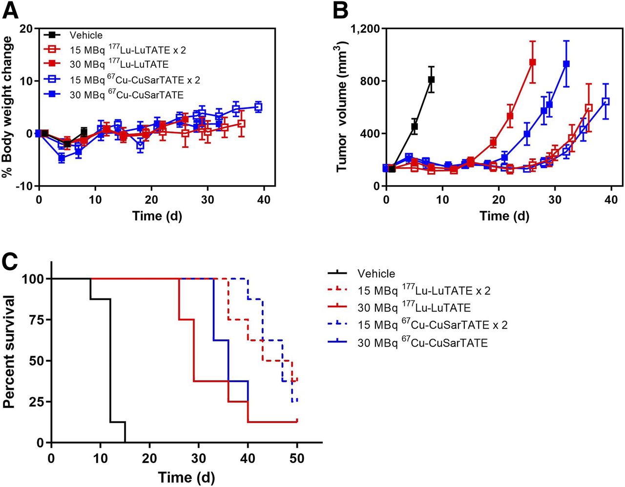
In a second study, the efficacy of fractionated delivery of PRRT was assessed. AR42J tumor–bearing mice were treated with a total of 30 MBq of 67Cu-CuSarTATE or 177Lu-LuTATE, either as a single intravenous injection or as two 15-MBq fractions 2 wk apart. All treatments were well tolerated (Fig. 4A) and induced tumor stasis, with reinitiation of tumor growth seen earlier in the single-fraction–treated groups (Fig. 4B). Furthermore, treatment of tumors with 2 fractions significantly improved survival when compared with delivery as a single fraction (67Cu-CuSarTATE: 47 vs. 36 d [P = 0.036]; 177Lu-LuTATE: 46 vs. 29 d [P = 0.00]; Fig. 4C). Equivalent efficacy was observed between 67Cu-CuSarTATE and 177Lu-LuTATE after treatment on both the single and the fractionated schedules (P = not statistically significant).

Enhanced efficacy of fractionated 67Cu-CuSarTATE PRRT. AR42J tumor–bearing mice were treated with vehicle on day 1, 30 MBq of 177Lu-LuTATE on day 1, 30 MBq of 67Cu-CuSarTATE on day 2, 15 MBq of 177Lu-LuTATE on day 1, and 15 or 15 MBq of 67Cu-CuSarTATE on days 2 and 16. Percentage body weight change from baseline (A) and tumor volumes were determined every 3–4 d. Data are shown until first mouse from group was removed from study. Data are expressed as mean ± SEM; n = 8 mice per group. (C) Kaplan–Meier survival analysis of data in B, where survival was defined as time to tumor volume ≥ 1,200 mm3.
DISCUSSION
A sarcophagine cage amine ligand, MeCOSar (5-(8-methyl-3,6,10,13,16,19-hexaaza-bicyclo[6.6.6]icosan-1-ylamino)-5-oxopentanoic acid) conjugated to (Tyr3)-octreotate (64Cu-CuSarTATE) was demonstrated to be an effective imaging agent and potential prospective dosimetry tool in 10 patients with neuroendocrine tumors (30). In this study, we aimed to investigate the therapeutic potential of 67Cu-CuSarTATE.
The previous preclinical evaluation of 64Cu-CuSarTATE for PET imaging of somatostatin-positive tumors (29) used a tumor model in which somatostatin receptor 2 is overexpressed through viral transfection (A427-7) (33). In this work, a different tumor cell line with endogenous expression of somatostatin receptor 2 (AR42J) was used. This cell line is commonly used as a model of neuroendocrine tumor because of high somatostatin receptor 2 expression and reliable growth as a xenograft. This cell line was derived from a spontaneous pancreatic tumor in rats. The effective tumor targeting of 64Cu-CuSarTATE in the AR42J model was confirmed by PET imaging before we proceeded with therapeutic evaluation of the ligand labeled with 67Cu. High tumor uptake was evident in the PET images at 1 h after injection, and retention of this uptake led to images at 4 h after injection with excellent tumor-to-background ratios (Fig. 2). The high tumor uptake was confirmed by an ex vivo biodistribution study (62 ± 2 %IA/g, 4 h after injection).
Administration of either 5 or 30 MBq, either as a single or a fractionated dose, of 67Cu-CuSarTATE demonstrated efficacy similar to that of the same activity of 177Lu-LuTATE. Both agents significantly reduced tumor volume and increased life span (Figs. 3 and 4). Fractionated-dose protocols can lead to reductions in tumor burden with decreased toxicity by delivering a high cumulative dose of activity to the tumor while allowing nontarget tissue to recover (21). It is also possible that cells in different phases of the cell cycle may be differentially sensitive to radiation and that, by fractionating doses, cells at different stages of the cell cycle may be more effectively targeted. Supporting the current clinical practice of performing several spaced cycles of PRRT, administration of a total of 30 MBq of 67Cu-CuSarTATE or 177Lu-LuTATE, as two 15-MBq fractions 2 wk apart, significantly improved survival over delivery as a single fraction of 30 MBq.
The increased fraction of γ-emission of 67Cu, when compared with 177Lu, leads to 3.2 times higher γ-exposure per decay with a slightly less penetrating mean energy and will result in a greater radiation cross dose to healthy tissues. This γ-cross exposure, however, has not been linked to adverse effects in 177Lu therapies, for which regions with primary uptake and exposure to β-electrons are most strongly implicated in tissue effects. The higher fraction of γ-emission for 67Cu may be beneficial in terms of SPECT imaging, potentially allowing dose verification after treatment by performance of SPECT with CT attenuation correction (34). By comparison, 131I has approximately 12-fold increased γ-emission per decay compared with 177Lu and, coupled with a greater mean γ-energy, raises several radiation protection considerations. In the United States, the Nuclear Regulatory Commission guidance indicates that up to 14 GBq of 67Cu can be administered on an outpatient basis if the external dose rate at 1 m is less than 0.22 mSv/h (35). Extrapolation of measured patient dose rate data has suggested no issue with release of patients who have received up to 5 GBq of 67Cu-CuSarTATE (36).
Importantly, 67Cu-CuSarTATE displayed efficacy similar to that of 177Lu-LuTATE. The energy of the β−-emissions from 67Cu is similar to the β−-emissions from 177Lu, but the significantly shorter t1/2 of 67Cu (2.58 vs. 6.71 d) provides a higher dose-rate. The ability to perform prospective dosimetry is an advantage for agents that use the 64Cu/67Cu theranostic pair, especially in patients with a large tumor burden (37) or impaired renal function and in the pediatric setting, where PRRT is an emerging treatment for advanced neuroblastoma (38). It is acknowledged that diagnostic imaging and therapy are often performed using different quantities of administered peptide, and this difference needs to be considered because biodistribution can change depending on the amount of peptide injected. However, initial data on the biodistribution and radiation dosimetry of 64Cu-CuSarTATE and 67Cu-CuSarTATE in meningioma patients showed similar tumor clearance for the 2 agents and consistent organ dose estimations (39,40). In terms of the potential translation of the 64Cu/67Cu theranostic pair to clinical studies, it is pertinent that 64Cu is produced on a cyclotron and its t1/2 enables distribution to sites without on-site radiochemistry facilities and permits imaging at later times points than is possible with 68Ga-based agents. In addition, because 67Cu can be produced with linear accelerators at high specific activity (>150 Ci/mg) and at radionuclide purity higher than 99%, its production is not reliant on nuclear reactors.
CONCLUSION
As anticipated, 67Cu-CuSarTATE is well tolerated in BALB/c nude mice and highly efficacious against AR42J tumors in vivo. Administration of 67Cu-CuSarTATE and 177Lu-LuTATE divided into 2 fractions over 2 wk was more efficacious than administration of a single fraction. The antitumor activity of 67Cu-CuSarTATE in the AR42J tumor model suggests that this novel agent warrants clinical assessment for the treatment of somatostatin-expressing neuroendocrine tumors.
DISCLOSURE
Charmaine Jeffery, Ellen van Dam, and Matthew Harris were or are employed by Clarity Pharmaceuticals, the licensee of the intellectual property for SarTATE. Charmaine Jeffery and Paul Donnelly have potential financial interests in Clarity Pharmaceuticals. Paul Donnelly is an inventor of intellectual property, in this area of research, which has been licensed from the University of Melbourne to Clarity Pharmaceuticals. Paul Donnelly serves on the Scientific Advisory Board of Clarity Pharmaceuticals. Unrelated to this project, Rodney Hicks has share options in Telix Radiopharmaceuticals that are held on behalf of the Peter MacCallum Cancer Centre. This study was partially funded by Clarity Pharmaceuticals. Paul Donnelly received funding from the Australian Research Council and the National Health and Medical Research Council (Australia), which funded aspects of this research. Rodney J. Hicks is the recipient of a National Health and Medical Research Council Practitioner Fellowship (APP1108050), which supported this work. The Australian Cancer Research Foundation provided a grant to fund the PET/CT scanner used in these studies. No other potential conflict of interest relevant to this article was reported.
KEY POINTS
QUESTION: Is PRRT with 67Cu-CuSarTATE efficacious against a somatostatin-positive xenograft model?
PERTINENT FINDINGS: 67Cu-CuSarTATE was well tolerated in BALB/c nude mice and was highly efficacious against AR42J tumors in vivo. The efficacy of 67Cu-CuSarTATE is similar to that of 177Lu-LuTATE.
IMPLICATIONS FOR PATIENT CARE: 64Cu-CuSarTATE offers the potential for diagnostic PET imaging to support prospective dosimetry for treatment with 67Cu-CuSarTATE.
Acknowledgments
We thank Rachael Walker and Jeannette Schreuders (Peter MacCallum Cancer Centre) for expert technical assistance and Peter Eu (Peter MacCallum Cancer Centre) for synthesizing 177Lu-LuTATE. We also thank Jon Stoner (ISU Idaho Accelerator Center) for providing 67Cu.
Footnotes
Published online May 15, 2020.
- © 2020 by the Society of Nuclear Medicine and Molecular Imaging.
REFERENCES
- Received for publication February 13, 2020.
- Accepted for publication April 16, 2020.


