


Abstract
Mesothelin is a cell-surface glycoprotein restricted to mesothelial cells overexpressed in several types of cancer, including triple-negative breast cancer not responding to trastuzumab or hormone-based therapies. Mesothelin-targeting therapies are currently being developed. However, the identification of patients potentially eligible for such a therapeutic strategy remains challenging. The objective of this study was to perform the radiolabeling and preclinical evaluation of 99mTc-A1 and 99mTc-C6, two antimesothelin single-domain antibody (sdAb)–derived imaging agents. Methods: A1 and C6 were radiolabeled with 99mTc and evaluated in vitro on recombinant protein and cells, as well as in vivo in xenograft mouse models of the triple-negative breast cancer cell lines HCC70 (mesothelin-positive) and MDA-MB-231 (mesothelin-negative). Results: Both 99mTc-A1 and 99mTc-C6 bound mesothelin with high affinity in vitro, with 99mTc-A1 affinity being 2.4-fold higher than that of 99mTc-C6 (dissociation constant, 43.9 ± 4.0 vs. 107 ± 16 nM, P < 0.05). 99mTc-A1 and 99mTc-C6 remained stable in vivo in murine blood (>80% at 2 h) and ex vivo in human blood (>90% at 6 h). In vivo 99mTc-A1 uptake (percentage injected dose) in HCC70 tumors was 5-fold higher than in MDA-MB-231 tumors and 1.5-fold higher than that of 99mTc-C6 (2.34% ± 0.36% vs. 0.48% ± 0.18% and 1.56% ± 0.43%, respectively, P < 0.01) and resulted in elevated tumor-to-background ratios. In vivo competition experiments demonstrated the specificity of 99mTc-A1 uptake in HCC70 tumors. Conclusion: Mesothelin-positive tumors were successfully identified by SPECT using 99mTc-A1 and 99mTc-C6. Considering its superior characteristics, 99mTc-A1 was selected as the most suitable tool for further clinical translation.
Triple-negative breast cancer (TNBC) is an invasive carcinoma that lacks expression of estrogen receptor, human epidermal growth factor receptor 2, and progesterone receptor. About 10%–20% of breast cancer cases are found to be TNBC. Patients with TNBC have an aggressive clinical course, with a high recurrence rate and a short survival outcome (1). TNBC lacks effective targeted therapies, and efforts are focused on the identification of new potential targets such as the cell-surface glycoprotein mesothelin (2,3). The mesothelin gene encodes a precursor protein of 71 kDa, processed into a shed form (megakaryocyte-potentiating factor) and a 40-kDa glycosylphosphatidylinisotol-anchored membrane protein, mesothelin (4). Mesothelin constitutive expression is restricted to mesothelial cells lining the pericardium, peritoneum, and pleura. However, mesothelin overexpression has been observed in 100% of epithelial mesotheliomas, most pancreatic and ovarian adenocarcinomas, and 34%–67% of TNBCs (5–7). Its biologic function is still poorly understood, and no detectable abnormalities have been observed in mesothelin-deficient mice (8). However, in pathologic conditions, several studies have recently indicated a potential role for mesothelin in cell survival, cell proliferation, tumor progression, and chemoresistance (9). Mesothelin expression has been correlated with a poorer patient outcome in several types of human cancer (10,11), and mesothelin is associated with lymph node tumoral infiltration and a decreased overall survival in breast cancer (12). Among patients with TNBC, those with a mesothelin-positive tumor were found to develop more distant metastases and to have lower overall and disease-free survival (13).
The limited expression of mesothelin in normal human tissues and its overexpression in several types of aggressive human cancer make it an attractive candidate for therapy, including for TNBC (7,14). Therefore, mesothelin-targeted therapies are currently undergoing clinical trials (15). In this setting, the identification of patients who could benefit from these targeted therapies remains challenging: an elevation of serum mesothelin has been shown in patients with mesothelioma and ovarian cancer but not in pancreatic cancer despite an overexpression of the membrane-bound mesothelin in these tumors (16,17). Moreover, because of inter- and intratumor heterogeneity, tumor phenotype cannot be accurately assessed by biopsy. Nuclear imaging is a highly sensitive noninvasive imaging modality that might address this challenge. Prantner et al. developed and performed the in vitro characterization of two antimesothelin single-domain antibodies (sdAbs), A1 and C6, with high specificities and nanomolar affinities for the 40-kDa mesothelin form (18). In the present study, in vitro and in vivo investigations were performed after 99mTc labeling of these sdAbs (99mTc-A1 and 99mTc-C6) to evaluate their potential as imaging probes for the nuclear imaging of mesothelin-positive TNBC.
MATERIALS AND METHODS
Cell Lines and Culture Conditions
The HCC70 cell line was cultured using RPMI 1640 medium supplemented with 10% fetal bovine serum and 1% penicillin–streptomycin. MDA-MB-231 cells were cultured with Dulbecco modified Eagle medium supplemented with 10% fetal bovine serum and 1% penicillin–streptomycin.
Western Blot Analysis
Total proteins from HCC70 and MDA-MB-231 cells were extracted with radioimmunoprecipitation assay buffer. Proteins were separated by 4%–15% denaturing sodium dodecyl sulfate polyacrylamide gel electrophoresis and transferred onto a nitrocellulose membrane. The membrane was incubated with the antimesothelin antibody (Boster immunoleader). Actin was detected with anti–β-actin antibody (Beckton Dickinson). Revelation was assessed using the chemiluminescence enhanced chemiluminescence kit (Biorad).
Production and Purification of sdAbs
Production and purification of the anti-mesothelin sdAbs were done as previously published (18).
Radiolabeling with 99mTc
A heating step was necessary over the course of the radiolabeling procedure. Thermal unfolding of A1 or C6 was therefore monitored by circular dichroism. A1, C6, and an irrelevant control sdAb were then radiolabeled with 99mTc using the tricarbonyl method and the C-terminal hexahistidine-tag as previously described (19). The radiochemical purity of all 3 sdAbs was determined immediately after labeling and 6 h after labeling by radio–high-performance liquid chromatography (Shimadzu).
Lipophilicity
The lipophilicity of 99mTc-A1 and 99mTc-C6 was evaluated using an octanol phosphate-buffered saline (PBS) distribution study. The partition coefficient (log P) was calculated as the radioactivity ratio of the organic phase to the aqueous phase using the following formula: log P = log (total counts in 1-octanol/total counts in PBS buffer).
Distribution and Stability in Murine and Human Blood
The distribution and stability of 99mTc-sdAbs were evaluated after in vitro incubation in human blood and after in vivo injection into mice. A blood sample was centrifuged (4,000g, 2 min) at the selected time points to separate the plasma from the blood cells. Trichloroacetic acid (10%, 5 μL) was then added to the plasma fraction and the sample was centrifuged at 11,000g for 3 min to separate large proteins (>>15 kD) from the remaining plasma. Blood distribution was expressed as a percentage of total blood tracer activity contained in blood cells, large proteins, and remaining plasma fractions. To determine the stability of each radiolabeled sdAb, the plasma fraction was analyzed using radio–high-performance liquid chromatography.
Saturation Binding Experiments
Saturation binding was determined on 96-well plates coated with human mesothelin recombinant protein (100 ng/well; R&D Systems) or formalin-fixed HCC70 or MDA-MB-231 cells (1.5 × 10⁵ cells/well). Serial dilutions of 99mTc-A1 and 99mTc-C6 were incubated for 1 h at room temperature before being washed 5 times with PBS–polysorbate 0.1%. The radioactivity in each well was then determined using a γ-counter (Wizard2; Perkin Elmer) and corrected for unspecific binding. 99mTc-A1 and 99mTc-C6 binding curves were fitted using a nonlinear regression equation (specific binding: y = Bmax × x/(KD + x), with x being the radioligand concentration, KD being the dissociation constant, and Bmax being the maximum number of binding sites, or receptor density) (GraphPad Prism, version 6, software), to determine KD values. Uptake was normalized to Bmax for graphic representation of 3 independent experiments.
In Vitro Competition Studies
HCC70 and MDA-MB-231 cells (200,000) were rinsed with PBS, detached using 5 mM ethylenediaminetetraacetic acid, and incubated with 99mTc-A1 or 99mTc-C6 at their respective KD for 1 h at 4°C in the absence or presence of a 200-fold excess of unlabeled A1 or C6, respectively. After 5 washes in cold PBS, the radioactivity was determined using a Wizard2 γ-counter.
Tumor Model
All procedures performed were in accordance with the institutional guidelines and approved by the animal care and use committees of Grenoble University. To evaluate 99mTc-A1 and 99mTc-C6 biodistribution and tumor uptake, female BALB/c nu/nu mice (5 wk old) were subcutaneously inoculated into the flank with either HCC70 cells (3.5 × 10⁶, n = 32) or MDA-MB-231 cells (2 × 10⁶, n = 6), in 2:1 (v/v) PBS/Matrigel (Corning). The tumors were allowed to grow for 3–4 wk to reach about 400 mm3.
SPECT/CT Imaging
The mice were divided into 4 groups: HCC70 tumor–bearing mice injected with either 99mTc-A1 (HCC70-A1, n = 8), 99mTc-C6 (HCC70-C6, n = 7), or irrelevant 99mTc-CTL (HCC70-CTL, n = 7), and MDA-MB-231 tumor–bearing mice injected with 99mTc-A1 (MDA-MB-231, n = 6). SPECT/CT acquisitions were performed 1 h after intravenous injection of 49.1 ± 13.7 MBq of 99mTc-sdAbs. For competition studies, HCC70 tumor–bearing mice were injected with 17.6 ± 5.3 MBq of 99mTc-A1 with or without a 150-fold excess of unlabeled A1 sdAb (n = 5 per condition). SPECT/CT acquisitions were performed using a dedicated system (nanoSPECT/CT; Mediso). Images were quantified after correction for decay and normalization to the injected dose. SPECT quantification was performed on the basis of the CT data, with the volume of interest corresponding to a sphere of 50 mm3 drawn at the center of the tumor on the CT image. 99mTc-sdAb uptake was expressed as percentage injected dose (%ID)/cm3.
Postmortem Analysis
Two hours after injection and immediately after the SPECT/CT image acquisition, the anesthetized mice were euthanized using CO2 and tumors were harvested along with other organs. Tissues were weighed, and tracer activity was determined with a Wizard2 γ-counter. The results were corrected for decay, injected dose, and organ weight and were expressed as %ID/g. Tumor-to-muscle (T/M) and tumor-to-blood (T/B) activity ratios were computed. Immunohistochemistry was performed on tumor slices using A1 sdAb (20 μg/mL) or a commercial antimesothelin antibody (0.5 μg/mL; Boster immunoleader).
Statistics
Data were expressed as mean ± SD and were compared using an unpaired Mann–Whitney test for intergroup analysis. The significance of linear correlations was assessed with a Pearson test. P values lower than 0.05 were considered significant.
RESULTS
99mTc Radiolabeling and Lipophilicity
No thermal unfolding was detected by circular dichroism up to 65°C (Supplemental Fig. 1 [available at http://jnm.snmjournals.org]). This temperature was therefore used for the radiolabeling procedure. A1 and C6 were successfully radiolabeled with 99mTc. The radiochemical purity was higher than 99% immediately after radiolabeling, with a 16.1- and 16.0-min retention time for 99mTc-A1 and 99mTc-C6, respectively (Figs. 1A and 1B). Moreover, 99mTc-A1 and 99mTc-C6 remained stable for 6 h after labeling, with a radiochemical purity higher than 98%. Log P values were −1.8 ± 0.5 for 99mTc-A1 and −2.3 ± 0.8 for 99mTc-C6 (Supplemental Table 1).

Radiochromatograms of 99mTc-A1 and 99mTc-C6 immediately after radiolabeling (A and B, respectively) and 2 h after injection into mice (C and D, respectively).
99mTc-A1 and 99mTc-C6 Affinity and Specificity for Mesothelin
Western blot analysis indicated that mesothelin was expressed by HCC70 cells but not by MDA-MB-231 cells (Fig. 2A). Figure 2B shows the specific binding of 99mTc-A1 and 99mTc-C6 on mesothelin recombinant protein. The affinity of 99mTc-A1 for mesothelin was more than 2-fold higher than that of 99mTc-C6 (KD = 43.9 ± 4.0 nM and 107.3 ± 15.9 nM, respectively, Fig. 2B). Similar results were obtained on HCC70 cells (Fig. 2C). Moreover, competition studies were performed to assess the specificity of binding (Fig. 2D). Binding of 99mTc-A1 and 99mTc-C6 to mesothelin-positive HCC70 cells was, respectively, 7.9- and 4.6-fold higher than that observed to mesothelin-negative MDA-MB-231 cells. Radiolabeled sdAb binding was displaced by a 200-fold excess of unlabeled sdAb, with a significant 6-fold and 3.5-fold decrease in 99mTc-A1 and 99mTc-C6 binding, respectively. This result indicates that radiolabeled sdAbs retain their specific binding activity on mesothelin (P < 0.001).

High-affinity and high-specificity binding of 99mTc-A1 to mesothelin. (A) As determined by Western blot, HCC70 were mesothelin-positive whereas MDA-MB-231 were mesothelin-negative. (B) Saturation binding on human recombinant mesothelin (MSLN): 99mTc-A1 and 99mTc-C6 KD was 44 ± 4 and 107 ± 16 nM, respectively. (C) Saturation binding on HCC70 cells: 99mTc-A1 and 99mTc-C6 KD was 45 ± 11 and 135 ± 37 nM, respectively. (D) Binding of 99mTc-A1 and 99mTc-C6 to HCC70 cells was significantly inhibited by 200-fold excess of unlabeled A1 or C6 sdAb. ***P < 0.001 vs. HCC70 + competition. Experiments were performed in triplicate.
Blood Distribution Patterns and Stability In Vitro in Human Blood
Similar distribution patterns were observed over time (Supplemental Fig. 2). Both sdAbs were predominantly localized in the plasma fraction (63%–75%), where they remained highly stable, with a radiochemical purity higher than 98% from 0 to 6 h (Supplemental Table 2).
In Vivo Stability in Mouse Blood
Radiochromatograms of 99mTc-A1 and 99mTc-C6, before and 2 h after injection, are shown in Figures 1C and 1D. A single peak corresponding to the radiolabeled sdAb was observed for both tracers. The absence of alternative radioactive products on the profiles confirmed the in vivo stability of 99mTc-A1 and 99mTc-C6, with radiochemical purities of 91.3% ± 5.2% and 95.4% ± 4.8%, respectively.
Immunohistochemistry
Mesothelin expression was evaluated by immunohistochemistry on HCC70 and MDA-MB-231 xenografts using a commercial antibody or A1 sdAb (Supplemental Fig. 3). In accordance with observations in cell cultures, HCC70 tumor xenografts expressed mesothelin whereas MDA-MB-231 did not.
SPECT/CT Imaging
Figure 3A shows sagittal, coronal, and transversal views of fused SPECT/CT images. 99mTc-A1 and 99mTc-C6 uptake in mesothelin-positive HCC70 tumors was readily identifiable, whereas a weak signal was observed with the irrelevant control sdAb in HCC70 tumors and with 99mTc-A1 in mesothelin-negative MDA-MB-231 tumors. Interestingly, uptake of 99mTc-C6 in HCC70 tumors was visually lower than that of 99mTc-A1. Furthermore, liver accumulation was observed for 99mTc-C6 but not for 99mTc-A1. Nonspecific kidney elimination was observed in all groups.
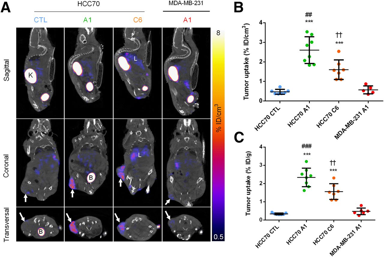
In vivo biodistribution of 99mTc-A1 and 99mTc-C6 in HCC70 and MDA-MB-231 tumor xenografts. (A) Representative sagittal, coronal, and transversal views of fused SPECT/CT images of HCC70 and MDA-MB-231 tumor–bearing mice 1 h after intravenous injection of 99mTc-A1 or 99mTc-C6. K = kidneys; B = bladder; L = liver. (B) In vivo quantification of 99mTc-A1 and 99mTc-C6 tumor uptake from SPECT images. (C) Ex vivo quantification of 99mTc-A1 and 99mTc-C6 tumor uptake from postmortem analysis. Results are expressed as %ID/g of tumor. ##P < 0.01 vs. MDA-MB-231 A1. ###P < 0.001 vs. MDA-MB-231 A1. ***P < 0.001 vs. HCC70 control sdAb. ††P < 0.01 vs. HCC-70 A1.
These observations were confirmed by image quantification showing that 99mTc-A1 activity was 5-fold higher than 99mTc-CTL activity in HCC70 tumor–bearing mice (2.6 ± 0.7 vs. 0.5 ± 0.13 %ID/g, respectively, P < 0.01). In addition, 99mTc-A1 uptake in HCC70 tumors was 4-fold higher than that observed in MDA-MB-231 tumors (0.6 ± 0.2 %ID/g, P < 0.001) (Fig. 3B). HCC70 99mTc-C6 uptake (1.4 ± 0.3 %ID/g) was also significantly higher than that of 99mTc-CTL (P < 0.001) but remained about 2-fold lower than that of 99mTc-A1 (P < 0.01). These results were confirmed by ex vivo γ-well counting quantification (Fig. 3C). The agreement between results from in vivo and ex vivo quantification was further confirmed by linear regression analysis (y = 1.08x + 0.09, r2 = 0.97, P < 0.001) (Supplemental Fig. 4).
Biodistribution of 99mTc-A1 and 99mTc-C6
The results from 2-h biodistribution studies after 99mTc-A1, 99mTc-C6, or 99mTc-CTL intravenous injection are summarized in Table 1. High kidney activity (>200 %ID/g) was observed for all sdAbs. 99mTc-A1 uptake was less than 1 %ID/g for all investigated organs with the exception of kidney and tumor. In comparison, 99mTc-C6 uptake was more than 1 %ID/g in liver and significantly higher than that of 99mTc-A1 and 99mTc-CTL in stomach, liver, and intestine (P < 0.01). The blood activity of all 3 sdAbs was less than 0.5 %ID/g. T/B and T/M ratios were determined for each group. The T/B ratio of the HCC70-A1 group was 10-fold higher than that of the HCC70-CTL group (10.3 ± 4.4 vs. 1.1 ± 0.7, P < 0.001). Similarly, the T/M ratio of the HCC70-A1 group was 5-fold higher than that of the CTL group (22.5 ± 3.4 vs. 4.0 ± 1.8, P < 0.001). The T/M and T/B ratios of the HCC70-C6 group were also found to be higher than those of the HCC70-CTL group (P < 0.01) but remained significantly lower than those of the HCC70-A1 group (P < 0.05 for both ratios).
Biodistribution of 99mTc-A1 and 99mTc-C6 in Athymic Nude Mice Bearing HCC70 or MDA-MB-231 Xenografts
In Vivo Competition Study
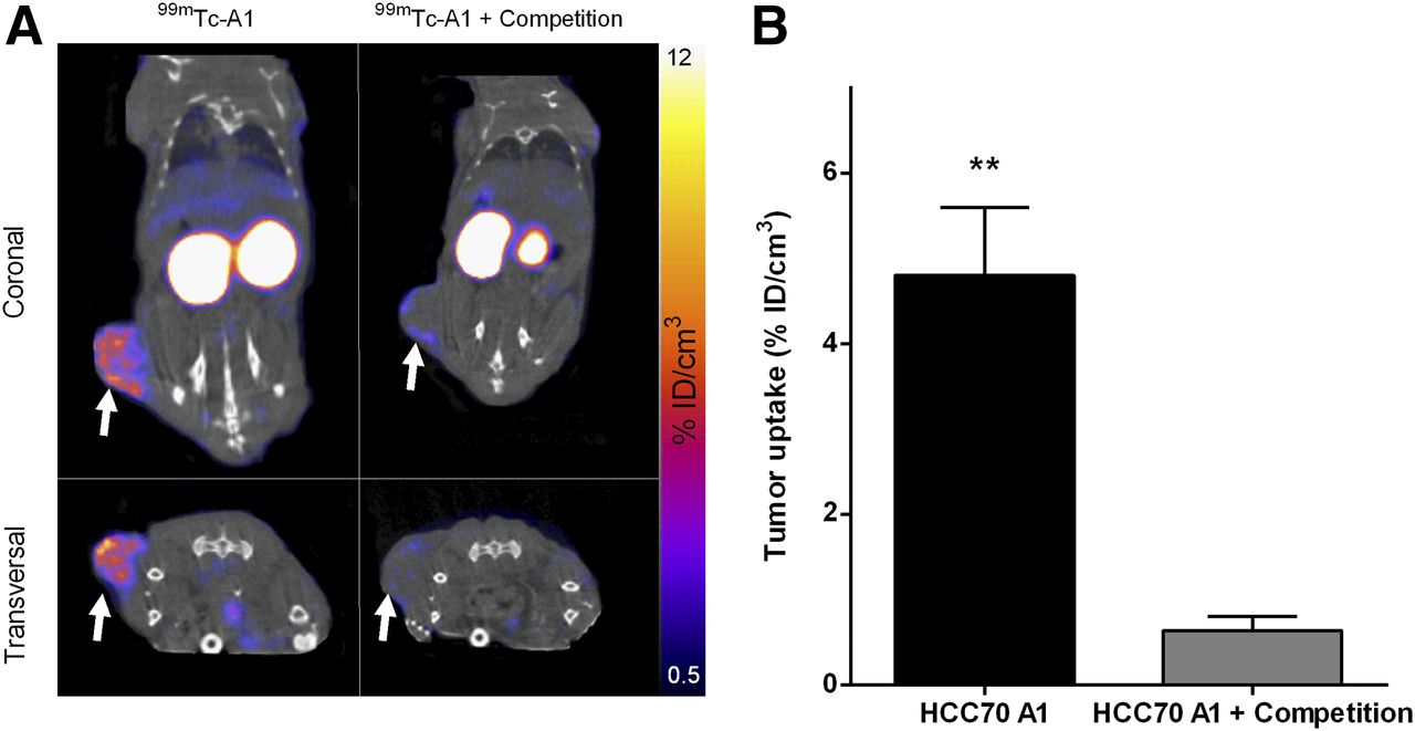
The in vivo coinjection of a 150-fold excess of unlabeled A1 induced an approximately 7-fold decrease in 99mTc-A1 uptake in HCC70 tumors as determined by SPECT image quantification (0.6 ± 0.2 %ID/g for HCC70-A1 + competition vs. 4.8 ± 0.8 %ID/g for HCC70-A1 alone, P < 0.01, Fig. 4). Ex vivo γ-well counting further confirmed this finding (0.5 ± 0.1 %ID/g with competitor vs. 4.2 ± 0.8 %ID/g without, P < 0.01) (Table 2). With the exception of kidney, 99mTc-A1 uptake in all other investigated organs did not significantly change. Consequently, a significant decrease was observed in T/B and T/M ratios (P < 0.01 for both).

In vivo competition study. (A) Representative SPECT/CT images of HCC70 tumor–bearing mice injected with 99mTc-A1 alone or with 150-fold excess of unlabeled A1 (competition). Arrows indicate tumor location (B) SPECT image quantification. Results are expressed as %ID/cm3 of tissue. **P < 0.01 vs. HCC70 A1 + competition.
Effect of Competitor on 99mTc-A1 Biodistribution in Athymic Nude Mice Bearing HCC70 Xenografts
DISCUSSION
Most cases of TNBC have an aggressive clinical course characterized by a higher recurrence rate, more distant metastases, and an overall lower survival than other forms of breast cancer (1). TNBCs are treated with chemotherapy or radiation therapy. However, because some are chemotherapy-resistant, other therapies, such as immunotherapy, are being evaluated (20).
Potential targets have been identified, including mesothelin (2,3). Mesothelin is a 40-kDa glycosylphosphatidylinisotol-anchored membrane glycoprotein with limited normal-tissue expression and frequent overexpression in most types of aggressive cancer, such as pancreatic cancer, ovarian adenocarcinoma, mesothelioma, and TNBC (5,7). Several therapies targeting mesothelin-expressing tumors have been developed and are currently undergoing clinical translation (15). Preclinical studies using RG7787, a recombinant immunotoxin, in combination with chemotherapy showed complete remission of mesothelin-expressing tumor xenografts in mice (21). Phase I clinical trials of RG7787 for treatment of mesothelioma have opened (22). Moreover, on the basis of studies showing that mesothelin could elicit a CD8-positive T-cell response in patients, tumor vaccines are under clinical investigations (23).
Patients with mesothelin-expressing tumors can be identified using biopsy and blood testing with the serum mesothelin-related peptide (16,24). However, discordance in the expression of tumor markers is often observed between the primary tumor and the metastases, which are not always accessible to biopsy. In addition, this marker is not increased in pancreatic cancer despite an overexpression of the mesothelin-membrane form (17). By allowing noninvasive tumoral phenotyping, nuclear imaging might help to overcome the limitations of biopsies and blood testing mentioned above.
The objective of the present study was to perform the in vivo nuclear imaging of TNBC xenografts with 99mTc-A1 and 99mTc-C6, two antimesothelin sdAbs, using the two TNBC cell lines HCC70 and MDA-MB-231. Mesothelin expression was observed on HCC70 cells but not on MDA-MB-231 cells, which were therefore used as a negative control. 99mTc-A1 exhibited a high affinity for mesothelin (KD = 43 nM), as demonstrated in vitro on recombinant mesothelin human protein and HCC70 cells. In vitro competition experiments on HCC70 cells confirmed the 99mTc-A1 binding specificity. In accordance with the results of Prantner et al. (18) using unlabeled compounds, the affinity of 99mTc-C6 for human recombinant mesothelin was found to be 3-fold lower than that of 99mTc-A1, thereby indicating the suitability of the radiolabeling method. Both radiotracers remained stable over time after in vitro incubation with human blood and in vivo injection into mice. Moreover, 99mTc-A1 and 99mTc-C6 remained mostly in the plasma fraction, thereby allowing good in vivo bioavailability. 99mTc-A1 and 99mTc-C6 both enabled the noninvasive visualization of mesothelin-positive tumors by SPECT imaging. High accumulation of 99mTc-A1 and 99mTc-C6 was observed in mesothelin-positive HCC70 tumors, whereas no signal was found in mesothelin-negative MDA-MB-231 tumors. However, the signal of 99mTc-A1 in HCC70 tumors was higher than that of 99mTc-C6. SPECT image quantification further confirmed these results, with a 5-fold higher 99mTc-A1 uptake in HCC70 tumors than in MDA-MB-231 tumors. Moreover, the in vivo competition study demonstrated the specificity of 99mTc-A1 binding to mesothelin. Renal accumulation was observed with both tracers, in accordance with the general pattern of sdAb biodistribution (25). In fact, most sdAbs are eliminated through the kidneys, and reuptake by the megalin–cubulin complex is responsible for their renal retention. In addition to tumor, kidney was also shown to have decreased 99mTc-A1 retention by the competition studies, as might be explained by the saturation of the megalin–cubulin complex by the competitor. Such saturation of this complex has been applied by other groups using succinylated gelatin, a plasma substitute, resulting in a significant 40%–50% decrease in kidney retention of the evaluated sdAb (26). No signal was observed on in vivo SPECT images after 99mTc-A1 injection with the exception of tumor, kidney, and bladder, most likely as a result of, first, weak mesothelin expression limited to the pericardium, pleura, and peritoneum and, second, A1 affinity for human mesothelin. Interestingly, mild-intensity signals were also observed in liver and intestine after 99mTc-C6 injection, suggesting that the liver is involved in 99mTc-C6 elimination. Monoclonal antibodies radiolabeled with 64Cu or 89Zr have recently allowed the detection of mesothelin-expressing tumors in mouse models of xenograft pancreatic tumors (27,28). Nevertheless, the hepatic elimination and slow blood clearance of these radiolabeled monoclonal antibodies represent major limitations. The smaller size of sdAb-based imaging agents allows fast blood clearance and image acquisition with high target-to-background ratios as early as 1 h after administration. Specifically, the 99mTc-A1 T/B ratio of 14 that was observed in the present study at 2 h after injection compares favorably with the T/B ratio of 1–4 that was previously observed at 24 h after injection for 64Cu and 89Zr-labeled antibodies (27,28).
Because 20% of women with breast cancer will develop distant metastases within 5 y of diagnosis, an ideal TNBC imaging agent should demonstrate high target-to-background ratios not only at the primary tumor site but also in pulmonary, hepatic, and bone lesions since these are the most frequent metastatic sites for TNBC. In the present study, minimal nonspecific uptake was observed in those organs with 99mTc-A1 but not with 99mTc-C6. Taken together with the higher affinity and higher absolute tumor uptake of 99mTc-A1, this additional result suggests that it would be better suited for metastasis imaging. We will therefore conduct further studies to evaluate the potential of 99mTc-A1 for the detection of breast cancer metastasis in mice. Although HCC70 cells share features of TNBC, few data on their metastatic abilities in mice are available. Other TNBC cells might therefore also be used.
CONCLUSION
The identification of mesothelin-expressing metastases would allow selection of patients who might benefit from mesothelin-targeted therapies. Two antimesothelin sdAbs were evaluated for their ability to detect mesothelin-expressing tumors in vivo. The results indicated that 99mTc-A1 is a promising tracer for the detection of mesothelin-expressing tumors. Further development of 99mTc-A1 will include 1,4,7,10-tetraazacyclododecane-1,4,7,10-tetraacetic acid chelation chemistry to allow either 68Ga or 177Lu radiolabeling for diagnosis and therapy, additional chemical engineering to minimize renal uptake, and additional characterizations such as internalization and tumor retention.
DISCLOSURE
This work was partly funded by grant ANR-11-INBS-0006 from France Life Imaging. No other potential conflict of interest relevant to this article was reported.
Acknowledgments
We thank Dr. Annie Molla (Université Grenoble Alpes, INSERM, CNRS, IAB, Grenoble), who kindly provided the HCC70 cell line.
Footnotes
↵* Contributed equally to this work.
Published online Mar. 23, 2018.
- © 2018 by the Society of Nuclear Medicine and Molecular Imaging.
REFERENCES
- Received for publication October 11, 2017.
- Accepted for publication November 28, 2017.


Jump to section
Related Articles
Cited By...
- No citing articles found.