


Abstract
The potential of receptor-mediated fluorescence-based image-guided surgery tracers is generally linked to the near-infrared emission profile and good-manufacturing-production availability of fluorescent dyes. Surprisingly, little is known about the critical interaction between the structural composition of the dyes and the pharmacokinetics of the tracers. In this study, a dual-modality tracer design was used to systematically and quantitatively evaluate the influence of elongation of the polymethine chain in a fluorescent cyanine dye on the imaging potential of a targeted tracer. Methods: As a model system, the integrin marker αvβ3 was targeted using arginylglycylaspartisc acid [RGD]–based vectors functionalized with a 111In-diethylenetriaminepentaacetic acid (DTPA) chelate and a fluorescent dye: (Cy3-(SO3)methyl-COOH [emission wavelength (λem), 580 nm], Cy5-(SO3)methyl-COOH [λem, 680 nm], or Cy7-(SO3)methyl-COOH [λem, 780 nm]). Tracers were analyzed for differences in photophysical properties, serum protein binding, chemical or optical stability, and signal penetration through tissue. Receptor affinities were evaluated using saturation and competition experiments. In vivo biodistribution (SPECT imaging and percentage injected dose per gram of tissue) was assessed in tumor-bearing mice and complemented with in vivo and ex vivo fluorescence images obtained using a clinical-grade multispectral fluorescence laparoscope. Results: Two carbon-atom-step variations in the polymethine chain of the fluorescent cyanine dyes were shown to significantly influence the chemical and photophysical characteristics (e.g., stability, brightness, and tissue penetration) of the hybrid RGD tracers. DTPA-Cy5-(SO3)methyl-COOH-c[RGDyK] structurally outperformed its Cy3 and Cy7 derivatives. Radioactivity-based evaluation of in vivo tracer pharmacokinetics yielded the lowest nonspecific uptake and highest tumor-to-background ratio for DTPA-Cy5-(SO3)methyl-COOH-c[RGDyK] (13.2 ± 1.7), with the Cy3 and Cy7 analogs trailing at respective tumor-to-background ratios of 5.7 ± 0.7 and 4.7 ± 0.7. Fluorescence-based assessment of tumor visibility revealed a similar trend. Conclusion: These findings underline that variations in the polymethine chain lengths of cyanine dyes have a profound influence on the photophysical properties, stability, and in vivo targeting capabilities of fluorescent imaging tracers. In a direct comparison, the intermediate-length dye (Cy5) yielded a superior c[RGDyK] tracer, compared with the shorter (Cy3) and longer (Cy7) analogs.
Within the nuclear medicine community, structural optimization of receptor-targeted radiotracers is a common good. Specific attention is paid to the influence of radiolabeling methods on the tracer pharmacokinetics (1). When the generation of fluorescence tracers is being pursued, however, the limited ability to quantify fluorescence tracer distribution (2) means that such studies are often ignored. As a result, fluorescent tracers are being transferred to clinical trials without knowledge or reporting of the whole-body kinetics (3–6). By including radiolabels on fluorescence tracers, so-called dual-modality (bimodal or hybrid) tracers are generated that allow quantitative mapping of the pharmacokinetic properties (7). This concept has been successfully applied to nanoparticles, antibody analogs, and peptides (8–13), as exemplified by the plurality of hybrid tracers in the literature (14–17). For example, for the arginylglycylaspartic acid (RGD) vector, it has been documented that the introduction of different pendant moieties on an otherwise stable conjugated system influences the pharmacokinetics (18–22).
The theoretic argument that near-infrared fluorescence emissions (emission wavelength [λem], ≥750 nm) provide superior tissue penetration has driven the use of reactive dyes such as indocyanine green (ICG)-OSu, ZW800, and IRDye 800CW (1,23,24). When these dyes are used to functionalize a targeting vector, their tendency to stack, chemical instability, low fluorescence brightness, or relatively large size are generally not considered (25–27). Preclinical and clinical studies indicate that the in-depth fluorescence imaging properties of slightly smaller far-red dyes (e.g., the cyanine dye Cy5; λem, 650–750 nm) also support in vivo applications (28–30), including fluorescence-guided surgery (3,31). In addition to providing an alternative to the use of near-infrared dyes, adoption of other fluorescence wavelengths also creates the potential for a multiparametric image-guidance process that supports the detection of two or more fluorescent features in one patient (29,32).
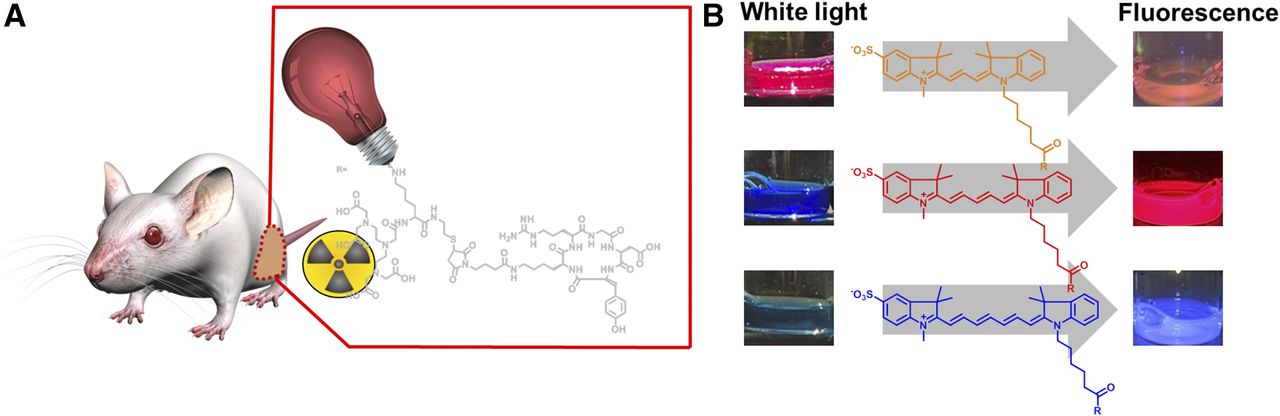
In the unique design of cyanine dyes, wherein the length of the polymethine chain that bridges the two indole units (being 3, 5, and 7 carbon atoms long in Cy3, Cy5, and Cy7, respectively) dictates the spectral properties of the dye. This feature allows the creation of fluorescent tracers of which the spectral properties can be specifically tuned for use alongside tracers of different wavelengths. Whereas dye optimization currently focuses on optimization of pendant moieties on cyanine dyes (18,33), the influence of alterations in the length of the polymethine chain has not yet been reported. To isolate the influence of the length of the polymethine chain on overall tracer performance, the performance of a previously optimized Cy5 dye (Cy5-(SO3)methyl-COOH) (18) was systematically compared with its Cy3 and Cy7 analogs with identical pendant moieties. A hybrid RGD-tracer design (Fig. 1) was used to directly link detailed evaluation of the chemical properties (synthesis and chemical stability) and photophysical properties (brightness and optical stability) of these three tracers to their respective biologic properties (in vitro, in vivo, and ex vivo) and imaging capabilities.

(A) Schematic visualization of mouse in which tumor is illuminated by hybrid c[RGDyK] tracer. (B) The 3 different cyanine dyes (Cy3 in orange, Cy5 in red, and Cy7 in blue) and their visible and fluorescence colorations as depicted by Karl Storz laparoscopic setup.
MATERIALS AND METHODS
The Cy3, Cy5, and Cy7 dyes and corresponding hybrid RGD peptides were prepared and photophysically analyzed in line with previous reports (18,34–37). Determination of photophysical properties and of stability (18,27,36,38), receptor affinity (15), and fluorescence confocal imaging (15) was performed as previously described.
The 4T1 tumor model was generated as previously described (15,18,39). All animal experiments were in accordance with Dutch welfare regulations and were approved by the local ethics committee of the Leiden University Medical Center.
Isotope-Based Assessment of In Vivo Tracer Distribution
Radiolabeling of the diethylenetriaminepentaacetic acid (DTPA) functionalized tracers DTPA-Cy3-(SO3)methyl-COOH-c[RGDyK], DTPA-Cy5-(SO3)methyl-COOH-c[RGDyK], and DTPA-Cy7-(SO3)methyl-COOH-c[RGDyK] with 111InCl3 and subsequent assessment of labeling accuracy was performed as described previously (18), using about 1 MBq for biodistribution (n = 4) and 10–20 MBq for SPECT imaging (n = 2), followed by ex vivo evaluation of the biodistribution (in total, 6 animals per tracer). Imaging of 4T1 tumor–bearing mice was performed on a U-SPECT scanner (MILabs) according to previously described methods (18,39).
Quantitative biodistribution (percentage injected dose per gram of tissue [%ID/g] and tumor-to-background ratio [T/B] [tumor-to-muscle %ID/g]) were measured 24 h after injection and analyzed with a relative performance scoring based on previously described procedures (18).
In Vivo Fluorescence Imaging
The mice were sacrificed after SPECT imaging, followed by fluorescence imaging performed using a preclinical IVIS Spectrum system (Caliper Life Sciences) and a clinical-grade laparoscope setup (Storz Endoskope GMBh) that allows multispectral imaging (30).
On the preclinical system, in vivo and ex vivo images of mice and excised tissues were acquired at Cy3 (excitation wavelength [λex], 535 nm; λem, 580 nm), Cy5 (λex, 640 nm; λem, 680 nm), and Cy7 (λex, 745 nm; λem, 780 nm) settings. Quantitative analysis of the fluorescence in the tissues (photons/s/cm2) and the T/B ratio was performed using the Living Image software, version 3.2 (Caliper Life Sciences). For imaging with the fluorescence laparoscope, mice were placed on top of an adjustable lift and imaged with a setup identical to that used to evaluate the signal penetration of the fluorescent emissions (28–30). Images were acquired using a clinical-grade IMAGE 1 S camera equipped with a 0° laparoscope. Excitation was achieved using a D-Light C light source (autofluorescence/fluorescein), a Cy5-modified D-Light C light source (Cy5), and a prototype D-Light P light source (ICG) (all Karl Storz). Built-in emission filters for Cy3 (autofluorescence mode) and Cy7 (ICG mode; catalog no. 26003AGA; Karl Storz) were used for detection of either Cy3 or Cy7. Cy5 was detected by placement of an additional standard eyepiece adaptor (catalog no. 20100034; Karl Storz) between the camera and the laparoscope (30).
Statistical Data Analysis
Analytic data were expressed as mean ± SD as calculated using Microsoft Excel software. The significance of 2 mean values was calculated using a Student t test. The level of significance was set at a P value of 0.05.
RESULTS
Synthesis of CyX-(SO3)Methyl-COOH Dyes and DTPA-CyX-c[RGDyK] Tracers
The ease of synthesis was highest for Cy5-(SO3)methyl-COOH. Although Cy7-(SO3)methyl-COOH could also be synthesized using a good-manufacturing-production–compatible solid support (see supplemental materials, available at http://jnm.snmjournals.org), reduced reactivity of the reagent for the elongated polymethine chain was observed. The preparation of Cy3-(SO3)methyl-COOH was most complex, probably because of the increased tendency of Cy3 (or trimethine) dyes to exist in the cis-state. In thermodynamic terms, that causes unfavorable steric influences (40). Conjugation of the dyes to the DTPA-containing dipeptide again was most efficient for Cy5; a 25% yield was achieved for DTPA-Cy5-(SO3)methyl-COOH-c[RGDyK], whereas DTPA-Cy5-(SO3)methyl-COOH-c[RGDyK] and DTPA-Cy7-(SO3)methyl-COOH-c[RGDyK] conveyed only a 10% and 19% yield, respectively.
Photophysical Properties of the Dyes and Complete Tracers
Differences in photophysical properties were observed between the hybrid c[RDGyK] tracers (Table 1 (36,38); Supplemental Fig. 1) and between the hybrid tracers and the unconjugated dyes (Fig. 2; Supplemental Fig. 2; Supplemental Table 1). Absorption and emission were slightly lower for Cy7-(SO3)methyl-COOH than for the reference dye ICG (Supplemental Fig. 3A).
Photophysical Properties of Hybrid c[RGDyK] Tracers

Photophysical properties of (SO3)methyl-COOH dyes. Absorption and emission spectra of Cy3-(SO3)methyl-COOH (orange), Cy5-(SO3)methyl-COOH (red), and Cy7-(SO3)methyl-COOH (blue) in phosphate-buffered saline (A) and in 200 mg/mL solution of albumin in water (B). Dotted lines reveal red-shift in emission after addition of albumin. Photophysical properties of hybrid tracers are provided in Supplemental Figure 1.
Perhaps most striking was the variation in the quantum yield, in which the order of superiority was DTPA-Cy5-(SO3)methyl-COOH-c[RGDyK] (19%) > DTPA-Cy7-(SO3)methyl-COOH-c[RGDyK] (11%) > DTPA-Cy3-(SO3)methyl-COOH-c[RGDyK] (3%), as well as Cy5-(SO3)methyl-COOH-SH (13%) > Cy7-(SO3)methyl-COOH-SH (9%) > Cy3-(SO3)methyl-COOH-SH (1%). The steric promotion to the less favorable cis-isomer significantly impaired the quantum yield of DTPA-Cy3-(SO3)methyl-COOH-SH and Cy3-(SO3)methyl-COOH-SH (Fig. 2; Supplemental Figs. 1 and 2; Table 1; Supplemental Table 1) (41).
Serum Protein Binding
Serum protein binding studies with the unconjugated dyes and the hybrid c[RGDyK] tracers (Fig. 3A) revealed a 40% decrease in serum binding for DTPA-Cy5-(SO3)methyl-COOH-c[RGDyK] compared with the unconjugated Cy5 derivative (36% vs. 76%). A similar effect was observed for DTPA-Cy7-(SO3)methyl-COOH-c[RGDyK], for which serum binding decreased by 34% within the hybrid construct (55% vs. 89%). For DTPA-Cy3-(SO3)methyl-COOH-c[RGDyK] (55%), no clear differences were seen between the hybrid tracer and the unconjugated Cy3 derivative (58%).
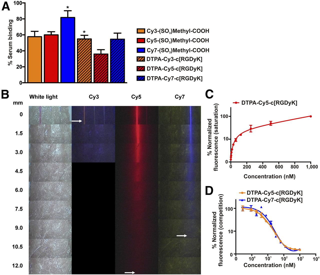
Protein binding, tissue penetration, and receptor affinity. (A) Protein binding in serum of free CyX-(SO3)methyl-COOH dyes and DTPA-CyX-(SO3)methyl-COOH-c[RGDyK] tracers (DTPA-CyX-c[RGDyK], dashed bars; *P < 0.005). (B) Tissue penetration of Cy3-(SO3)methyl-COOH (orange), Cy5-(SO3)methyl-COOH (red), and Cy7-(SO3)methyl-COOH (blue), diluted in albumin and measured with clinical-grade laparoscopic fluorescence camera. (C) Saturation binding curve of DTPA-Cy5-(SO3)methyl-COOH-c[RGDyK]. (D) Competition binding curves of DTPA-Cy3-(SO3)methyl-COOH-c[RGDyK] and DTPA-Cy7-(SO3)methyl-COOH-c[RGDyK].
Stability of Unconjugated Dyes and Hybrid Tracers
Assessment of the fluorescence intensity before and after 30 min of excitation revealed a good photostability for both DTPA-Cy3-(SO3)methyl-COOH-c[RGDyK] and DTPA-Cy5-(SO3)methyl-COOH-c[RGDyK], as the intensity loss for both tracers remained below 14% (Supplemental Fig. 4). For DTPA-Cy7-(SO3)methyl-COOH-c[RGDyK], the fluorescence intensity decreased by 50%, indicating that the length of the polymethine chain is related to dye stability.
Although the dyes and hybrid constructs all proved to be stable under nucleophilic attack, a 57% decrease in absorbance and fluorescence signal in serum was seen for DTPA-Cy7-(SO3)methyl-COOH-c[RGDyK] (Supplemental Figs. 5–7). No change in absorbance was seen for DTPA-Cy3-(SO3)methyl-COOH-c[RGDyK] or DTPA-Cy5-(SO3)methyl-COOH-c[RGDyK]. Regarding the fluorescence emission, the fluorescence of DTPA-Cy3-(SO3)methyl-COOH-c[RGDyK] was not altered, and only a 2% decrease in fluorescence signal was seen for DTPA-Cy5-(SO3)methyl-COOH-c[RGDyK].
Signal Penetration of Fluorescence Emissions
The in-depth detectability of the dyes was studied using videorate recordings from a clinical-grade fluorescence laparoscope (Fig. 3B) (29,30). Cy5-(SO3)methyl-COOH was shown to have signal penetration of up to 12 mm (as visibly detectable on screen). This visibility was superior to that of the near-infrared dyes Cy7-(SO3)methyl-COOH (9 mm) and ICG (7.5 mm; Supplemental Fig. 3B) and the dye Cy3-(SO3)methyl-COOH (detectable only when not covered by tissue).
Receptor Affinity and Binding Localization
With receptor affinities of 37.8 ± 9 nM for Cy3, 39.0 ± 11 nM for Cy5, and 48 ± 9.5 nM for Cy7, affinity for ανβ3 integrin was similar for all three hybrid tracers (Fig. 3C). Fluorescence confocal microscopy revealed that focal cytoplasmic uptake of DTPA-Cy3-(SO3)methyl-COOH-c[RGDyK] and DTPA-Cy5-(SO3)methyl-COOH-c[RGDyK] overlapped with the localization of lysosomes (Supplemental Fig. 8). Unfortunately, the Cy7 dye in DTPA-Cy7-(SO3)methyl-COOH-c[RGDyK] could not be effectively excited by the lasers incorporated within the available microscopy setups (maximum excitation, 670 nm), underlining the complexity of performing in vitro studies with near-infrared dyes.
In Vivo Comparison of Biodistribution of 111In-DTPA-Cy3-c[RGDyK], 111In-DTPA-Cy5-c[RGDyK], and 111In-DTPA-Cy7-c[RGDy]
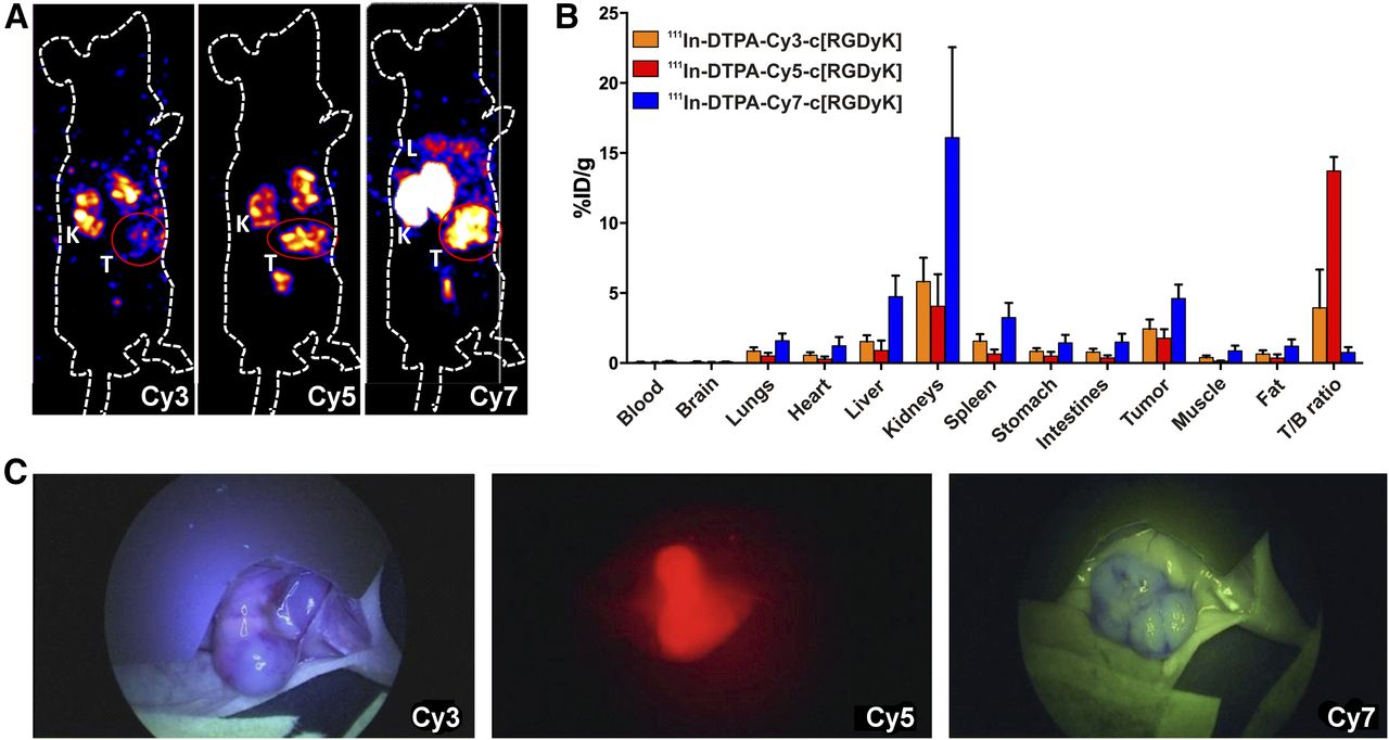
All three hybrid tracers enabled in vivo lesion discrimination using SPECT (Fig. 4A). Uptake of radioactivity in the tumor was 2.6 ± 0.8, 1.7 ± 0.6, and 5.2 ± 0.5 %ID/g for, respectively, 111In-DTPA-Cy3-(SO3)methyl-COOH-c[RGDyK], 111In-DTPA-Cy5-(SO3)methyl-COOH-c[RGDyK], and 111In-DTPA-Cy7-(SO3)methyl-COOH-c[RGDyK]. Together with the %ID/g uptake in the adjoining muscle tissue, a corresponding T/B ratio of 13.2 ± 1.7 was found for 111In-DTPA-Cy5-(SO3)methyl-COOH-c[RGDyK], 5.7 ± 0.7 for 111In-DTPA-Cy3-(SO3)methyl-COOH-c[RGDyK], and 4.7 ± 0.7 for 111In-DTPA-Cy7-(SO3)methyl-COOH-c[RGDyK] (P < 0.001; Fig. 4B).

Comparison of in vivo biodistribution of hybrid c[RGDyK] tracers with fluorescent components with polymethine chains of different lengths. (A) In vivo SPECT image of 111In-DTPA-Cy3-(SO3)methyl-COOH-c[RGDyK] (Cy3), of 111In-DTPA-Cy5-(SO3)methyl-COOH-c[RGDyK] (Cy5), and of 111In-DTPA-Cy7-(SO3)methyl-COOH-c[RGDyK] (Cy7) at 24 h after tracer injection. K = kidney; L = liver; T = tumor (encircled in red). (B) Quantified biodistribution based on %ID/g of excised tissue (radioactivity-based) for 111In-DTPA-Cy3-(SO3)methyl-COOH-c[RGDyK] (orange), 111In-DTPA-Cy5-(SO3)methyl-COOH-c[RGDyK] (red), and 111In-DTPA-Cy3-(SO3)methyl-COOH-c[RGDyK] (blue). Determination of T/B was based on %ID/g in tumor and adjoining muscle tissue. (C) Accompanying in vivo fluorescence images obtained using clinical-grade fluorescence camera at 2-cm distance. Imaging and biodistribution analyses were performed at 24 h after injection. Images obtained with preclinical fluorescence scanner are provided in Supplemental Fig. 9.
In vivo fluorescence imaging and fluorescence-based evaluation of the T/B ratio (Supplemental Fig. 9) revealed a comparable Cy5 > Cy3 > Cy7 trend, although with a greater difference between values (10.89 ± 1.2 > 1.28 ± 0.3 > 0.99 ± 0.6, respectively; P < 0.001). These differences in T/B ratio agreed with the intraoperative visibility of the tumors using a laparoscopic setup (Fig. 4C).
Quantitative scoring of the relative performance of the Cy3 and Cy7 derivatives, compared with 111In-DTPA-Cy5-(SO3)methyl-COOH-c[RGDyK] (Supplemental Tables 2 and 3), further underlined the superiority of the Cy5 derivate.
DISCUSSION
Underlining and extending previous reports that alterations in the pendant moieties on cyanine dyes affect tracer pharmacokinetics, the presented data reveal similar effects on tracer kinetics after the length of the polymethine chain was varied in cyanine dyes with identical pendant moieties. This finding provides yet another feature to consider when pursuing the utility of a tracer in fluorescence-guided surgery.
Alteration of the length of the polymethine chain in otherwise identical cyanine dyes affected many features. The influence of alteration on rotational freedom around the polymethine bond within the cyanine dye (41) was reflected by variation in the photophysical properties of both the unconjugated dyes and the hybrid c[RGDyK] tracers (Table 1; Supplemental Table 1; Supplemental Fig. 4). The five atoms in the polymethine chain of the Cy5 analog seemed to produce the optimal degree of freedom, resulting in the highest quantum yield and both good chemical stability and good optical stability.
The chemical variation among the dyes affected the interaction with albumin and serum, as revealed by differences in quantum yield (Fig. 3A; Table 1; Supplemental Table 1), but also altered the in vivo pharmacokinetics (Fig. 4), as is attributable to the differences in protein binding among the tracers. While overall binding to albumin increased the quantum yield of both the unconjugated dyes (Supplemental Table 1) and the hybrid constructs (Table 1), the degree of nonspecific tissue uptake increased with increasing levels of protein binding (Cy5 < Cy3 < Cy7; Figs. 3A and 3B) (15). Together, these findings underline that interaction of dye with albumin affects the biodistribution, bioactivity, and metabolism of a compound (42–44). However, as the level of protein binding of the Cy3 and Cy7 conjugates was shown to be highly similar, the difference in biodistribution between these tracers cannot be solely explained through this interaction. The increased dye lipophilicity of Cy7 compared with Cy3 (Table 1; Supplemental Table 1), however, seems to reveal an amplifying effect on the level of nonspecific uptake (45).
The hybrid nature of the tracers allowed imaging using both radioactivity and fluorescence, as well as quantitative %ID/g assessments using radioactivity (Fig. 4; Supplemental Fig. 9; Supplemental Tables 2 and 3) (46,47). Interestingly, whereas radioactivity-based assessment revealed a 2.3- to 2.8-fold higher T/B ratio for 111In-DTPA-Cy5-(SO3)methyl-COOH-c[RGDyK] than for the Cy3 and Cy7 derivatives (Fig. 4B), fluorescence-based assessment revealed an 8.4- to 9.1-fold higher ratio (Supplemental Fig. 9); only the Cy5 analog yielded a T/B of more than 2. The 3-fold difference between improvements determined using a radioactive readout and improvements determined using a fluorescence readout seems to be related to a combination of the superior brightness of the Cy5 dye (Table 1; Supplemental Table 1), the tissue penetration of the Cy5 fluorescence signal (Fig. 3B), and the compatibility of this dye with the fluorescence imaging modality used. The ability to sensitively detect Cy5 and Cy7 with the prototype Karl Storz fluorescence laparoscope (48) provides a promising extension of its previously proven ability to image fluorescein and ICG in a clinical setting ([29]).
In addition to the analysis steps described above, translation from molecule to man also favors the use of Cy5, because such far-red dyes are compatible with preclinical flow cytometers, fluorescence (confocal) microscopes, and clinical and preclinical fluorescence imaging systems (Fig. 3C; Supplemental Figs. 8 and 9B) (47). A lack of compatibility of near-infrared dyes with, for example, common confocal microscopes can be considered a limiting factor during the in vitro stages of tracer evaluation.
Combined, the reported findings advocate careful selection of dye during the development of receptor-targeted fluorescence-guided surgery applications. The fact that modest chemical variation induces significant effects on tracer performance also underlines the assumption that there is no such thing as a one-dye-suits-all solution to cover the complexity and plurality of targeting vectors explored for image-guided surgery (18). Our findings also underline the need for accurate (and quantitative) analysis of tracer pharmacokinetics, something that is made possible using hybrid tracer designs. Given the clinical potential demonstrated by hybrid tracers such as ICG-99mTc-nanocolloid (10,46,49–51), adhering to such designs may also help improve clinical tracer implementation in image-guided surgery trials.
CONCLUSION
The length of the polymethine chain in cyanine dyes exerts a direct influence on the chemical, photophysical, and biologic properties of a tracer. In a c[RGDyK]-based model, the Cy5 analog proved to be superior to the Cy3 (visible fluorescence) and Cy7 (near-infrared fluorescence) analogs in all respects.
DISCLOSURE
This research was financially supported by STW-VIDI grant BGT11272 from the Netherlands Organization for Scientific Research, grant 2012-306890 from the European Research Council under the European Union’s Seventh Framework Program (FP7/2007-2013), and the 2015–2016 Post-Doctoral Molecular Imaging Scholar Program Grant from the Society of Nuclear Medicine and Molecular imaging and the Education and Research Foundation for Nuclear Medicine and Molecular Imaging. No other potential conflict of interest relevant to this article was reported.
Footnotes
Published online Feb. 15, 2018.
- © 2018 by the Society of Nuclear Medicine and Molecular Imaging.
REFERENCES
- Received for publication November 17, 2017.
- Accepted for publication January 12, 2018.


Jump to section
Related Articles
Cited By...
- Hybrid Tracers Based on Cyanine Backbones Targeting Prostate-Specific Membrane Antigen: Tuning Pharmacokinetic Properties and Exploring Dye-Protein Interaction
- Trending: Radioactive and Fluorescent Bimodal/Hybrid Tracers as Multiplexing Solutions for Surgical Guidance
- New Developments in Dual-Labeled Molecular Imaging Agents
- Synthesis and Preclinical Characterization of the PSMA-Targeted Hybrid Tracer PSMA-I&F for Nuclear and Fluorescence Imaging of Prostate Cancer
- Multispectral-Fluorescence Imaging as a Tool to Separate Healthy from Disease-Related Lymphatic Anatomy During Robot-Assisted Laparoscopy