


Abstract
Elevated expression of the c-Met receptor plays a crucial role in cancers. In non–small cell lung cancer (NSCLC), aberrant activation of the c-Met signaling pathway contributes to tumorigenesis and cancer progression and may mediate acquired resistance to epidermal growth factor receptor–targeted therapy. c-Met is therefore emerging as a promising therapeutic target for NSCLC, and methods for noninvasive in vivo assessment of c-Met expression would improve NSCLC treatment and diagnosis. Methods: We developed a new c-Met–binding peptide (cMBP) radiotracer, 99mTc-hydrazine nicotinamide (HYNIC)-cMBP, for SPECT imaging. Cell uptake assays were performed on 2 NSCLC cell lines with different c-Met expressions: H1993 (high expression) and H1299 (no expression). In vivo tumor specificity was assessed by SPECT imaging in tumor-bearing mice at 0.5, 1, 2, and 4 h after injection of the probe. Blocking assays, biodistribution, and autoradiography were also conducted to determine probe specificity. Results: 99mTc-HYNIC-cMBP was prepared with high efficiency and showed higher uptake in H1993 cells than in H1299 cells. Biodistribution and autoradiography also showed significantly higher percentages of the injected dose for 99mTc-HYNIC-cMBP in H1993 tumors than in H1299 tumors at 0.5 h (4.74 ± 1.43%/g and 1.00 ± 0.37%/g, respectively; P < 0.05). H1993 tumors were clearly visualized at 0.5 h in SPECT images, whereas H1299 tumors were not observed at any time. The specificity of 99mTc-HYNIC-cMBP for c-Met was demonstrated by a competitive block with an excess of nonradiolabeled peptide. Conclusion: For c-Met–targeted SPECT imaging of NSCLC, we developed 99mTc-HYNIC-cMBP, a tracer that specifically binds to c-Met with favorable pharmacokinetics in vitro and in vivo.
The c-Met receptor is a tyrosine kinase receptor encoded by a protooncogene (1). On binding of hepatocyte growth factor, active c-Met stimulates various signaling pathways implicated in cellular processes, including cell proliferation, motility, and apoptosis (2). Aberrantly high expression of c-Met resulting from gene amplification and protein overexpression increases c-Met activation and oncogenic transformation (3). Elevated c-Met expression has been observed in many types of cancer, including thyroid, pancreatic, and lung (4).
Lung cancer has the highest incidence and mortality rates worldwide (5), and non–small cell lung cancer (NSCLC) accounts for about 80% of lung cancer cases (6). High expression of c-Met has been detected in many cases of NSCLC and is thought to play an important role in promoting tumorigenesis and cancer progression (7). Activation of hepatocyte growth factor/c-Met downstream signaling by the phosphatidylinositol 3 kinase/protein kinase B and mitogen-activated protein kinase pathways can promote tumor growth, metastasis, angiogenesis, and apoptosis inhibition, which contribute to invasiveness and poor prognosis in NSCLC. Furthermore, c-Met gene amplification is a significant mechanism mediating resistance to therapies targeting the epidermal growth factor receptor in NSCLC (8,9).
Studies suggest that c-Met is an effective drug target for treating NSCLC (10), and combination therapies targeting c-Met and the epidermal growth factor receptor have attracted attention in the clinical setting (11). However, limitations in early detection challenge the overall efficacy of cancer treatment. Typically, the methods of screening for lung cancer, such as CT, detect tumors mostly at middle or advanced stages (12). Diagnoses for lung cancer currently rely on biopsies, with multiple disadvantages such as a risk of pleural metastasis, patient intolerance, and sampling inaccuracy due to tumor heterogeneity (13). It is therefore desirable to develop more precise and less invasive methods to diagnose lung cancer, particularly at early stages.
Molecular imaging is a noninvasive method to obtain qualitative and quantitative information on tumor-associated molecular targets in vivo. Over decades, c-Met has increasingly been used as a biomarker for molecular imaging. Despite significant advances in c-Met–targeted imaging, problems remain. For instance, an anti–c-Met monoclonal antibody, DN30 (∼150 kDa), labeled with 89Zr provided quantitative images with high uptake in gastric cancer. However, the tracer had prolonged circulation and slow clearance, reducing tumor-to-background contrast and leading to late optimal imaging times (3–5 d) (14). An anti–c-Met diabody (∼55 kDa) labeled with 89Zr achieved acceptable images but also showed slow clearance (15). These examples highlight the disadvantages of antibody-based tracers: long biologic half-life and slow clearance.
More recent imaging strategies using peptide targeting have received greater attention by overcoming most of the limitations of antibody-based tracers. Indeed, peptide-based tracers have a smaller size, simpler synthesis, faster clearance, and lower risk of immunogenicity (16). GE-137 (∼4.2 kDa), a c-Met–targeted peptide labeled with cyanine dye, produced encouraging results in detecting malignant polyps in high-risk colon cancer patients by optical imaging (17). Based on same targeted peptide to GE-137, 18F-AH113804 also successfully assessed locoregional recurrence of breast cancer (18).
99mTc is widely used in nuclear medicine. To our knowledge, 99mTc-labeled peptides targeting c-Met have not been studied in NSCLC. A c-Met–binding peptide (cMBP) (peptide KSLSRHDHIHHH) identified through phage display screening was previously labeled with a fluorescent dye to detect c-Met in U87MG glioblastoma (19,20). Here, cMBP was modified with a bifunctional chelator, hydrazine nicotinamide (HYNIC), for labeling with 99mTc (half-life, 6.02 h) (99mTc-HYNIC-cMBP) and used to assess c-Met expression in NSCLC by SPECT.
MATERIALS AND METHODS
Radiosynthesis of 99mTc-HYNIC-cMBP
ChinaPeptides Co., Ltd., synthesized HYNIC-cMBP, which was radiolabeled with 99mTc as previously described (21). Briefly, 5 μg of HYNIC-cMBP were added to ethylenediamine-N,N′-diacetic acid (10 mg)/tricine (20 mg) in 1 mL of phosphate-buffered saline (PBS, pH 6–7), followed by 0.5 mL of 99mTcO4 solution (370 MBq) and 20 μL of tin-II solution (10 mg of SnCl2 in 10 mL of 0.1N HCl). All reagents were purchased from Sigma. The mixture was heated (85°C, 15 min, nitrogen protection) and then cooled to room temperature. The reaction mixture was purified with a Sep-Pak C18 Plus cartridge (Waters), and the radiolabeled peptide was collected.
The partition coefficient of 99mTc-HYNIC-cMBP was measured in an octanol and saline system. A 7.4 MBq activity of 99mTc-HYNIC-cMBP was mixed with the octanol (0.5 mL) and saline (0.5 mL) system for 1 h. Then, the mixture was centrifuged (3,000 rpm, 5 min), and activity from each layer (samples of 100 μL) was counted using a γ-counter (PerkinElmer 2480).
Cell Culture
Two NSCLC cell lines were selected—H1993 (high c-Met expression) and H1299 (no c-Met expression)—and cultured as previously reported (22).
Determination of Dissociation Constant (Kd)
H1993 cells with high c-Met expression were used in a saturation binding experiment to determine the Kd of 99mTc-HYNIC-cMBP (23). Briefly, H1993 cells were seeded in two 24-well plates at 0.2 × 10⁶ cells per well 24 h beforehand. A stock solution of 99mTc-HYNIC-cMBP was diluted with medium to 6 concentrations (1, 10, 40, 80, 100, and 160 nM). Increasing concentrations of tracer were added to the wells of one plate to assess cell-bound activity, and nonspecific binding was determined in the other plate by adding nonradiolabeled cMBP (100 μM). After incubation for 0.5 h at 4°C, the tracer was removed. The cells were washed with cold PBS, and activity was measured. Specific binding was calculated by subtracting nonspecific binding from cell-bound activity. Kd was determined from specific binding curves by using nonlinear regression curve fits (GraphPad Prism software, version 5).
Cellular Uptake and Internalization
99mTc-HYNIC-cMBP cellular uptake and blocking with nonradiolabeled cMBP were performed at 15, 30, 60, 120, and 240 min as previously reported (22).
c-Met–positive H1993 cells were used to evaluate whether 99mTc-HYNIC-cMBP was internalized (24). Briefly, H1993 cells were seeded in 12-well plates (5 × 10⁵ cells). After 24 h, 99mTc-HYNIC-cMBP was added (37 kBq/mL) and incubated for different times. Free 99mTc-HYNIC-cMBP was removed, and the cells were washed with ice-cold PBS followed by 0.2 M glycine buffer in 4 M urea (pH 2.5) for 5 min on ice to determine membrane-bound activity. Next, the cells were harvested using 1N NaOH to determine internalized activity. The results are presented as the internalized percentage of cell-associated activity (membrane-bound + internalized).
Animal Xenograft Models
Female BALB/c nude mice (5–6 wk old, weighing 19–23 g) were implanted subcutaneously with H1993 or H1299 (5 × 10⁶ cells) in the right shoulder. Tumor volume was measured by calipers until reaching about 500 mm3 (∼5–6 wk). All animal experiments were approved by the Harbin Medical University Animal Ethics Committee in accordance with Chinese legislation and followed relevant guidelines. The institutional review board approved this study on mice with NSCLC tumor xenografts.
In Vitro and In Vivo Stability
The radiochemical purity of 99mTc-HYNIC-cMBP incubated in either mouse serum or PBS at 37°C was measured for 0, 0.5, 1, 2, and 4 h. 99mTc-HYNIC-cMBP was collected and diluted with PBS at baseline to 0.37 MBq in 0.5 mL of solution A (99.9:0.1 water:trifluoroacetic acid), and radiochemical purity was measured by radio–high-performance liquid chromatography (HPLC). At subsequent time points, the probe samples were added to 0.5 mL of dimethyl formamide to precipitate protein. After centrifugation, the supernatant was diluted with 0.5 mL of solution A and analyzed with HPLC.
In vivo stability was determined with a procedure described previously (25). Briefly, H1993 tumor–bearing mice were injected with 99mTc-HYNIC-cMBP (11.1 MBq in 150 μL of saline) and euthanized 1 h afterward. Blood and urine were collected and immediately centrifuged to remove blood cells. Plasma and urine were added to 0.5 mL of 1% Triton X-100 (Sigma) in dimethyl formamide for further centrifugation. Then, supernatant portions of each sample were diluted with 0.5 mL of solution A for radio-HPLC analysis. Tumors and liver were homogenized with 0.5 mL of 1% Triton X-100 in dimethyl formamide and then centrifuged. The supernatants were diluted with 0.5 mL of solution A for radio-HPLC analysis. Radio-HPLC (GX-281; Gilson) used a C18 column (10 μm, 19 × 250 mm; Waters) with a mobile phase of 50% ethanol at a flow rate of 5 mL/min. The eluted fractions were collected for 30 s/tube to measure radioactivity, and the resultant radio-HPLC chromatogram was plotted.
In Vivo SPECT Imaging
SPECT scanning of tumor-bearing mice (injected with 8.51 MBq of 99mTc-HYNIC-cMBP) was performed with a clinical SPECT scanner (Discovery 670H3100NA; GE Healthcare), equipped with low-energy, high-resolution collimators with parallel pinholes. All images were obtained through detector 1 for anterior static scanning (100 counts), with a zoom factor of 1.00, and stored at a resolution of 1,024 × 1,024. The energy window setting was 140 keV. Image analysis was performed with a Xeleris functional imaging workstation (version 3.1; GE Healthcare). The ratio of counts in the tumor to counts in the contralateral tissue (T/C) was calculated over the regions of interest.
Biodistribution and Pharmacokinetics
The tumor-bearing mice were injected with 1.85 MBq of 99mTc-HYNIC-cMBP and euthanized at serial points for biodistribution testing (22).
The pharmacokinetics of 99mTc-HYNIC-cMBP was evaluated as previously described (26), with modifications. The mice were euthanized, and blood (100 μL) was drawn by cardiac puncture at 3, 9, 15, 30, 45, 60, and 120 min after injection and centrifuged to collect the plasma. The plasma (extracted at 30 min) was mixed with 10% sulfosalicylic acid to precipitate protein. The activity of the protein precipitate and supernatant was measured, and a time-logarithmic–versus–plasma activity curve was plotted. Tracer elimination followed first-order reaction kinetics, and the elimination rate constant was calculated as the negative slope of the linear part of the plotted curve. The elimination half-life was calculated as ln(2)/elimination rate constant.
Blocking Assays
For blocking assays in SPECT imaging and biodistribution, nonradiolabeled cMBP was preinjected into H1993 xenografts at 100 mg/kg of body weight 1 h before injection of the 99mTc-HYNIC-cMBP.
Autoradiography
Tumor-bearing mice were euthanized at 0.5 h after injection. Tumors were excised, embedded within optimal-cutting-temperature compound, and frozen at −80°C. Autoradiography, hematoxylin and eosin staining, and immunofluorescence staining were performed on consecutive tumor sections (10 μm). Sections were exposed to photoreceptive film for 24 h and then imaged with a Cyclone Plus storage phosphor system (PerkinElmer).
Western Blots and Immunofluorescence
Western blots and immunofluorescence experiments were performed as previously reported (22). The concentration of anti–c-Met primary antibody (Abcam-ab51067) was 1:1,000 for Western blots and 1:100 for immunofluorescence.
Statistical Analysis
All data are expressed as mean ± SD (n = 3). Statistical analyses were performed using Prism software. Statistical differences between the groups were estimated using the t test. A P value of less than 0.05 was considered statistically significant.
RESULTS
Radiosynthesis and Characterization of 99mTc-HYNIC-cMBP
The process of radiochemical synthesis and specific binding to c-Met of 99mTc-HYNIC-cMBP is illustrated in Supplemental Figures 1A and 1B (supplemental materials are available at http://jnm.snmjournals.org). The product was obtained with a yield of at least 75% and a radiochemical purity of more than 98% after purification. The logP value was −3.003 ± 0.48, indicating that the probe was water-soluble.
Determination of Kd
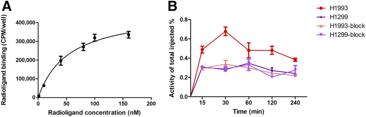
The binding affinity of 99mTc-HYNIC-cMBP to H1993 cells was determined by its specific binding curve (Fig. 1A) to have a Kd of 56.30 ± 2.11. According to a reported theory, the Kd value of 99mTc-HYNIC-cMBP exhibits moderate affinity (1 nM–1 μM) (27), suggesting that 99mTc-HYNIC-cMBP would be effective as a tumor c-Met radiotracer.

(A) Saturation curve for binding of 99mTc-HYNIC-cMBP to H1993 cells with Kd of 56.30 ± 2.11. (B) Cell uptake and block-uptake of 99mTc-HYNIC-cMBP in NSCLC cells. CPM = counts/min.
Cellular Uptake and Blocking Assays
Cellular uptake and blocking assays for H1993 and H1299 cells are shown in Figure 1B. There was rapid uptake of 99mTc-HYNIC-cMBP, which peaked at 30 min in H1993 cells. The tracer subsequently decreased gradually at 60, 120, and 240 min. In contrast, visibly lower cellular uptake was noted in the H1299 cells, with no obvious uptake trend. The accumulation of 99mTc-HYNIC-cMBP in H1993 cells reached its highest level (0.68% ± 0.06% of total input activity) at 30 min, about 2.4-fold higher than in H1299 cells (P < 0.01). In blocking assays with an excess of nonradiolabeled cMBP, uptake of 99mTc-HYNIC-cMBP in H1993 cells at 30 min was lower (0.33% ± 0.06%, P < 0.01), and uptake in H1299 cells remained unchanged. These results suggest that 99mTc-HYNIC-cMBP binds specifically to the c-Met expressed in H1993 cells.
Internalization results (Supplemental Fig. 2) indicated that 99mTc-HYNIC-cMBP was internalized in c-Met–positive H1993 cells. The tracer exhibited a trend toward an increasing internalization rate, with a maximum of 10.21% ± 0.46% cell-associated activity at 120 min followed by a plateau.
In Vitro and In Vivo Stability Assays
According to radio-HPLC analyses, the retention time of 99mTc-HYNIC-cMBP was 6.5 min, and more than 98% of the tracer remained intact during a 0–4 h incubation in mouse serum and PBS (Supplemental Figs. 3A and 3B). The results showed that 99mTc-HYNIC-cMBP was highly stable in vitro. In vivo stability measured by radio-HPLC under identical conditions is shown in Figure 2. At 1 h, intact tracer in plasma and tumor was 98% and 96%, respectively, and intact tracer in liver and urine was 40% and 54%, respectively. These results suggest excellent in vivo stability for 99mTc-HYNIC-cMBP, with expected degradation through the hepatobiliary and urinary systems.
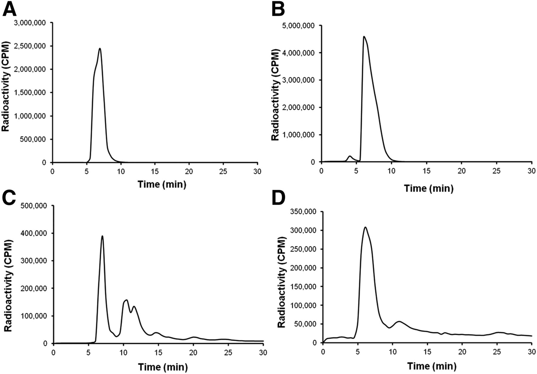
In vivo stability of 99mTc-HYNIC-cMBP as indicated by radiochemical purity in samples of plasma (A), tumor (B), liver (C), and urine (D) at 1 h. CPM = counts/min.
In Vivo SPECT Imaging and Blocking Assays
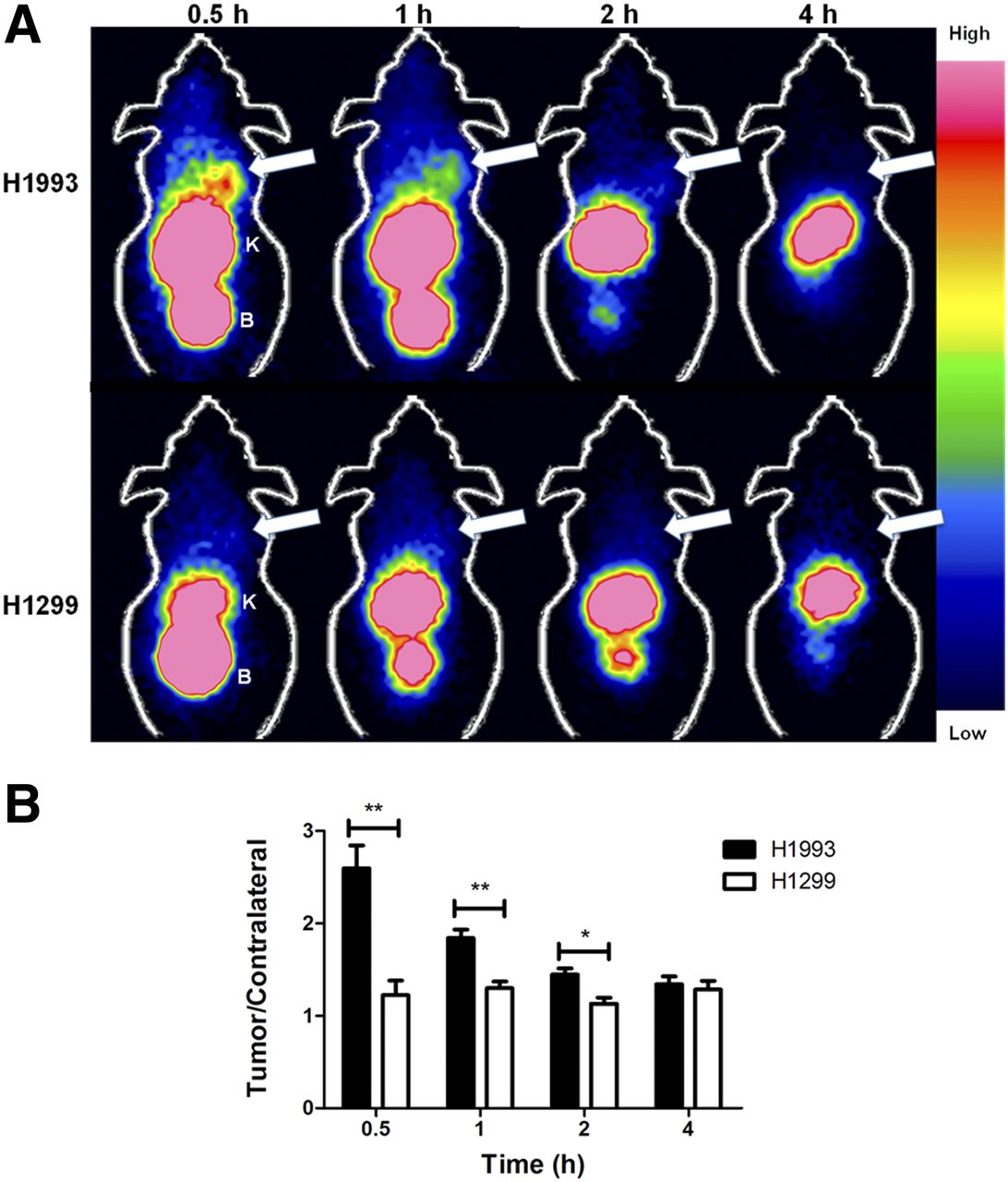
99mTc-HYNIC-cMBP provided effective SPECT imaging of c-Met–positive H1993 tumors (Fig. 3A, top), with H1299 tumors serving as a negative control (Fig. 3A, bottom). Quantitative analyses of the region of interest expressed as T/C ratios are shown in Figure 3B.

(A) Representative coronal SPECT images of H1993 and H1299 xenografts 0–4 h after injection of 99mTc-HYNIC-cMBP. Arrows = tumor; B = bladder; K = kidney. (B) Quantitative analysis of images through counts in region of interest.*P < 0.05.**P < 0.01.
H1993 xenografts were clearly visualized by SPECT imaging at 0.5 h, with the highest T/C ratio being 2.60 ± 0.35, whereas H1299 xenografts were not detected (T/C ratio, 1.22 ± 0.22). H1993 xenografts remained detectable at 1 h, but the T/C ratio decreased to 1.90 ± 0.13. 99mTc-HYNIC-cMBP cleared from tumors within 2–4 h, likely because the tracer lacked lipophilicity. Thus, 0.5 h was the optimum imaging time for 99mTc-HYNIC-cMBP in NSCLC. In H1299 xenografts, no obvious tumor activity was detected over time, with T/C ratios ranging from 1.13 ± 0.09 to 1.29 ± 0.10, consistent with negligible c-Met expression. Activity was highest in the kidney and bladder, indicating that 99mTc-HYNIC-cMBP is cleared primarily via the urinary system.
Representative SPECT images and quantitative analyses for blocking experiments are shown in Figures 4A and 4B, respectively. After pretreatment with an excess of nonradiolabeled cMBP, H1993 xenografts were difficult to discern in images. Consistently, 99mTc-HYNIC-cMBP accumulation in pretreated H1993 tumors was significantly lower (T/C ratio, 1.29 ± 0.11). Collectively, these results support that 99mTc-HYNIC-cMBP specifically accumulates in c-Met–positive H1993 xenografts in vivo.

(A) Representative coronal SPECT images for H1993 and block-H1993 xenografts at 0.5 h. Arrows = tumor; B = bladder; K = kidney. (B) Quantitative analysis of images through counts in region of interest. **P < 0.01.
Biodistribution and Pharmacokinetics
To confirm the SPECT imaging results, the biodistribution of 99mTc-HYNIC-cMBP was assessed in mice bearing H1993 and H1299 xenografts. As shown in Supplemental Tables 1 and 2 and in Figure 5, the highest percentage injected dose (%ID) in H1993 xenografts was at 0.5 h (4.74 ± 1.43 %ID/g), then decreasing at 1 h (2.84 ± 0.28 %ID/g) and further declining at 2 and 4 h (0.87 ± 0.13 and 0.75 ± 0.14 %ID/g, respectively). In contrast, uptake in H1299 xenografts was significantly lower at 0.5 and 1 h (1.00 ± 0.37 and 0.71 ± 0.13 %ID/g, respectively). Uptake in nontarget tissues was similar in H1993 and H1299 xenograft–bearing mice. The signal was highest in kidney at 0.5 h (252.66 ± 44.37 %ID/g in H1299 and 247.55 ± 40.52 %ID/g in H1993) and then decreased over time, further suggesting that 99mTc-HYNIC-cMBP is rapidly cleared through the urinary system. Blocking assays demonstrated that uptake was significantly reduced in H1993 xenografts (1.02 ± 0.43 %ID/g) with an excess of nonradiolabeled cMBP. Probe uptake for different organs did not differ (P > 0.05) between the H1993 and the H1993-block groups, excluding the tumors (P < 0.05).
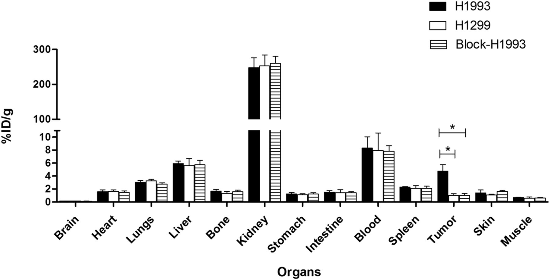
Biodistribution of 99mTc-HYNIC-cMBP in H1993, H1299, and block-H1993 groups at 0.5 h after injection. *P < 0.05.
Protein-to-plasma and supernatant-to-plasma activity, the protein-to-plasma binding ratio of 99mTc-HYNIC-cMBP to total plasma activity, was determined to be 0.10 ± 0.02 at 30 min. The time-logarithmic value of the plasma activity curve was plotted (Supplemental Fig. 4), and the elimination rate constant was calculated to be 0.02 ± 0.001/min. The elimination half-life was 32.80 ± 4.21 min.
Autoradiography
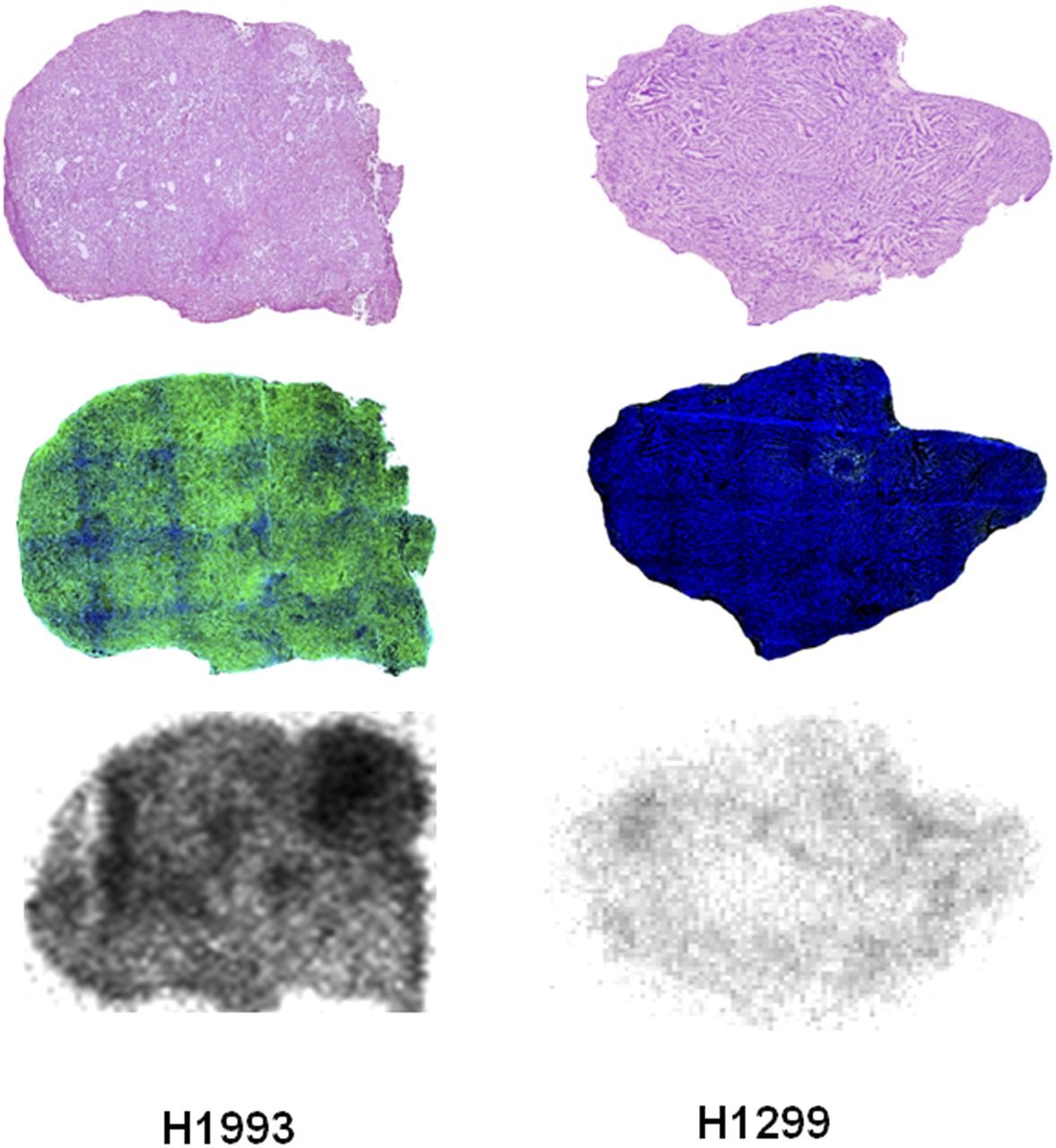
The radioactivity exposure, hematoxylin and eosin staining, and immunofluorescence staining of consecutive tumor sections are presented in Figure 6. The results revealed preferential accumulation of 99mTc-HYNIC-cMBP in H1993 tumors, whereas no accumulation other than background signal was found in H1299 tumors, demonstrating that 99mTc-HYNIC-cMBP specifically binds to c-Met–positive tumors. Importantly, the intratumoral distribution of 99mTc-HYNIC-cMBP in H1993 tumor sections reflected the heterogeneity of NSCLC.

Comparison of hematoxylin and eosin staining (top), immunofluorescence staining (middle), and autoradiography images (bottom) of H1993 and H1299 tumor sections at 0.5 h after injection of 99mTc-HYNIC-cMBP. Green fluorescence indicates high expression of c-Met.
In Vitro Assays
Confirmed by Western blots, high c-Met expression was detected in H1993 cells and xenografts but not in H1299 cells and xenografts (Supplemental Figs. 5A and 5B). Similarly, a significantly more intense fluorescence signal was observed by immunostaining in H1993 cells, whereas signal was negligible in H1299 cells (Supplemental Fig. 5C).
DISCUSSION
Over the decades, c-Met has emerged as a promising therapeutic tumor target (28). Various molecular imaging strategies evaluating c-Met expression in cancer have been reported (29)—mainly strategies involving antibody-based or peptide-based imaging agents. Although antibody-based c-Met–targeted imaging has been widely researched, inherent limitations such as large size, weak tissue penetration, prolonged clearance, and potential immunogenicity have diminished its utility (30).
Low-molecular-weight peptides offer many advantages in molecular imaging, including more favorable pharmacokinetics and tissue penetration, a lower risk of immunogenicity and toxicity, and flexibility in chemical modification (16,31). To date, the only c-Met–targeted probe in clinical trials is GE-137, a fluorescently labeled c-Met–specific peptide that has been used successfully to detect malignant polyps in patients at high risk of colon cancer (17). However, optical imaging is applied mainly to detect superficial tissue externally or in endoscopic procedures, and nuclear imaging is more useful in whole-body imaging.
In this study, the feasibility of using 99mTc-HYNIC-cMBP to detect NSCLC by SPECT imaging was investigated. As a way to mediate resistance to epidermal growth factor receptor–targeted therapy in NSCLC (7,8), c-Met imaging might provide a new strategy for NSCLC diagnosis and treatment.
With a molecular weight of only 1.6 kDa, cMBP is significantly smaller than monoclonal antibodies (∼150 kDa) or derivatives (19,20). Moreover, as compared with other positron radionuclide–labeled tracers, radiolabeling with 99mTc was simple, with high yield and low cost. Our results indicate that 99mTc-HYNIC-cMBP is a promising radiotracer for targeting c-Met in vivo and in vitro. When compared with other previously reported tracers targeting c-Met (e.g., 18.1 ± 4.5 %ID/g for 89Zr-DN30 in GTL-16 gastric cancer and 3.4 ± 0.3 %ID/g for 89Zr-DFO-H2cys-diabody in Hcc827-GR NSCLC when there is optimal image contrast) (14,15), the smaller 99mTc-HYNIC-cMBP showed moderate uptake (4.74 ± 1.43 %ID/g). However, H1993 tumors were clearly visualized in SPECT images at as early as 0.5 h. Additionally, 99mTc-HYNIC-cMBP cleared rapidly via the renal system, as is consistent with water-soluble low-molecular-weight probes, albeit unattractive for imaging tumors of the urinary system (32,33). Nevertheless, rapid clearance is a desirable property for diagnostic tracers, as it reduces tissue exposure to radiation, background noise, and long delays between contrast treatment and image readout for receptor-targeted imaging. Although it is difficult to directly compare c-Met–targeted tracers because of intrinsic differences in tumor models, peptide-based tracers tend to produce high-contrast images at earlier times than antibody-based tracers, as there is better tissue penetration and more rapid elimination.
Despite these advantages, 99mTc-HYNIC-cMBP had limitations, such as a short retention time in tumors and a low resolution of anatomic structures in images. Furthermore, the optimal imaging time of 99mTc-HYNIC-cMBP, 30 min, may not allow sufficient time for preparation, and it may be preferable to extend this imaging time to approximately 1 h, as is the case for 18F-FDG (34). Thus, in future studies, the efficacy of 99mTc-HYNIC-cMBP might be improved by introducing an amino octanoic acid to increase peptide lipophilicity (35) or by creating dimers, such as dimeric arginine-glycine-aspartic acid peptide, to increase binding affinity (36,37). Small-animal SPECT imaging may appropriately provide not only anatomic information with higher resolution but also dynamic imaging with better quantitative analysis and pharmacokinetics modeling.
CONCLUSION
This study provided proof of the concept of using a novel peptide-based tracer targeting c-Met for SPECT imaging. 99mTc-HYNIC-cMBP specifically bound to c-Met in vitro and in vivo. Although further modification and optimization are warranted, cMBP-based radiotracers may be translated into the clinic for the selection and monitoring of NSCLC patients to receive c-Met–responsive therapy.
DISCLOSURE
This work was supported partly by the National Natural Science Foundation of China (81471724, 81627901, 31210103913, 81101088, and 81130028), the National Basic Research Program of China (2015CB931800), the Heilongjiang Province Foundation for Returned Overseas Chinese Scholars, and the Key Laboratory of Molecular Imaging Foundation (College of Heilongjiang Province). No other potential conflict of interest relevant to this article was reported.
Acknowledgments
We thank Po Yang, Yingxun Zhang, Yali Cui, Yongyi Wu, Feng Chen, and Tengchuang Ma for technical support.
Footnotes
↵* Contributed equally to this work.
Published online May 18, 2018.
- © 2018 by the Society of Nuclear Medicine and Molecular Imaging.
REFERENCES
- Received for publication December 6, 2017.
- Accepted for publication May 8, 2018.


