
Main menu

  • Home
  • Content
    • Current
    • Ahead of print
    • Past Issues
    • JNM Supplement
    • SNMMI Annual Meeting Abstracts
    • Continuing Education
    • JNM Podcasts
  • Subscriptions
    • Subscribers
    • Institutional and Non-member
    • Rates
    • Journal Claims
    • Corporate & Special Sales
  • Authors
    • Submit to JNM
    • Information for Authors
    • Assignment of Copyright
    • AQARA requirements
  • Info
    • Reviewers
    • Permissions
    • Advertisers
  • About
    • About Us
    • Editorial Board
    • Contact Information
  • More
    • Alerts
    • Feedback
    • Help
    • SNMMI Journals
  • SNMMI
    • JNM
    • JNMT
    • SNMMI Journals
    • SNMMI

User menu

  • Subscribe
  • My alerts
  • Log in
  • My Cart

Search

  • Advanced search
Journal of Nuclear Medicine
  • SNMMI
    • JNM
    • JNMT
    • SNMMI Journals
    • SNMMI
  • Subscribe
  • My alerts
  • Log in
  • My Cart
Journal of Nuclear Medicine

Advanced Search

  • Home
  • Content
    • Current
    • Ahead of print
    • Past Issues
    • JNM Supplement
    • SNMMI Annual Meeting Abstracts
    • Continuing Education
    • JNM Podcasts
  • Subscriptions
    • Subscribers
    • Institutional and Non-member
    • Rates
    • Journal Claims
    • Corporate & Special Sales
  • Authors
    • Submit to JNM
    • Information for Authors
    • Assignment of Copyright
    • AQARA requirements
  • Info
    • Reviewers
    • Permissions
    • Advertisers
  • About
    • About Us
    • Editorial Board
    • Contact Information
  • More
    • Alerts
    • Feedback
    • Help
    • SNMMI Journals
  • View or Listen to JNM Podcast
  • Visit JNM on Facebook
  • Join JNM on LinkedIn
  • Follow JNM on Twitter
  • Subscribe to our RSS feeds
Research ArticleRadiobiology/Dosimetry

New Fetal Radiation Doses for 18F-FDG Based on Human Data

Paolo Zanotti-Fregonara and Michael G. Stabin
Journal of Nuclear Medicine November 2017, 58 (11) 1865-1866; DOI: https://doi.org/10.2967/jnumed.117.195404
Paolo Zanotti-Fregonara
1Houston Methodist Research Institute, Houston, Texas; and
  • Find this author on Google Scholar
  • Find this author on PubMed
  • Search for this author on this site
Michael G. Stabin
2Vanderbilt University, Nashville, Tennessee
  • Find this author on Google Scholar
  • Find this author on PubMed
  • Search for this author on this site
  • Article
  • Figures & Data
  • Info & Metrics
  • PDF
Loading

Abstract

Current standard values of fetal dosimetry deriving from 18F-FDG injection in pregnant women are estimated from animal data. The present communication offers a revision of fetal dosimetry values calculated from recently published human data, in which fetal 18F-FDG uptake was directly observed in vivo. The final doses were obtained from the observed time-integrated activity coefficients and a new generation of anthropomorphic voxel-based pregnancy phantoms.

  • PET
  • radiation safety
  • radiobiology/dosimetry
  • dosimetry
  • FDG
  • pregnancy

Fetal radiation dose estimates for administration of 18F-FDG were originally proposed in 1997 by Russell et al. (1), but with no information about possible placental crossover. In 2004, Stabin (2) amended the dose estimates to include placental crossover data observed in primates (3). Recently, Zanotti-Fregonara et al. (4) have quantified fetal uptake in 19 fetuses from 5 to 34 wk of pregnancy. Four women were in the early weeks of pregnancy, and the dose to the uterus was used as a proxy for the fetal dose. Starting from 10 wk, however, the fetus was more clearly visible inside the uterus, and therefore the 18F-FDG uptake in fetal tissues could be calculated directly in 15 cases.

By extrapolating with mathematic modeling from the data of Zanotti-Fregonara et al. (4) and by using the realistic voxel-based phantoms of Shi et al. (5), this brief communication recommends the best standardized values for estimating the dose to the fetus at any stage of pregnancy.

MATERIALS AND METHODS

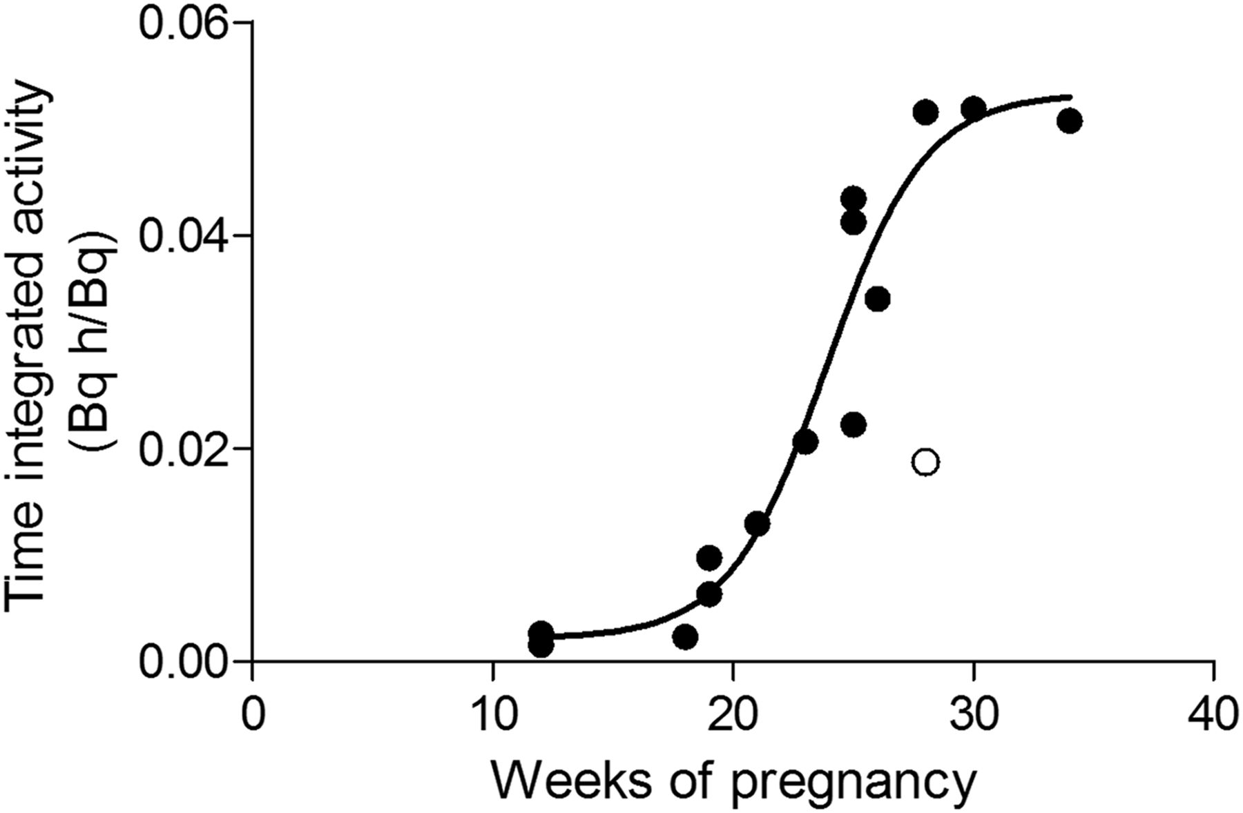
The fetal time-integrated activity coefficients of the 15 cases reported by Zanotti-Fregonara et al. (4) were fitted with the following logistic function:Embedded ImageThe function is characterized by these parameters: a (bottom value) = 0.00216 Bq h/Bq; b (slope factor) = 0.2128 wk−1; c (inflection point) = 23.9 wk; and d (top value) = 0.05328 Bq h/Bq. The fitting was performed after automatic elimination of one outlier (ROUT [robust regression and outlier removal] Q coefficient, 1.0%) (Fig. 1).

FIGURE 1.
  • Download figure
  • Open in new tab
  • Download powerpoint
FIGURE 1.

Sigmoid fitting through observed time-integrated activity data points reported by Zanotti-Fregonara et al. (4). Automatic detection of outliers removed one point (in white).

The logistic function was chosen because it fitted the data points better than either a monoexponential or a quadratic function, in terms of Akaike score. Moreover, a runs test for randomness was not significant for the logistic fitting (P = 0.79) but was significant for the exponential (P = 0.007) and the quadratic curve (P = 0.025). Time–activity integrals for activity in the mother’s organs were taken from publication 128 of the International Commission on Radiological Protection (Table 1) (6).

View this table:
  • View inline
  • View popup
TABLE 1

Time-Integrated Activity Coefficients (6)

The final doses were calculated using the realistic voxel-based phantoms of Shi et al. (5), which are implemented in the forthcoming new version of OLINDA/EXM. The activity coefficient at 5 wk was assigned to the uterus of the nongravid female phantom. The coefficients at 13, 26, and 39 wk were assigned to the fetus of the first-, second-, and third-trimester pregnant phantom, respectively.

RESULTS

The interpolated time-integrated activity coefficients at different stages of gestation, as predicted by the logistic fitting, are reported in Table 2. The resulting dose estimates, calculated with OLINDA/EXM 2.0, are reported in Table 3. For comparison, Table 3 also reports the values previously suggested by Russell et al. (1) and Stabin (2), as well as the present dose estimates calculated with Cristy and Eckerman’s stylized phantoms (7), included in the first version of OLINDA/EXM (8). Differences between the values estimated with the two versions of the OLINDA/EXM software are mainly due to the assumed fetal masses. This is especially true for the 3-mo fetus, whose mass is 460 g in the OLINDA/EXM 1.1 phantom and 85 g in the newer ones of OLINDA 2.

View this table:
  • View inline
  • View popup
TABLE 2

Time-Integrated Activity Coefficients at Different Stages of Gestation

View this table:
  • View inline
  • View popup
TABLE 3

Fetal Dose per Megabecquerel of 18F-FDG Administered to Mother

DISCUSSION

Fetal dose estimates for 18F-FDG were originally calculated with no information on placental crossover and fetal uptake (1) and were later amended using placental crossover data observed in primates (2). With the recent publication of several case reports and small series of women who underwent a PET scan while pregnant (4), it has become possible to calculate 18F-FDG fetal uptake directly in living human subjects.

The observed time-integrated activity coefficients were well fitted by a logistic function, which performed better in terms of goodness of fit than either a monoexponential curve or a quadratic curve. The parameters of this function allow extrapolating the time-integrated activity coefficients, and hence the dosimetry values, at any week of pregnancy. Future case reports may further refine these estimates, especially if full dynamic data become available.

Because of sigmoid fitting, activity coefficients plateaued after 34 wk. It is possible that some increase may occur also in the last weeks of pregnancy, but the data suggest that this increase would be small, especially when compared with the uncertainties in dose estimate (9). Moreover, a final plateau is present in many, although not all, fetal growth curves (10,11). In all likelihood, an accurate fetal dosimetry in the very last weeks of pregnancy has probably little clinical relevance: if the PET scan cannot be postponed until delivery a couple of weeks later, labor can be induced before the beginning of diagnostic and clinical work-up.

According to our estimates, an administered activity of 200 MBq would deliver a fetal dose ranging from 5.2 mSv in early pregnancy to 1.4 mSv in late pregnancy. Considering the uncertainties in all internal dose values for radiopharmaceuticals, conservatism and rounding in general should be applied to reported values. However, these values are well below the threshold for deterministic effects and in a range where stochastic effects have never been observed (12).

CONCLUSION

Using the new activity coefficients published by Zanotti-Fregonara et al. (4) and the realistic voxel-based phantoms of Shi et al. (5), updated fetal doses are recommended: 2.6E−02 mGy/MBq in early pregnancy, 1.9E−02 mGy/MBq at 3-mo gestation, 1.4E−02 mGy/MBq at 6-mo gestation, and 6.9E−03 mGy/MBq at 9-mo gestation.

DISCLOSURE

No potential conflict of interest relevant to this article was reported.

Footnotes

  • Published online Jun. 1, 2017.

  • © 2017 by the Society of Nuclear Medicine and Molecular Imaging.

REFERENCES

  1. 1.↵
    1. Russell JR,
    2. Stabin MG,
    3. Sparks RB,
    4. Watson E
    . Radiation absorbed dose to the embryo/fetus from radiopharmaceuticals. Health Phys. 1997;73:756–769.
    OpenUrlCrossRefPubMed
  2. 2.↵
    1. Stabin MG
    . Proposed addendum to previously published fetal dose estimate tables for 18F-FDG. J Nucl Med. 2004;45:634–635.
    OpenUrlAbstract/FREE Full Text
  3. 3.↵
    1. Benveniste H,
    2. Fowler JS,
    3. Rooney WD,
    4. et al
    . Maternal-fetal in vivo imaging: a combined PET and MRI study. J Nucl Med. 2003;44:1522–1530.
    OpenUrlAbstract/FREE Full Text
  4. 4.↵
    1. Zanotti-Fregonara P,
    2. Chastan M,
    3. Edet-Sanson A,
    4. et al
    . New fetal dose estimates from 18F-FDG administered during pregnancy: standardization of dose calculations and estimations with voxel-based anthropomorphic phantoms. J Nucl Med. 2016;57:1760–1763.
    OpenUrlAbstract/FREE Full Text
  5. 5.↵
    1. Shi CY,
    2. Xu XG,
    3. Stabin MG
    . SAF values for internal photon emitters calculated for the RPI-P pregnant-female models using Monte Carlo methods. Med Phys. 2008;35:3215–3224.
    OpenUrlCrossRefPubMed
  6. 6.↵
    ICRP. Radiation dose to patients from radiopharmaceuticals: a compendium of current information related to frequently used substances—ICRP publication 128. Ann ICRP. 2015;44(2S).
  7. 7.↵
    1. Cristy M,
    2. Eckerman KF
    . Specific Absorbed Fractions of Energy at Various Ages from Internal Photon Sources. Oak Ridge, TN: Oak Ridge National Laboratory; 1987:1–110. ORNL/TM-8381.
  8. 8.↵
    1. Stabin MG,
    2. Sparks RB,
    3. Crowe E
    . OLINDA/EXM: the second-generation personal computer software for internal dose assessment in nuclear medicine. J Nucl Med. 2005;46:1023–1027.
    OpenUrlAbstract/FREE Full Text
  9. 9.↵
    1. Stabin MG
    . Uncertainties in internal dose calculations for radiopharmaceuticals. J Nucl Med. 2008;49:853–860.
    OpenUrlAbstract/FREE Full Text
  10. 10.↵
    1. Olsen IE,
    2. Groveman SA,
    3. Lawson ML,
    4. Clark RH,
    5. Zemel BS
    . New intrauterine growth curves based on United States data. Pediatrics. 2010;125:e214–e224.
    OpenUrlAbstract/FREE Full Text
  11. 11.↵
    1. Oken E,
    2. Kleinman KP,
    3. Rich-Edwards J,
    4. Gillman MW
    . A nearly continuous measure of birth weight for gestational age using a United States national reference. BMC Pediatr. 2003;3:6.
    OpenUrlCrossRefPubMed
  12. 12.↵
    1. Brent RL
    . Saving lives and changing family histories: appropriate counseling of pregnant women and men and women of reproductive age, concerning the risk of diagnostic radiation exposures during and before pregnancy. Am J Obstet Gynecol. 2009;200:4–24.
    OpenUrlCrossRefPubMed
  • Received for publication April 27, 2017.
  • Accepted for publication May 26, 2017.
PreviousNext
Back to top

In this issue

Journal of Nuclear Medicine: 58 (11)
Journal of Nuclear Medicine
Vol. 58, Issue 11
November 1, 2017
  • Table of Contents
  • Table of Contents (PDF)
  • About the Cover
  • Index by author
Print
Download PDF
Article Alerts
Sign In to Email Alerts with your Email Address
Email Article

Thank you for your interest in spreading the word on Journal of Nuclear Medicine.

NOTE: We only request your email address so that the person you are recommending the page to knows that you wanted them to see it, and that it is not junk mail. We do not capture any email address.

Enter multiple addresses on separate lines or separate them with commas.
New Fetal Radiation Doses for 18F-FDG Based on Human Data
(Your Name) has sent you a message from Journal of Nuclear Medicine
(Your Name) thought you would like to see the Journal of Nuclear Medicine web site.
Citation Tools
New Fetal Radiation Doses for 18F-FDG Based on Human Data
Paolo Zanotti-Fregonara, Michael G. Stabin
Journal of Nuclear Medicine Nov 2017, 58 (11) 1865-1866; DOI: 10.2967/jnumed.117.195404

Citation Manager Formats

  • BibTeX
  • Bookends
  • EasyBib
  • EndNote (tagged)
  • EndNote 8 (xml)
  • Medlars
  • Mendeley
  • Papers
  • RefWorks Tagged
  • Ref Manager
  • RIS
  • Zotero
Share
New Fetal Radiation Doses for 18F-FDG Based on Human Data
Paolo Zanotti-Fregonara, Michael G. Stabin
Journal of Nuclear Medicine Nov 2017, 58 (11) 1865-1866; DOI: 10.2967/jnumed.117.195404
Twitter logo Facebook logo LinkedIn logo Mendeley logo
  • Tweet Widget
  • Facebook Like
  • Google Plus One
Bookmark this article

Jump to section

  • Article
    • Abstract
    • MATERIALS AND METHODS
    • RESULTS
    • DISCUSSION
    • CONCLUSION
    • DISCLOSURE
    • Footnotes
    • REFERENCES
  • Figures & Data
  • Info & Metrics
  • PDF

Related Articles

  • This Month in JNM
  • PubMed
  • Google Scholar

Cited By...

  • 18F-FDG Fetal Dosimetry Calculated with PET/MRI
  • New-Generation Fetal Dose Estimates for Radiopharmaceuticals
  • Google Scholar

More in this TOC Section

Radiobiology/Dosimetry

  • Feasibility of Single-Time-Point Dosimetry for Radiopharmaceutical Therapies
  • γ-Tocotrienol–Loaded Liposomes for Radioprotection from Hematopoietic Side Effects Caused by Radiotherapeutic Drugs
  • Dose–Effect Relationships of 166Ho Radioembolization in Colorectal Cancer
Show more Radiobiology/Dosimetry

Basic

  • Dopamine D1 Receptor Agonist PET Tracer Development: Assessment in Nonhuman Primates
  • Optical Navigation of the Drop-In γ-Probe as a Means to Strengthen the Connection Between Robot-Assisted and Radioguided Surgery
  • Synthesis and Preclinical Evaluation of a 68Ga-Labeled Adnectin, 68Ga-BMS-986192, as a PET Agent for Imaging PD-L1 Expression
Show more Basic

Similar Articles

Keywords

  • PET
  • radiation safety
  • radiobiology/dosimetry
  • dosimetry
  • FDG
  • pregnancy
SNMMI

© 2025 SNMMI

Powered by HighWire