


Abstract
One mechanism of resistance to trastuzumab in human epidermal growth factor receptor-2 (HER2)–positive breast cancer (BC) is increased epidermal growth factor receptor (EGFR) expression. We have developed 111In-labeled bispecific radioimmunoconjugates (bsRICs) that bind HER2 and EGFR on BC cells by linking trastuzumab Fab fragments through a polyethylene glycol (PEG24) spacer to epidermal growth factor (EGF). We hypothesized that tumors coexpressing HER2 and EGFR could be treated by dual-receptor–targeted radioimmunotherapy with these bsRICs labeled with the β-particle emitter 177Lu or the Auger electron-emitter 111In. Methods: The binding of 177Lu-DOTA-Fab-PEG24-EGF to tumor cells (MDA-MB-231, SK-OV-3, MDA-MB-231/H2N, or TrR1) coexpressing HER2 and EGFR was assessed in competition assays. The clonogenic survival of these cells was measured after exposure to 177Lu-DOTA-Fab-PEG24-EGF or 111In-DTPA-Fab-PEG24-EGF or to monospecific 177Lu- or 111In-labeled trastuzumab Fab or EGF. The tumor and normal tissue biodistribution of 177Lu-DOTA-Fab-PEG24-EGF was studied at 48 h after injection in athymic mice bearing subcutaneous MDA-MB-231/H2N tumors. Radiation-absorbed doses to tumors and normal tissues were estimated and compared for 111In- and 177Lu-labeled bsRICs. The maximum injected amount of 177Lu-DOTA-Fab-PEG24-EGF that caused no observable adverse effects (NOAEL) was identified in BALB/c mice. Athymic CD1 nu/nu mice bearing subcutaneous trastuzumab-sensitive MDA-MB-231/H2N or trastuzumab-resistant TrR1 tumors were treated with 177Lu-DOTA-Fab-PEG24-EGF or 111In-DTPA-Fab-PEG24-EGF at the NOAEL, or with unlabeled immunoconjugates or normal saline. Tumor growth was evaluated over a period of 49 d. Results: 177Lu-DOTA-Fab-PEG24-EGF bound specifically to HER2 and EGFR on tumor cells. Monospecific 177Lu- and 111In-labeled trastuzumab Fab or EGF killed tumor cells that predominantly expressed HER2 or EGFR, respectively, whereas bsRICs were cytotoxic to cells that displayed either HER2 or EGFR or both receptors. bsRICs were more effective than monospecific agents. 177Lu-DOTA-Fab-PEG24-EGF was more cytotoxic than 111In-DTPA-Fab-PEG24-EGF. The tumor uptake of 177Lu-DOTA-Fab-PEG24-EGF was 2-fold greater than 177Lu-DOTA-trastuzumab Fab or 177Lu-DOTA-EGF. The NOAEL for 177Lu-DOTA-Fab-PEG24-EGF was 11.1 MBq (10 μg). Trastuzumab-sensitive MDA-MB-231/H2N and trastuzumab-resistant TrR1 tumors were growth-inhibited by 177Lu-DOTA-Fab-PEG24-EGF or 111In-DTPA-Fab-PEG24-EGF. Unlabeled immunoconjugates had no effect on tumor growth. 177Lu-DOTA-Fab-PEG24-EGF inhibited tumor growth more effectively than 111In-DTPA-Fab-PEG24-EGF because of a 9.3-fold-higher radiation-absorbed dose (55.0 vs. 5.9 Gy, respectively). Conclusion: These results are encouraging for further development of these bsRICs for dual-receptor–targeted radioimmunotherapy of BC coexpressing HER2 and EGFR, including trastuzumab-resistant tumors.
Overexpression of the human epidermal growth factor receptor-2 (HER2) occurs in about 20%–25% of breast cancers (BCs) (1) and is the therapeutic target for trastuzumab (Herceptin; Roche Pharmaceuticals), pertuzumab (Perjeta), and trastuzumab-emtansine (T-DM1; Kadcycla) (2). Although these HER2-targeted therapies combined with chemotherapy have improved the outcome for women with HER2-positive metastatic BC, not all patients respond and many patients with HER2-positive BC develop resistance within a year (3). Pertuzumab combined with trastuzumab and docetaxel has improved patient survival compared with trastuzumab and docetaxel alone (4). Trastuzumab combined with the HER2 tyrosine kinase inhibitor lapatinib improved survival in patients with progressive HER2-positive BC (5). Recently, T-DM1 has been shown to be more effective than lapatinib combined with capecitabine for treatment of BC resistant to trastuzumab and taxanes (6). Despite these encouraging results, trastuzumab resistance remains a challenge. The reasons for tumor resistance are not completely understood but one mechanism is the upregulation of other human epidermal growth factor receptor (EGFR) family members (e.g., EGFR and HER3) (7–9). Our group has developed 111In-labeled bispecific radioimmunoconjugates (bsRICs) that recognize EGFR or HER3 expressed alone or coexpressed with HER2 on BC cells (10,11). These bsRICs may be useful for molecular imaging or radioimmunotherapy of trastuzumab-resistant tumors that coexpress these receptors. These bsRICs were constructed by linking trastuzumab Fab fragments that bind HER2 through a 24-mer polyethylene glycol (PEG24) spacer to human epidermal growth factor (EGF) or to heregulin-β, which are the natural ligands for EGFR and HER3, respectively. Micro-SPECT/CT imaging demonstrated specific accumulation of these bsRICs in tumor xenografts in mice that displayed HER2 or EGFR or HER3 or HER2 coexpressed with EGFR or HER3 (10,11).
Our hypothesis in the current study was that 177Lu- or 111In-labeled bsRICs that bind HER2 and EGFR would cause cytotoxicity in vitro to BC cells expressing these receptors and tumor growth inhibition in vivo in athymic mice bearing trastuzumab-sensitive or -resistant BC xenografts coexpressing HER2 and EGFR. 177Lu (half-life, 6.7 d) emits moderate energy β-particles (maximum beta energy, 0.50 MeV [78.6%]; 0.38 MeV [9.1%]; 0.18 MeV [12.2%]) useful for radioimmunotherapy with a maximum range in tissues of 2 mm, as well as 2 low abundance γ-photons (energy of gamma photon, 113 [3%] and 210 keV [11%]) that can be exploited for SPECT imaging. 111In (half-life, 2.8 d) emits a cascade of 15 low-energy (<25 keV) Auger electrons per decay and 2 high-abundance γ-photons (energy of gamma photon, 171 [90%] and 245 keV [94%]) for SPECT imaging. Auger electrons have subcellular nanometer–micrometer range but are lethally damaging to the DNA of cancer cells when emitted near the cell nucleus (12). 111In-labeled trastuzumab modified with nuclear translocation sequence (NLS) peptides (111In-NLS-trastuzumab) to promote its nuclear importation after internalization was highly effective for killing HER2-positive BC cells in vitro (13) and strongly inhibited tumor growth in vivo in athymic mice bearing human HER2-overexpressing BC xenografts (14).
MATERIALS AND METHODS
Cancer Cells
SK-OV-3 human ovarian cancer cells and MDA-MB-231 human BC cells were purchased from the American Type Culture Collection. The MDA-MB-231/H2N cell line was derived from EGFR-positive MDA-MB-231 cells that were stably transfected to overexpress c-erbB-2 (HER2) (15). TrR1 cells are a subclone of MDA-MB-231/H2N cells with acquired trastuzumab resistance but that continue to express HER2 (15). Both MDA-MB-231/H2N and TrR1 cells were provided by Dr. Robert S. Kerbel (Sunnybrook Health Sciences Centre). SK-OV-3 cells were cultured in RPMI-1640 medium (Sigma-Aldrich) supplemented with 10% fetal bovine serum (Invitrogen). MDA-MB-231, MDA-MB-231/H2N, and TrR1 cells were cultured in Dulbecco modified Eagle medium supplemented with 10% fetal bovine serum. All cells were cultured in 5% CO2 at 37°C. BC cells were selected on the basis of their reported HER2 or EGFR expression (11,16,17), but these receptor densities were confirmed by direct (saturation) radioligand binding assays with 111In-labeled trastuzumab Fab fragments or 111In-labeled EGF. Because MDA-MB-231 cells exhibited low HER2 expression (5.4 × 10⁴ receptors/cell) and moderate EGFR density (5.2 × 10⁵ receptors/cell), these cells were designated as HER2low/EGFRmod. MDA-MB-231/H2N cells exhibited moderate HER2 and EGFR expression (4.5 × 10⁵ and 4.8 × 10⁵ receptors/cell, respectively) and were designated as HER2mod/EGFRmod. TrR1 cells displayed 4.4 × 10⁵ HER2/cell and 4.6 × 10⁵ EGFR/cell and were designated as HER2mod/EGFRmod. SK-OV-3 cells exhibited high HER2 but low EGFR expression (9.6 × 10⁵ and 8 × 10⁴ receptors/cell, respectively) (18) and were designated as HER2high/EGFRlow. The HER2 and EGFR density measured by radioligand binding assays in these cells was in agreement with the receptor expression assessed by Western blot (15,19).
bsRICs
Fab-PEG24-EGF bispecific immunoconjugates (bsICs) recognizing HER2 and EGFR were constructed as reported by cross-linking trastuzumab Fab fragments (molecular weight, 50 kDa) produced by proteolytic digestion of trastuzumab IgG to human EGF (molecular weight, 6.2 kDa; Peprotech) through a PEG24 spacer (11). Insertion of this PEG24 spacer improved the binding of analogous 111In-diethylenetriaminepentaacetic acid (DTPA)-trastuzumab Fab-heregulin bsRICs to HER2 and HER3 on BC cells (10) and preserved the HER2 and EGFR binding of 111In-DTPA-trastuzumab-PEG24-EGF bsRICs (11). The bsICs were reconcentrated to 1.0 mg/mL in phosphate-buffered saline (pH 7.0) on a Microcon centrifugal device (molecular weight cut-off, 30 kDa; Millipore) and then derivatized with a 10-fold molar excess of DOTA-N-hydroxysuccinimide ester (NHS-DOTA; Macrocyclics Inc.) for labeling with 177Lu. DOTA conjugation efficiency was measured by analysis of a sample of the unpurified reaction mixture trace-labeled with 177Lu by instant thin-layer silica gel chromatography (Pall Life Sciences) developed in 100 mM sodium citrate (pH 5.0). The conjugation efficiency was multiplied by the 10-fold molar excess of DOTA in the reaction to determine the number of DOTA chelators per molecule of bsICs. The bsICs were purified on a Sephadex G25 (Sigma-Aldrich) minicolumn eluted with 400 mM ammonium acetate buffer, pH 5.0, to remove unconjugated DOTA. Purified DOTA-Fab-PEG24-EGF (200 μg; 100 μL) was labeled by incubation with 20 MBq (5 μL) of 177LuCl3 (PerkinElmer) for 90 min at 42°C. 111In-DTPA-Fab-PEG24-EGF bsRICs were synthesized as reported (11). 111In-DTPA-trastuzumab Fab, 177Lu-DOTA-trastuzumab Fab, 111In-DTPA-EGF, and 177Lu-DOTA-EGF were synthesized for comparison. There were 0.8 ± 0.3 DTPA or 0.7 ± 0.4 DOTA chelators per molecule of EGF, respectively. Fab fragments were derivatized with 2.3 ± 0.5 DOTA or 3.6 ± 0.7 DTPA per molecule. The final radiochemical purity of all radioimmunoconjugates was greater than 90% measured by instant thin-layer silica gel chromatography.
The HER2 and EGFR binding of 177Lu-labeled bsRICs was evaluated by competition radioligand binding assays using SK-OV-3 (HER2high/EGFRlow), MDA-MB-231 (HER2low/EGFRmod), or MDA-MB-231/H2N (HER2mod/EGFRmod) cells. Approximately 1.5 × 10⁵ cells were seeded into 24-well plates and cultured overnight. The cells were then exposed to 10 nM (1.0 MBq/μg) of 177Lu-DOTA-Fab-PEG24-EGF in phosphate-buffered saline in the presence of 500 nM of unlabeled Fab, EGF, or both competitors. After incubation for 3 h at 4°C, unbound radioactivity was removed and the cells were solubilized in 1 N NaOH and transferred to γ-counting tubes, and the cell bound radioactivity was measured in a γ-counter. The binding of 177Lu-DOTA-trastuzumab Fab or 177Lu-DOTA-EGF to MDA-MB-231 cells (HER2low/EGFRmod) or MDA-MB-231/H2N cells (HER2mod/EGFRmod) was determined in the presence or absence of an excess of trastuzumab Fab (69 nmol/L) or EGF (1,659 nmol/L). The HER2- and EGFR-binding properties of 111In-labeled bsRICs, 111In-DTPA-trastuzumab Fab, or 111In-DTPA-EGF were previously reported (11).
Clonogenic Survival (CS) Assays
The CS of SK-OV-3 (HER2high/EGFRlow), MDA-MB-231 (HER2low/EGFRmod), MDA-MB-231/H2N (HER2mod/EGFRmod), or trastuzumab-resistant TrR1 (HER2mod/EGFRmod) cells exposed to 177Lu-DOTA-Fab-PEG24-EGF (350 ± 14.3 MBq/mg), 177Lu-DOTA-trastuzumab Fab (350 ± 9.4 MBq/mg), or 177Lu-DOTA-EGF (350 ± 13.5 MBq/mg) was determined. Approximately 1.5 × 10⁵ cells were seeded into 24-well plates and cultured overnight. The cells were then exposed for 24 h at 37°C to 500 μL of serum-free growth medium containing 2.5 × 10⁻⁷ mol/L of 177Lu-DOTA-Fab-PEG24-EGF, 177Lu-DOTA-trastuzumab Fab, or 177Lu-DOTA-EGF. Controls consisted of cells exposed to growth medium alone or cells exposed to equivalent concentrations of unlabeled Fab-PEG24-EGF, trastuzumab Fab, or EGF. For comparison, the CS was evaluated in these cells exposed to the same concentrations of 111In-DTPA-trastuzumab Fab (350 ± 19.4 MBq/mg), 111In-DTPA-EGF (349 ± 15.2 MBq/mg), or 111In-DTPA-Fab-PEG24-EGF (350 ± 12.8 MBq/mg). After treatment, the cells were recovered by trypsinization and rinsed twice with medium, and sufficient cells were seeded in triplicate into 6-well plates to obtain a measurable number of colonies after culturing for 5 d. Surviving colonies were stained with methylene blue, and those with 30 cells or more were counted. The plating efficiency was determined by dividing the number of colonies formed by the number of cells seeded. The CS was calculated by dividing the plating efficiency for treated cells by that for untreated cells.
Biodistribution Studies
The tumor and normal tissue biodistribution of the 177Lu-DOTA-PEG24-EGF bsRICs was compared with 177Lu-DOTA-trastuzumab Fab or 177Lu-DOTA-EGF in female athymic CD1 nu/nu mice (Charles River) bearing subcutaneous MDA-MB-231/H2N (HER2mod/EGFRmod) BC xenografts at 48 h after injection. This time point was selected on the basis of an earlier study that evaluated the biodistribution of 111In-DTPA-Fab-PEG24-EGF from 4 to 72 h after injection in mice with MDA-MB-231//H2N tumors and determined that maximum tumor uptake occurred at 48 h after injection (11). Mice were inoculated subcutaneously in the thigh with 1 × 10⁷ cells in 200 μL of a 1:1 mixture of Matrigel (BD Biosciences) and serum-free growth medium. After 4–6 wk, groups of 3 tumor-bearing mice were injected intravenously (tail vein) with 177Lu-DOTA-Fab-PEG24-EGF, 177Lu-DOTA-trastuzumab Fab, or 177Lu-DOTA-EGF (10 μg, 3–5 MBq per mouse) in 100 μL of normal saline. Mice were sacrificed, and the tumor and samples of selected normal tissues including blood were collected and weighed and their radioactivity measured in a γ-counter. Tumor and normal tissue uptake were expressed as percentage injected dose per gram (%ID/g). All animal studies were conducted under a protocol (no. 989.13) approved by the Animal Care Committee at the University Health Network in accordance with guidelines of the Canadian Council on Animal Care.
Radiation Dosimetry Estimates
The radiation-absorbed doses to the tumor and normal tissues in CD1 athymic nu/nu mice with subcutaneous MDA-MB-231/H2N xenografts after injection of 111In-DTPA-Fab-PEG24-EGF or 177Lu-DOTA-Fab-PEG24-EGF were estimated on the basis of the reported biodistribution of 111In-DTPA-PEG24-EGF (11) in this same tumor xenograft mouse model, assuming that these 2 analogous bsRICs would exhibit comparable tumor and normal tissue uptake. The cumulative radioactivity in source organs was calculated and the absorbed doses estimated using published S values for mice (20) and OLINDA/EXT radiation dose assessment software, as described in the supplemental information (supplemental materials are available at http://jnm.snmjournals.org) (21).
Normal-Tissue Toxicity Studies
Groups of 5 female non–tumor-bearing BALB/c mice were injected intravenously (tail vein) with 3.7, 11.1, or 18.5 MBq of 177Lu-DOTA-Fab-PEG24-EGF (10 μg, 100 μL). Control mice received injections of normal saline. Body weight was monitored every 2–4 d for 14 d. The mice were then sacrificed by cervical dislocation under anesthesia and samples of blood collected into ethylenediaminetetraacetic acid–coated microtubes for biochemistry (serum alanine aminotransferase [ALT] and creatinine [Cr]) and hematology analyses. A complete blood cell count as well as hematocrit and hemoglobin were measured on a HemaVet 950FS (Drew Scientific) instrument.
Radioimmunotherapy Studies
The tumor growth–inhibitory properties of 177Lu-DOTA-Fab-PEG24-EGF and 111In-DTPA-Fab-PEG24-EGF were compared in groups of 5 female athymic CD1 nu/nu mice implanted subcutaneously with trastuzumab-sensitive MDA-MB-231/H2N (HER2mod/EGFRmod) or trastuzumab-resistant TrR1 (HER2mod/EGFRmod) xenografts. Mice bearing 2- to 5-mm-diameter tumors received a single intraperitoneal injection of 11.1 MBq (10 μg) of 177Lu-DOTA-Fab-PEG24-EGF or 111In-DTPA-Fab-PEG24-EGF. Control mice received an intraperitoneal injection of unlabeled DOTA-Fab-PEG24-EGF (10 μg) or normal saline. We have previously found that 111In-labeled monoclonal antibodies are rapidly absorbed after intraperitoneal injection, with an absorption half-life of 1.9 h that provides a bioavailability of 70% compared with intravenous injection (14). Others have similarly found equivalent blood concentrations at 24 h after intraperitoneal or intravenous injection of radiolabeled antibodies (22). The tumor length and width were measured using calipers. The tumor volume was calculated as (length × width2) multiplied by 0.5, and the tumor growth index (TGI) was calculated by dividing the tumor volume at each time point by the initial tumor volume. The mean TGI was plotted versus the time from the start of treatment to obtain the tumor growth curves. Treatment experiments were terminated when tumor size exceeded a mean diameter of 12 mm or at the planned end of the study (49 d).
Statistical Analysis
Statistical comparisons were made using a 2-tailed Student t test (P < 0.05).
RESULTS
bsRICs
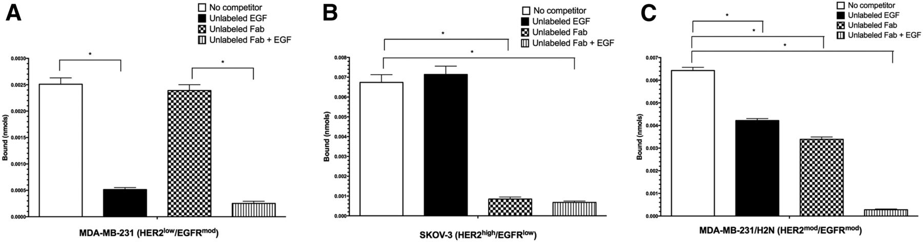
The synthesis and characterization of 111In-DTPA-Fab-PEG24-EGF bsRICs were previously reported (11). 177Lu-DOTA-Fab-PEG24-EGF bsRICs were constructed by conjugating trastuzumab Fab fragments modified with NHS-PEG24-maleimide to EGF functionalized with 2-iminothiolane (Traut reagent) and then derivatizing these bsICs with NHS-DOTA for labeling with 177Lu (Supplemental Fig. 1). There were 3.2 ± 0.8 DOTA chelators substituted per molecule of bsICs. The binding of 177Lu-DOTA-Fab-PEG24-EGF to SK-OV-3 cells (HER2high/EGFRlow) was reduced to 12.4% ± 2.6%, 105.0% ± 10.7%, and 10.1% ± 1.6% by coincubation with a 100-fold molar excess of trastuzumab Fab, EGF, or both ligands, respectively, compared with no competition (Fig. 1A). The binding of 177Lu-DOTA-Fab-PEG24-EGF to MDA-MB-231 cells (HER2low/EGFRmod) was reduced to 95.2% ± 7.5%, 20.3% ± 2.8%, and 9.9% ± 2.9% by trastuzumab Fab, EGF, or both ligands, respectively (Fig. 1B). The binding to MDA-MB-231/H2N cells (HER2mod/EGFRmod) was reduced to 52.7% ± 2.8%, 65.6% ± 2.3%, and 4.4% ± 0.9%, respectively, by competition with trastuzumab Fab, EGF, or both ligands (Fig. 1C). An excess of unlabeled trastuzumab Fab inhibited the binding of 177Lu-DOTA-Fab to MDA-MB-231/H2N cells (HER2mod/EGFRmod) but not to MDA-MB-231 cells (HER2low/EGFRmod) whereas an excess of unlabeled EGF inhibited the binding of 177Lu-DOTA-EGF to both cell lines (Supplemental Fig. 2).

Binding of 177Lu-DOTA-Fab-PEG24-EGF to MDA-MB-231 (HER2low/EGFRmod), SKOV-3 (HER2high/EGFRlow), or MDA-MB-231/H2N (HER2mod/EGFRmod) cells in absence or presence of competitors (EGF, trastuzumab Fab, or combination of EGF and trastuzumab Fab). Values are mean ± SD (n = 3). *Significant differences (P < 0.05).
Clonogenic Survival (CS)
There was no effect on the CS of MDA-MB-231 (HER2low/EGFRmod) cells exposed for 24 h to 2.5 × 10⁻⁷ mol/L of unlabeled trastuzumab Fab, EGF, or Fab-PEG24-EGF compared with untreated cells (Fig. 2A). No significant decrease in survival was found for MDA-MB-231 cells treated with 111In-DTPA-trastuzumab Fab (CS, 94.5% ± 10.1%), but the survival of MDA-MB-231 cells exposed to 111In-DTPA-EGF was reduced by 2-fold (CS, 55.8% ± 3.0%; P < 0.01) compared with untreated cells. The survival of MDA-MB-231 cells treated with 111In-DTPA-Fab-PEG24-EGF was decreased by 1.4-fold compared with untreated cells, but this difference was not significant (CS, 70.1% ± 15.7% vs. 100.0% ± 19.7%; P = 0.09). The survival of MDA-MB-231 cells was significantly decreased by 2.6- to 3.3-fold by 177Lu-DOTA-EGF or 177Lu-DOTA-Fab-PEG24-EGF (CS, 38.4% ± 9.0%, P < 0.0001; and 30.2% ± 4.8%, P < 0.0001, respectively; Fig. 2A). There was no significant effect of 177Lu-DOTA-trastuzumab Fab on the CS of MDA-MB-231 cells (77.3% ± 19.2%; P = 0.06).

CS of MDA-MB-231 (HER2low/EGFRmod) (A), SKOV-3 (HER2high/EGFRlow) (B), MDA-MB-231/H2N (HER2mod/EGFRmod) (C), or TrR1 (HER2mod/EGFRmod) (D) cells after exposure to 177Lu- or 111In-labeled trastuzumab Fab, EGF, Fab-PEG24-EGF, unlabeled agents, or no treatment. Values are mean ± SD (n = 3). *Significant differences (P < 0.05).
The survival of SK-OV-3 (HER2high/EGFRlow) cells exposed to unlabeled trastuzumab Fab or Fab-PEG24-EGF was significantly decreased compared with untreated cells (71.9% ± 9.0%, P = 0.02; and 71.8% ± 2.2%, P = 0.01, respectively, vs. 100.0% ± 10.5%; Fig. 2B). Unlabeled EGF had no effect on the CS of these cells (94.4% ± 4.5%). The survival of SK-OV-3 cells treated with 111In-DTPA-trastuzumab Fab or 111In-DTPA-Fab-PEG24-EGF was decreased compared with untreated cells (CS, 65.9% ± 7.0%, P = 0.01; and 59.8% ± 4.3%, P = 0.003, respectively). The survival of SK-OV-3 cells treated with 111In-DTPA-EGF was significantly decreased (CS, 75.5% ± 7.5%; P = 0.02) compared with untreated cells. 177Lu-DOTA-trastuzumab Fab and 177Lu-DOTA-Fab-PEG24-EGF killed SK-OV-3 cells more efficiently than the corresponding 111In-labeled agents (CS, 35.5% ± 7.5% and 19.8% ± 4.2%, respectively; P < 0.001). 177Lu-DOTA-EGF significantly reduced the CS of SK-OV-3 cells to 65.5% ± 7.5% (P < 0.01).
The survival of MDA-MB-231/H2N cells (HER2mod/EGFRmod) was significantly reduced to 71.6% ± 5.6% by treatment with unlabeled trastuzumab Fab for 24 h (P = 0.01; Fig. 2C) but not by unlabeled EGF (79.1% ± 8.5%; P = 0.054) or Fab-PEG24-EGF (86.4% ± 8.0%; P = 0.14). The survival of MDA-MB-231/H2N cells was significantly decreased by 2.3- to 3.2-fold to 31.6% ± 3.4% (P < 0.001), 43.4% ± 5.9% (P = 0.001), and 43.8% ± 0.03% (P < 0.001) by exposure to 111In-DTPA-trastuzumab Fab, 111In-DTPA-EGF, or 111In-DTPA-Fab-PEG24-EGF, respectively. The CS of MDA-MB-231/H2N cells was reduced to 37.6% ± 7.9%, 25.6% ± 17.6%, and 12.2% ± 10.6% by 177Lu-DOTA-trastuzumab Fab, 177Lu-DOTA-EGF, or 177Lu-DOTA-Fab-PEG24-EGF, respectively. For MDA-MB-231/H2N cells, 177Lu-DOTA-trastuzumab Fab, 177Lu-DOTA-EGF, or 177Lu-DOTA-Fab-PEG24-EGF was not more cytotoxic than the corresponding 111In-labeled agents.
There was no effect of unlabeled trastuzumab Fab, EGF, or Fab-PEG24-EGF on the CS of TrR1 (HER2mod/EGFRmod) cells (Fig. 2D). TrR1 cells treated with 111In-DTPA-trastuzumab Fab and 111In-DTPA-EGF also showed no significant decrease in survival (CS, 92.6% ± 17.6%, P = 0.6; and 88.6% ± 0.6%, P = 0.09, respectively). However, exposure to 111In-DTPA-Fab-PEG24-EGF decreased the CS of these cells to 67.4% ± 11.9% (P = 0.01). Treatment of TrR1 cells with 177Lu-DOTA-trastuzumab Fab, 177Lu-DOTA-EGF, or 177Lu-DOTA-Fab-PEG24-EGF significantly reduced their CS to 51.7% ± 9.8% (P = 0.003), 41.5% ± 8.3% (P = 0.001), and 20.3% ± 5.6% (P < 0.001), respectively. 177Lu-DOTA-trastuzumab Fab, 177Lu-DOTA-EGF, and 177Lu-DOTA-Fab-PEG24-EGF were more cytotoxic than the corresponding 111In-labeled agents.
Biodistribution Studies
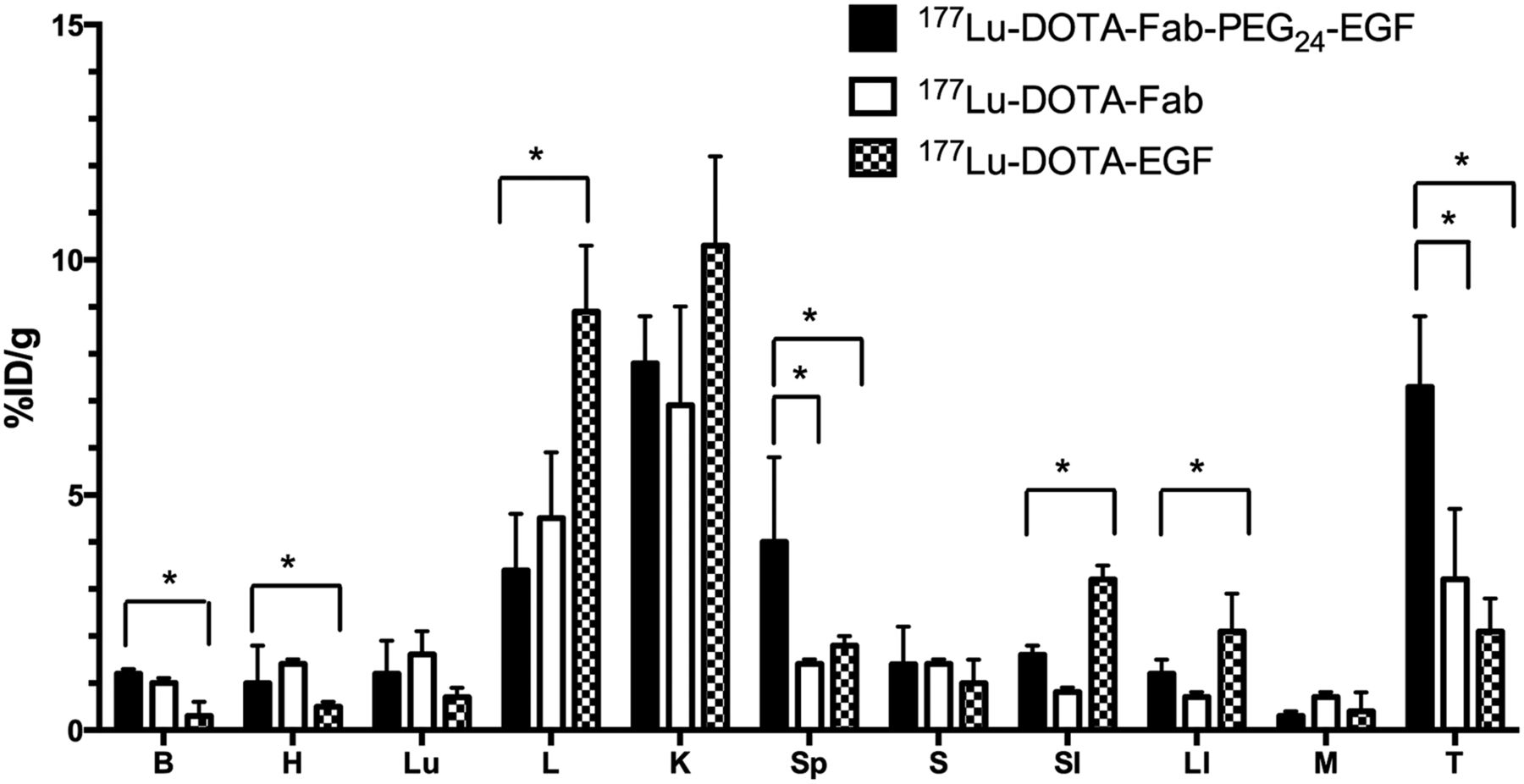
Tumor uptake at 48 h after injection in mice bearing subcutaneous MDA-MB-231/H2N (HER2mod/EGFRmod) BC xenografts was 2.3-fold significantly greater for 177Lu-DOTA-Fab-PEG24-EGF (7.3 ± 1.5 %ID/g) than 177Lu-DOTA-trastuzumab Fab (3.2 ± 1.5 %ID/g; P = 0.02) and 3.5-fold significantly higher than 177Lu-DOTA-EGF (2.1 ± 0.7 %ID/g; P < 0.01; Fig. 3). The blood concentration of radioactivity for 177Lu-DOTA-Fab-PEG24-EGF (1.2 ± 0.1 %ID/g) was 4-fold significantly higher than 177Lu-DOTA-EGF (0.3 ± 0.3 %ID/g; P < 0.01) but not greater than 177Lu-DOTA-trastuzumab Fab (1.0 ± 0.1 %ID/g; P = 0.07). Similar results were found for the heart. Spleen uptake of 177Lu-DOTA-Fab-PEG24-EGF (4.0 ± 1.8 %ID/g) was 2.8-fold significantly greater than 177Lu-DOTA-trastuzumab Fab (1.4 ± 0.1 %ID/g; P = 0.01) and 2.2-fold higher than 177Lu-DOTA-EGF (1.8 ± 0.2 %ID/g; P = 0.02). Liver uptake was 2.6-fold significantly lower for 177Lu-DOTA-Fab-PEG24-EGF (3.4 ± 1.2 %ID/g) than 177Lu-DOTA-EGF (8.9 ± 1.4 %ID/g; P < 0.01) but was not different from 177Lu-DOTA-trastuzumab Fab (4.5 ± 1.4 %ID/g; P = 0.4). There was lower intestinal uptake for 177Lu-DOTA-Fab-PEG24-EGF than 177Lu-DOTA-EGF but not compared with 177Lu-DOTA-trastuzumab Fab.

Tumor and normal-tissue biodistribution of 177Lu-DOTA-Fab-PEG24-EGF, 177Lu-DOTA-trastuzumab Fab, and 177Lu-DOTA-EGF at 48 h after intravenous injection in athymic mice bearing subcutaneous MDA-MB-231/H2N (HER2mod/EGFRmod) BC xenografts. Values are mean ± SD (n = 3). *Significant differences (P < 0.05). B = blood; H = heart; K = kidneys; L = liver; LI = large intestine; Lu = lungs; M = muscle; S = stomach; SI = small intestine; Sp = spleen; T = tumor.
Radiation Dosimetry Estimates
A comparison of radiation-absorbed doses estimated for the tumor and normal organs in CD1 athymic mice with subcutaneous MDA-MB-231/H2N xenografts and injected with 11.1 (10 μg) of 111In-DTPA-Fab-PEG24-EGF or 177Lu-DOTA-Fab-PEG24-EGF is shown in Table 1. The doses to normal organs were 1.5- to 8.2-fold higher for 177Lu-DOTA-Fab-PEG24-EGF than 111In-DTPA-Fab-PEG24-EGF. 177Lu-DOTA-Fab-PEG24-EGF deposited a 9.3-fold-higher radiation dose in the tumor than 111In-DTPA-Fab-PEG24-EGF.
Estimated Radiation-Absorbed Doses in CD1 Athymic Mice with MDA-MB-231/H2N Human Breast Cancer Xenografts Injected with 111In- or 177Lu-Labeled bsRICs
Normal-Tissue Toxicity Studies
A dose–escalation acute toxicity study was performed to select the dose of 177Lu-DOTA-Fab-PEG24-EGF for radioimmunotherapy studies. There was no significant change in body weight over 14 d for non–tumor-bearing BALB/c mice injected with 3.7–18.5 MBq (10 μg each) of 177Lu-DOTA-Fab-PEG24-EGF compared with normal saline-treated mice, but there was a trend toward decreased body weight for mice receiving 18.5 MBq (Fig. 4). There were significantly reduced leukocyte (white blood cell) counts in mice receiving 18.5 MBq of 177Lu-DOTA-Fab-PEG24-EGF compared with saline-treated mice (P = 0.015; Fig. 5A) but not for lower doses. Platelet counts were not significantly decreased in mice receiving 177Lu-DOTA-Fab-PEG24-EGF compared with normal saline mice (Fig. 5B). Erythrocyte (red blood cell) counts, hemoglobin, and hematocrit were significantly lower in mice receiving 18.5 MBq of 177Lu-DOTA-Fab-PEG24-EGF (P = 0.010, 0.05, and 0.013, respectively; Figs. 5C–5E) than normal saline-treated mice but not at lower doses. There was no significant effect on serum ALT or Cr at any dose of bsRICs (Fig. 6), but there was a trend toward higher Cr with increasing dose of 177Lu-DOTA-Fab-PEG24-EGF. On the basis of the overall normal-tissue toxicity profile, a dose of 11.1 MBq (10 μg) was defined as the no observable adverse effect level (NOAEL) and was selected for radioimmunotherapy studies.

Body weight normalized to initial body weight at different times after injection of 3.7, 11.1, or 18.5 MBq (10 μg) of 177Lu-DOTA-Fab-PEG24-EGF or for untreated mice. Values are mean ± SD (n = 5). There were no significant differences (P < 0.05) between groups.
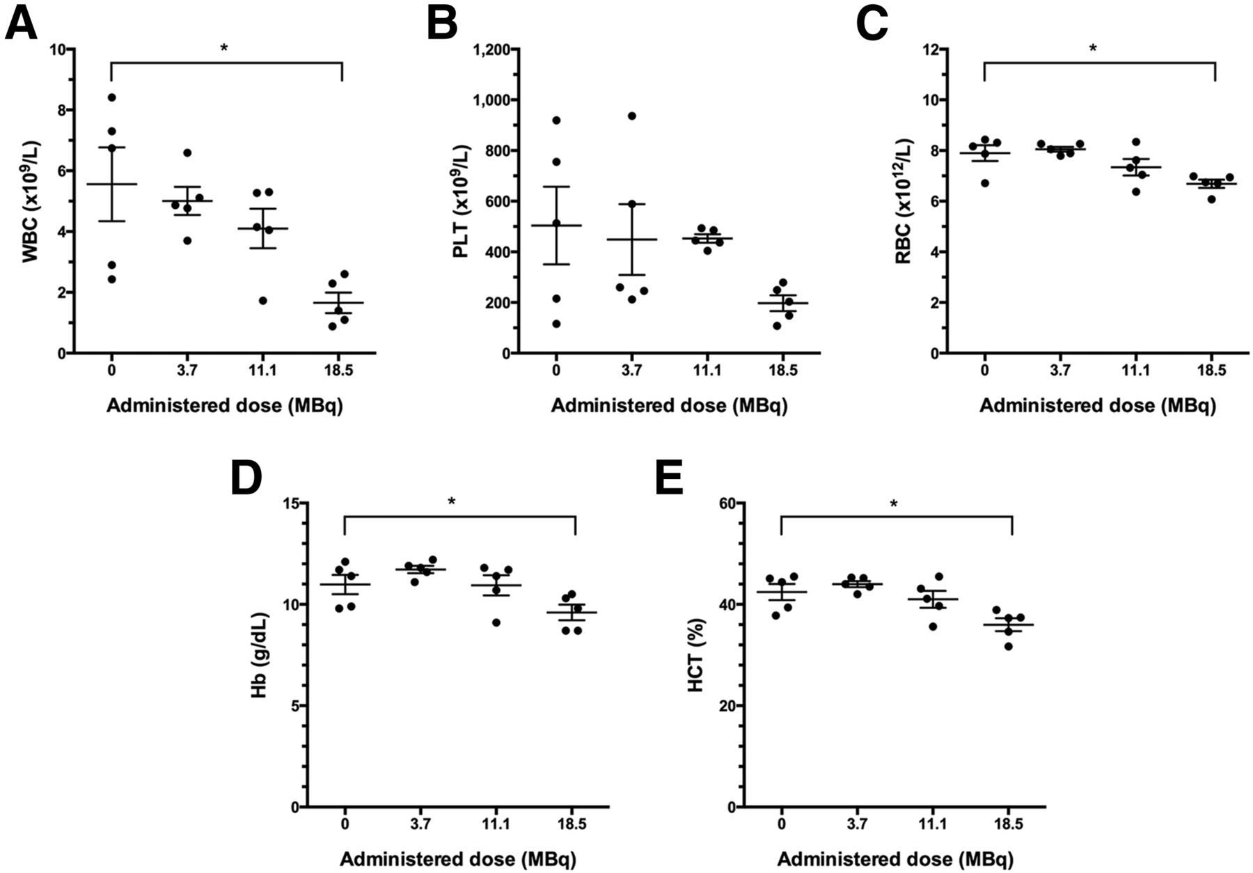
Blood cell counts (PLT = platelets; red blood cell [RBC] = erythrocytes; white blood cell [WBC] = leukocytes), hemoglobin (Hb), and hematocrit (HCT) in non–tumor-bearing BALB/c mice at 14 d after injection of 177Lu-DOTA-Fab-PEG24-EGF (3.7–11.1 MBq; 10 μg) or for untreated mice. Values shown are those for individual mice as well as mean ± SD (n = 5). *Significant differences (P < 0.05).
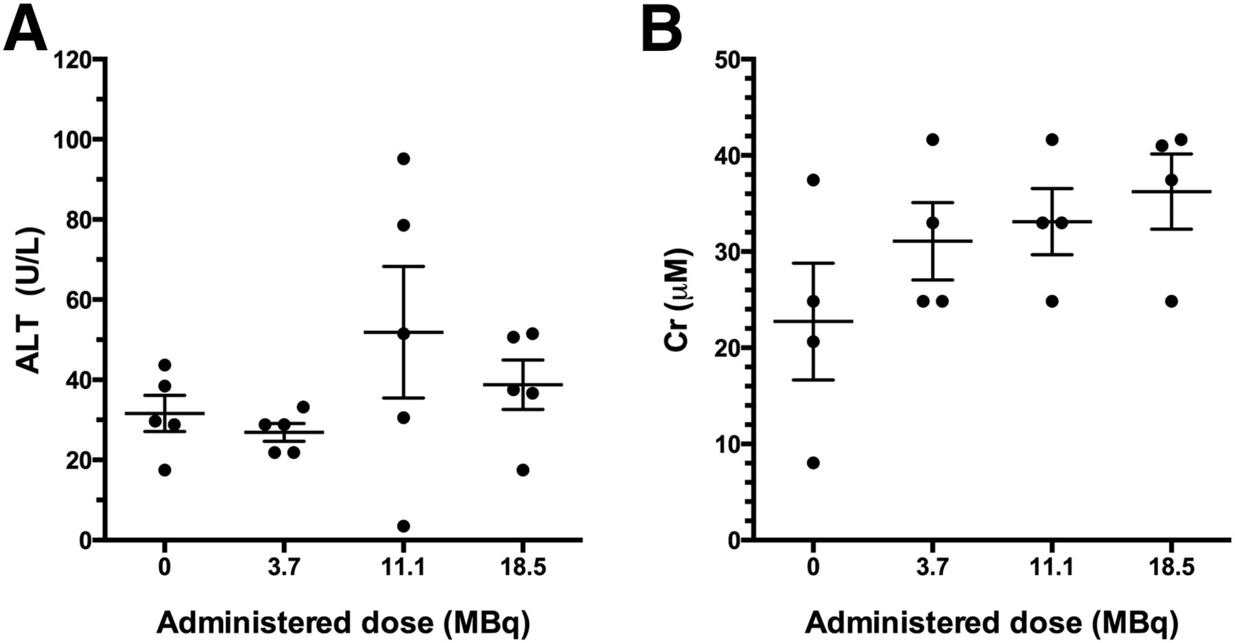
Serum ALT and Cr levels in non–tumor-bearing BALB/c mice at 14 d after injection of 177Lu-DOTA-Fab-PEG24-EGF (3.7–11.1 MBq; 10 μg) or for untreated mice. Values shown are those for individual mice as well as mean ± SD (n = 5). *Significant differences (P < 0.05).
Radioimmunotherapy Studies
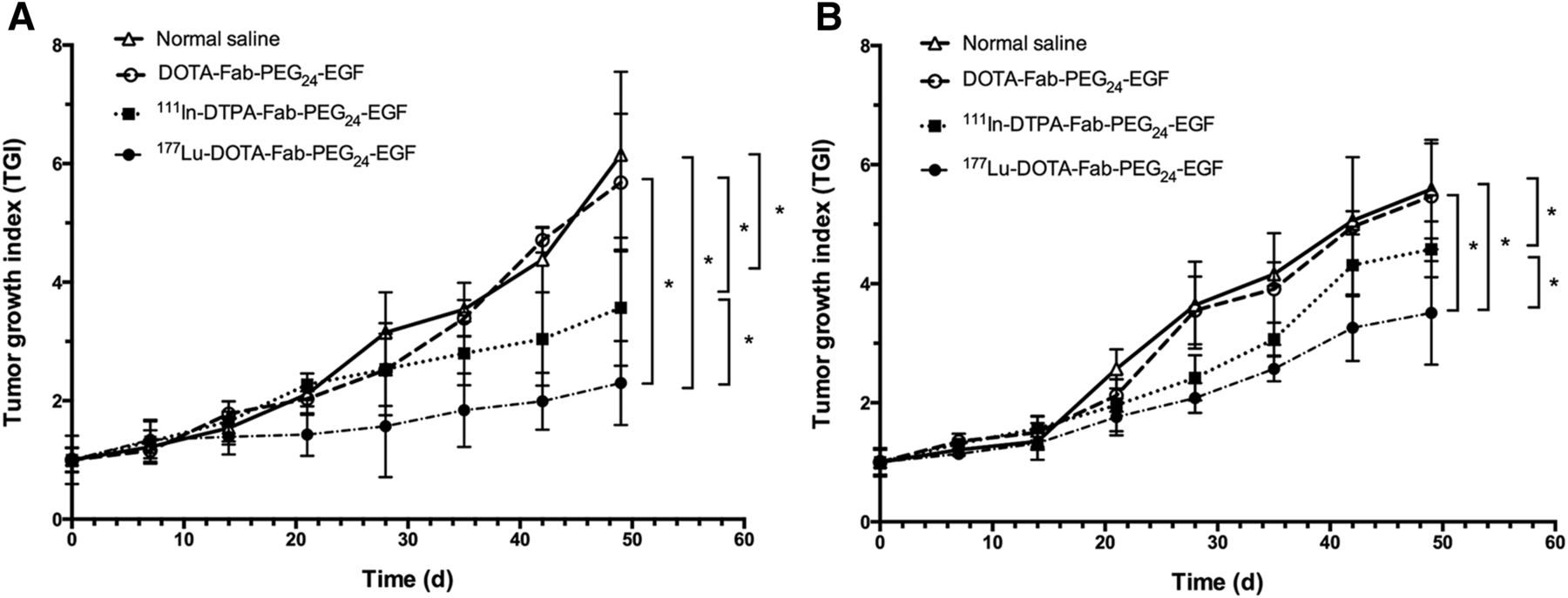
Radioimmunotherapy studies in mice with MDA-MB-231/H2N (HER2mod/EGFRmod) BC xenografts demonstrated strong tumor growth inhibition after a single injection of 11.1 MBq (10 μg) of 177Lu-DOTA-Fab-PEG24-EGF (Fig. 7A). The TGI at 49 d for 177Lu-DOTA-Fab-PEG24-EGF (2.3 ± 0.7) was 2.3- to 2.6-fold significantly lower than for mice treated with normal saline (6.2 ± 1.4; P < 0.001) or unlabeled DOTA-Fab-PEG24-EGF (5.7 ± 1.2; P < 0.001). 177Lu-DOTA-Fab-PEG24-EGF treatment was more effective than 111In-DTPA-Fab-PEG24-EGF (TGI at 49 d, 3.6 ± 1.0; P = 0.047). Unlabeled DOTA-Fab-PEG24-EGF had no significant effect on the growth of MDA-MB-231/H2N tumors. 177Lu-DOTA-Fab-PEG24-EGF treatment moderately inhibited the growth of trastuzumab-resistant TrR1 (HER2mod/EGFRmod) BC xenografts in mice (Fig. 7B). The TGI at 49 d after treatment with 177Lu-DOTA-Fab-PEG24-EGF (3.5 ± 0.9) was 1.6-fold significantly lower than treatment with normal saline (5.6 ± 0.8; P < 0.001) or unlabeled DOTA-Fab-PEG24-EGF (5.5 ± 0.9; P < 0.01). 177Lu-DOTA-Fab-PEG24-EGF was 1.3-fold significantly more effective at inhibiting the growth of TrR1 tumors than 111In-DTPA-Fab-PEG24-EGF (TGI, 4.6 ± 0.5; P = 0.042). Unlabeled DOTA-Fab-PEG24-EGF was not effective for inhibiting the growth of TrR1 tumors.

TGI for athymic mice bearing trastuzumab-sensitive MDA-MB-231/H2N (HER2mod/EGFRmod) tumor xenografts (A) or TrR1 (HER2mod/EGFRmod) trastuzumab-resistant tumor xenografts (B) treated with unlabeled DOTA-Fab-PEG24-EGF, 111In-DTPA-Fab-PEG24-EGF, or 177Lu-DOTA-Fab-PEG24-EGF or receiving no treatment. Values are mean ± SD (n = 5). *Significant differences (P < 0.05).
DISCUSSION
We previously reported that 111In-DTPA-Fab-PEG24-EGF bsRICs imaged subcutaneous tumor xenografts in athymic mice that expressed EGFR or HER2 or both receptors (11). We now extend these findings to radioimmunotherapy by complexing these bsRICs to the β-particle emitter 177Lu or by exploiting the Auger electron emissions of 111In. 177Lu-DOTA-Fab-PEG24-EGF bsRICs were constructed by linking trastuzumab Fab fragments to EGF through a PEG24 spacer and then derivatizing the bsICs with 3.2 ± 0.8 DOTA for labeling with 177Lu (Supplemental Fig. 1). DOTA is likely substituted predominantly onto the Fab domain because trastuzumab Fab presents 25 ε-amino groups on lysines for reaction with NHS-DOTA (23), whereas EGF contains only 2 lysines and 1 N-terminal amine for DOTA modification or thiolation with Traut's reagent (24). We previously reported that 111In-DOTA-trastuzumab Fab exhibited preserved HER2 binding affinity (25), and 68Ga-DOTA-EGF was reported to bind with high affinity to EGFR (26). The binding of 177Lu-DOTA-trastuzumab Fab or 177Lu-DOTA-EGF to MDA-MB-231 cells (HER2low/EGFRmod) and MDA-MB-231/H2N cells (HER2mod/EGFRmod) was inhibited by an excess of trastuzumab Fab or EGF in agreement with the receptor expression of these cells (Supplemental Fig. 2). Bivalent affibody molecules binding HER2 and EGFR have also been constructed (27). However, these were not radiolabeled and not studied for tumor imaging or radioimmunotherapy. The dissociation constant for binding of these bispecific affibody molecules to EGFR or HER2 was 35–86 and 6–219 nM, respectively. In competition receptor binding assays with EGF or trastuzumab Fab, we previously measured an effective concentration of 50% for displacement of the binding of 111In-DTPA-Fab-PEG24-EGF to EGFR of 25–36 and 18–24 nM for HER2 (11). These effective concentration of 50% values approximate the dissociation constant. The specificity of binding of 177Lu-DOTA-Fab-PEG24-EGF to EGFR and HER2 was confirmed by competition with unlabeled EGF or trastuzumab Fab using MDA-MB-231 (HER2low/EGFRmod), SK-OV-3 (HER2high/EGFRlow), MDA-MB-231/H2N (HER2mod/EGFRmod), or TrR1 (HER2mod/EGFRmod) cells (Fig. 1). The inability of EGF to compete with the binding of 177Lu-DOTA-Fab-PEG24-EGF to SK-OV-3 cells (HER2high/EGFRlow) may be due to predominant HER2 binding on these cells due to the 12-fold-greater density of HER2 than EGFR (9.6 × 10⁵ vs. 8 × 10⁴ receptors/cell, respectively).
Exposure to 177Lu-DOTA-Fab-PEG24-EGF killed all 4 cell types: MDA-MB-231 (HER2low/EGFRmod), SK-OV-3 (HER2high/EGFRlow), MDA-MB-231/H2N (HER2mod/EGFRmod), and TrR1 (HER2mod/EGFRmod) (Fig. 2). Monospecific 177Lu-DOTA-EGF killed MDA-MB-231 cells but was less effective for killing SK-OV-3 cells. Conversely, 177Lu-DOTA-trastuzumab Fab was more effective for killing SK-OV-3 cells than MDA-MB-231 cells. 111In-DTPA-Fab-PEG24-EGF bsRICs were less cytotoxic than 177Lu-DOTA-Fab-PEG24-EGF (Fig. 2). Although HER2 contains a putative NLS, the receptor is slowly internalized (28), which may limit the intracellular accumulation of the bsRICs. Moreover, the bsRICs were not modified with exogenous NLS peptides to promote more efficient nuclear importation after HER2-mediated internalization. Nuclear importation amplifies the DNA damage caused by the Auger electrons emitted by 111In (12). The longer range (2 mm) β-particles emitted by 177Lu do not require internalization or nuclear importation of the bsRICs for cytotoxicity. The bsRICs were more versatile than the monospecific agents because they were able to kill tumor cells displaying HER2 or EGFR or both receptors. This ability to target and kill tumor cells that are HER2- or EGFR-positive or that coexpress these 2 receptors may overcome intratumoral HER2 heterogeneity. Intratumoral heterogeneity in HER2 expression was found in 18% of HER2-positive BC, with HER2-amplified and HER2-nonamplified regions detected in the same specimen (29). HER2-negative cells may express EGFR (30). The bsRICs killed trastuzumab-resistant TrR1 cells that display both EGFR and HER2, whereas exposure to unlabeled Fab-PEG24-EGF or trastuzumab Fab had no effect on the survival of these cells (Fig. 2). These results confirm our previous report that Auger electron–emitting 111In-NLS-trastuzumab was cytotoxic to TrR1 cells, despite their resistance to trastuzumab (19).
Biodistribution studies at 48 h after injection in CD1 athymic mice with subcutaneous MDA-MB-231/H2N xenografts (Fig. 3) revealed 2-fold-significantly-greater tumor uptake of 177Lu-DOTA-Fab-PEG24-EGF than 177Lu-DOTA-trastuzumab Fab or 177Lu-DOTA-EGF. These results indicate that cross-linking trastuzumab Fab through a PEG24 spacer to EGF improved tumor localization compared with the monospecific agents. Tumor uptake of 177Lu-DOTA-Fab-PEG24-EGF at 48 h after injection (7.3 ± 3.5 %ID/g) was identical to 111In-DTPA-Fab-PEG24-EGF at this time point, indicating that substitution of DOTA did not affect tumor uptake of the bsRICs (11). A dose–escalation study was conducted in BALB/c mice to select a dose of 177Lu-DOTA-Fab-PEG24-EGF for radioimmunotherapy studies. An administered dose of 11.1 MBq (10 μg) caused no significant decrease in body weight (Fig. 4) or blood cell counts, hemoglobin, or hematocrit (Fig. 5) and no increase in serum ALT or Cr over 14 d (Fig. 6) and was therefore defined as the NOAEL. Interestingly, the hematologic toxicity of 177Lu-DOTA-Fab-PEG24-EGF was much lower than for comparable amounts of 177Lu-DTPA-trastuzumab administered to mice (31), which may be due to its more rapid elimination from the blood (1.2 ± 0.1 vs. 13.7 ± 0.8 %ID/g, respectively).
Radioimmunotherapy studies using a single NOAEL dose of 11.1 MBq (10 μg) of 177Lu-DOTA-Fab-PEG24-EGF in mice engrafted subcutaneously with trastuzumab-sensitive MDA-MB-231/H2N BC xenografts yielded strong tumor growth inhibition (Fig. 6) compared with mice treated with normal saline or unlabeled DOTA-Fab-PEG24-EGF. In agreement with the results of the in vitro cytotoxicity results, 177Lu-DOTA-Fab-PEG24-EGF was more effective for inhibiting tumor growth than 111In-DTPA-Fab-PEG24-EGF. Radiation dosimetry estimates (Table 1) revealed that 177Lu-DOTA-Fab-PEG24-EGF delivered a 9-fold-higher dose to the tumor than 111In-DTPA-Fab-PEG24-EGF, which explains its greater potency. However, normal-organ absorbed doses from 111In-DTPA-Fab-PEG24-EGF were 1.5- to 8-fold lower than 177Lu-DOTA-Fab-PEG24-EGF, suggesting that the administered amount of 111In-DTPA-Fab-PEG24-EGF could be increased to compensate for a lower potency. Radioimmunotherapeutic agents labeled with Auger electron emitters have been found to be more effective for treating tumors in mice than the same agents labeled with β-emitters when administered at equitoxic doses (32). Trastuzumab-resistant TrR1 tumors also responded to treatment with the bsRICs but were less sensitive than MDA-MB-231/H2N BC xenografts. 177Lu-DOTA-Fab-PEG24-EGF was the most effective for treating these tumors. 111In-DTPA-Fab-PEG24-EGF was not modified with NLS peptides to promote efficient nuclear translocation after HER2-mediated internalization (12) and HER2 is slowly internalized (28), which may have limited the effectiveness of the subcellular-range Auger electrons released by 1111In for radioimmunotherapy. The resistance of TrR1 tumors to treatment with 111In- or 177Lu-labeled bsRICs compared with MDA-MB-231/H2N tumors may be due to their 3-fold-higher expression of insulin growth factor-1 receptors (33). Increased insulin growth factor-1 receptors has been associated with radiation resistance (34). The resistance of TrR1 tumors could also be due to lower tumor accumulation of the bsRICs because this was not measured in biodistribution studies.
CONCLUSION
111In-DTPA-Fab-PEG24-EGF and 177Lu-DOTA-Fab-PEG24-EGF were effective for killing tumor cells in vitro that displayed HER2 or EGFR or both receptors, and a single dose of the bsRICs at the NOAEL yielded moderate to strong tumor growth inhibition in vivo in mice bearing subcutaneous trastuzumab-sensitive or -resistant human BC xenografts. 177Lu-DOTA-Fab-PEG24-EGF was more effective for inhibiting tumor growth than 111In-DTPA-Fab-PEG24-EGF, due to a 9-fold-higher radiation-absorbed dose in tumors. The lower normal-organ absorbed doses deposited by 111In-DTPA-Fab-PEG24-EGF suggest that the administered amount of these bsRICs could be increased to compensate for a lower potency. These results are encouraging for further development of these bsRICs for dual-receptor–targeted radioimmunotherapy of BC that coexpresses HER2 and EGFR, including trastuzumab-resistant tumors.
DISCLOSURE
The costs of publication of this article were defrayed in part by the payment of page charges. Therefore, and solely to indicate this fact, this article is hereby marked “advertisement” in accordance with 18 USC section 1734. This research was supported by a grant from the Ontario Institute for Cancer Research (Smarter Imaging Program) with funds from the Province of Ontario and a grant from the Canadian Institutes of Health Research (MOP130322). Eva J. Razumienko is the recipient of a Canadian Breast Cancer Foundation Doctoral Fellowship (Ontario Region). No other potential conflict of interest relevant to this article was reported.
Footnotes
Published online Oct. 1, 2015.
- © 2016 by the Society of Nuclear Medicine and Molecular Imaging, Inc.
REFERENCES
- Received for publication June 16, 2015.
- Accepted for publication September 21, 2015.


