


Abstract
Carbonic anhydrase IX (CA-IX), a transmembrane enzyme, mediates cell survival under hypoxic conditions and is overexpressed in solid malignancies. In this study, we synthesized four 18F sulfonamide derivatives and evaluated their potential for imaging CA-IX expression with PET. Methods: Azide derivatives of 2 carbonic anhydrase inhibitors, 4-(2-aminoethyl)benzenesulfonamide (AEBS) and 4-aminobenzensulfonamide (ABS), were coupled to radiosynthons with either 1 or 3 alkynes and a pendent ammoniomethyltrifluoroborate (AmBF3) to generate monovalent or trivalent enzyme inhibitors. Binding affinity to CA-IX and other CA isoforms was determined via a stopped-flow, CA-catalyzed CO2 hydration assay. Tracers were radiolabeled via 18F-19F isotope exchange reactions. Imaging/biodistribution studies were performed using HT-29 tumor–bearing immunocompromised mice. Results: Monomeric AmBF3-AEBS and AmBF3-ABS were obtained in 41% and 40% yields, whereas trimeric AmBF3-(AEBS)3 and AmBF3-(ABS)3 were obtained in 47% and 55% yields, respectively. Derivatives bound CA-I, -II, -IX, and -XII with good affinity (0.49–100.3 nM). 18F-labeled sulfonamides were obtained in 16.3%–36.8% non–decay-corrected radiochemical yields, with 40–207 GBq/μmol specific activity and greater than 95% radiochemical purity. Biodistribution/imaging studies showed that the tracers were excreted through both renal and hepatobiliary pathways. At 1 h after injection, HT-29 tumor xenografts were clearly visualized in PET images with modest contrast for all 4 tracers. Tumor uptake was 2-fold higher for monovalent tracers (∼0.60 percentage injected dose per gram [%ID/g]) than for trivalent tracers (∼0.30 %ID/g); however, tumor-to-background ratios were significantly better for 18F-AmBF3-(ABS)3. Preblocking with acetazolamide reduced more than 80% uptake of 18F-AmBF3-(ABS)3 in HT-29 tumors. Conclusion: Our data suggest that trimerization of an otherwise nonspecific CA inhibitor greatly enhances the selectivity for CA-IX in vivo and represents a promising strategy for creating multivalent enzyme inhibitors for selectively imaging extracellular enzyme activity by PET.
PET imaging plays an increasingly important role in noninvasively characterizing tumor subtypes to inform drug development and evaluate drug target expression, in vivo. Extracellular targets are typically imaged with radiolabeled peptides, aptamers, and antibodies. Small molecules (e.g., 18F-fluoromisonidazole, 18F-fluorothymidine, and a host of others) have been used to probe enzymatic activity that may be associated within a given cell type. Nevertheless, the application of small-molecule inhibitors as PET tracers can be challenged by several effects such as nonspecific uptake in cells, poor clearance from blood (1), and rapid metabolic defluorination with accompanying bone uptake (2,3). In addition, 18F labeling in high yield and at high specific activity often presents a significant challenge in the development of new PET tracers (4).
Yet even when conditions for clinically useful radiolabeling have been met, nonspecific uptake due to association with off-target enzyme isoforms of greater abundance can result in images marked by low tumor-to-background ratios. Lipophilicity can also enhance nonspecific uptake in some cases. We hypothesized that a multivalent enzyme inhibitor would restrict intracellular accumulation and enhance specific binding to an extracellular tumor target. To accomplish this, we used a radiosynthon that readily enables grafting of various ligands to a pentaerythritol core while allowing for facile radiolabeling at high specific activity with Curie levels of 18F activity. Recently, we communicated this method for linking both peptides and small-molecule inhibitors in a trivalent motif (5). Here we expand the application of this method to address the imaging potential of 2 related enzyme inhibitors and report detailed findings on how this method can be used to reduce nonspecific intracellular accumulation.
Toward these ends, we sought to image carbonic anhydrase IX (CA-IX), which is differentially expressed in tumors under hypoxia and therefore of great clinical interest (6,7). This transmembrane enzyme represents a considerable challenge because of ubiquitously expressed isoforms that otherwise compete with known inhibitors, leading to high background and often low if not unobserved tumor uptake (vide infra). Regulated by hypoxia-inducible factor 1α, CA-IX is involved in, among others, maintaining an alkaline intracellular pH while acidifying the extracellular environment. The latter process facilitates cell migration and invasion and potentiates metastatic activity in primary lesions (8,9). Recently, CA-IX has been shown to mediate growth and expansion of breast cancer stem cells within hypoxic niches (10). Clinically, the expression of CA-IX is negatively correlated with patient prognosis and survival (11). In addition to breast cancer, CA-IX is overexpressed in many other solid malignancies including lung, colon, cervix, ovary, head and neck, bladder, and renal cancers (11). With limited expression in normal tissues (12–14), CA-IX represents an attractive target for therapy, with a sulfonamide inhibitor in early phase I clinical trial (ClinicalTrails.gov: safety study of SLC-0111 in subjects with advanced solid tumors).
Compared with therapeutics, the development of imaging agents to quantify expression of CA-IX is still in its early stages. Monoclonal antibodies and small-molecule inhibitors have been explored for nuclear imaging. 124I-cG250, a monoclonal antibody targeting the PG-like domain of CA-IX, advanced to phase III clinical trial for the diagnosis of clear cell renal cell carcinoma (marketed as RENAREX by WILEX AG) (15). Because of concerns with tumor penetrance within hypoxic regions and nonideal delayed imaging, research groups have sought alternatives to monoclonal antibody–based imaging. Indeed, one coumarin and several sulfonamides have been radiolabeled for targeting CA-IX (Fig. 1) (16–24). Although these tracers are potent CA-IX inhibitors, those that were evaluated in vivo (18–21) suffered from low tumor uptake, selectivity, or stability (Table 1).

Reported radiolabeled small-molecule inhibitors for CA-IX imaging. Compounds A–M are sulfonamide-based derivatives, whereas compound N is a coumarin-based inhibitor.
Summary of Biologic Evaluation Data of Radiolabeled CA-IX Imaging Probes Shown in Figure 1
Our previous attempt using 18F-U-104 (compound H) resulted in high uptake in blood (13.97 ± 3.07 percentage injected dose per gram [%ID/g] at 1 h after injection) (21) presumably because of the binding of 18F-U-104 to intracellular off targets CA-I and CA-II that are abundant in erythrocytes (25). We hypothesized that cell-impermeant tracers would disfavor binding to intracellular CA isoforms and in turn provide high-contrast images. To achieve cell impermeabilitiy, we sought multivalent inhibitors to increase both avidity and molecular weight. In the present study, we synthesized and evaluated 2 monomeric (as controls) and 2 trimeric 18F-labeled sulfonamides using 4-(2-aminoethyl)benzenesulfonamide (AEBS) and 4-aminobenzensulfonamide (ABS) as our pharmacophores for imaging CA-IX expression with PET. In terms of a radioprosthetic group, we chose the zwitterionic organotrifluoroborate (5), the polarity of which would likely further reduce lipophilicity and membrane permeability.
MATERIALS AND METHODS
Information for reagents, instruments, affinity measurements, radiochemistry, LogD7.4 measurements, plasma stability studies, PET imaging experiments, and data analysis are provided in the supplemental materials (supplemental materials are available at http://jnm.snmjournals.org). All animal studies were performed in accordance with the Canadian Council on Animal Care guidelines and approved by the Animal Care Committee of the University of British Columbia.
RESULTS
Chemistry and Radiochemistry
The preparation of nonradioactive 19F-AmBF3-AEBS, 19F-AmBF3-ABS, 19F-AmBF3-(AEBS)3, and 19F-AmBF3-(ABS)3 followed synthetic schemes as shown in Figure 2. Azidoacetyl-AEBS 1 was obtained in 74% yield by coupling AEBS with azidoacetic acid, whereas azidoacetyl-ABS 2 was prepared in 60% yield by the displacement of the chloro group of 2-chloro-N-(4-sulfamoyl-phenyl)-acetamide with azide. The coupling of 1 and 2 to AmBF3-conjugated alkyne 3 via the Cu+-catalyzed click reaction afforded the desired AmBF3-AEBS and AmBF3-ABS in 41% and 40% yields, whereas coupling to alkyne 4 generated AmBF3-(AEBS)3 and AmBF3-(ABS)3 in 47% and 55% yields, respectively.
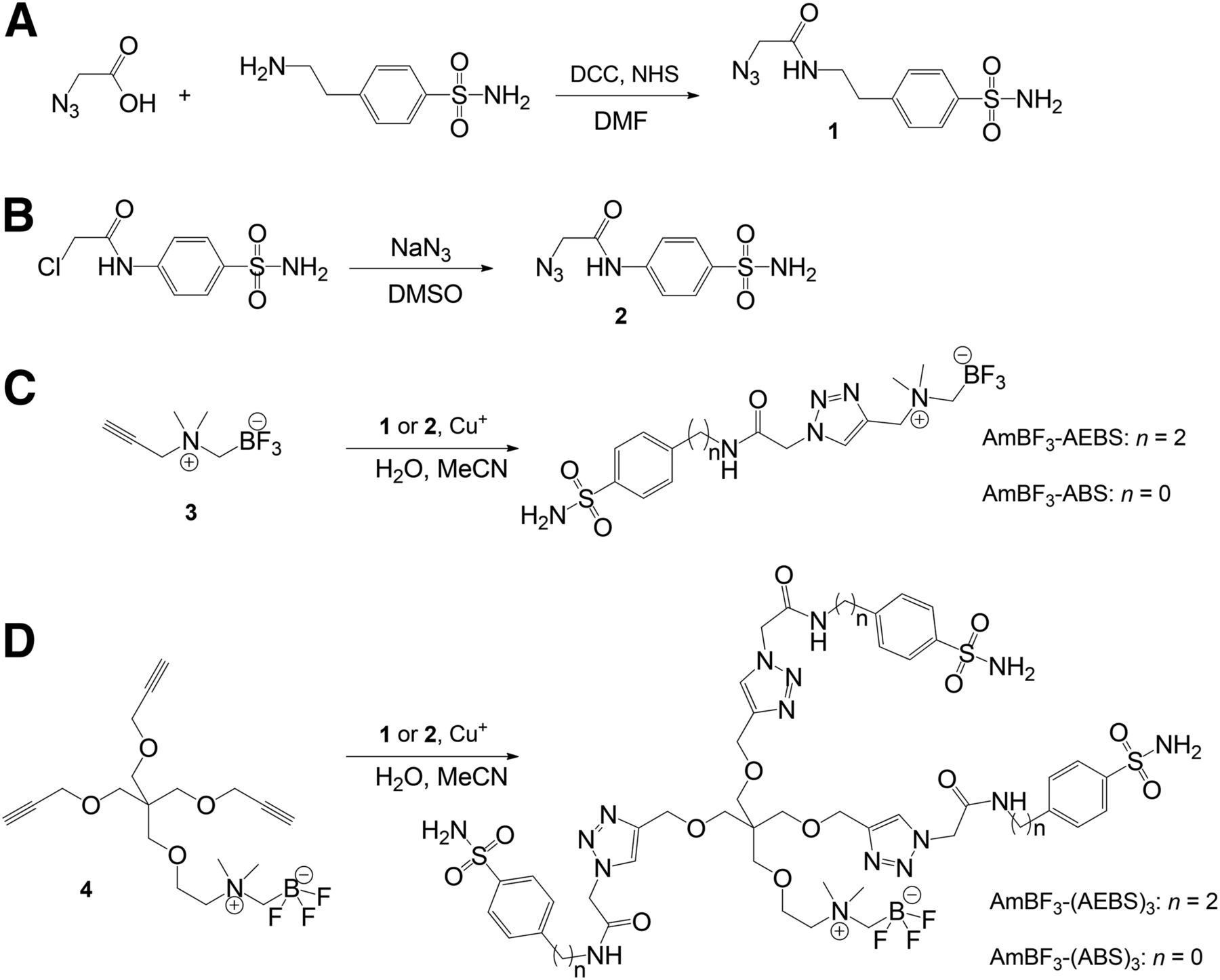
Synthesis of azidoacetyl-AEBS 1 (A), azidoacetyl-ABS 2 (B), AmBF3-AEBS and AmBF3-ABS (C), and AmBF3-(AEBS)3 and AmBF3-(ABS)3 (D). DCC = N,N′-dicyclohexylcarbodiimine; NHS = N-hydroxysuccinimde.
The 18F-labeling reactions were performed via 18F-19F isotope exchange as depicted in Figure 3. Starting with 23.3–38.5 GBq of 18F-fluoride, 18F-AmBF3-AEBS, 18F-AmBF3-ABS, 18F-AmBF3-(AEBS)3, and 18F-AmBF3-(ABS)3 were obtained in 24.8% ± 2.2%, 26.4% ± 10.2%, 28.0% ± 3.5%, and 26.9% ± 5.6% non–decay-corrected radiochemical yields (n = 3) with corresponding specific activities of 185 ± 22, 141 ± 11, 66 ± 9, and 49 ± 12 GBq/μmol, respectively. After C18 Sep-Pak (Waters) purification, more than 95% radiochemical purity was obtained for all tracers based on high-performance liquid chromatography radiochromatograms.
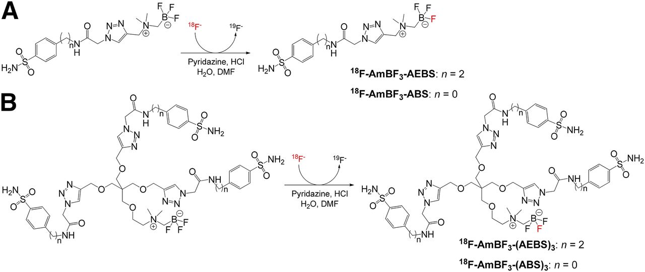
Radiosynthesis of 18F-AmBF3-AEBS and 18F-AmBF3-ABS (A) and 18F-AmBF3-(AEBS)3 and 18F-AmBF3-(ABS)3 (B) via 18F-19F isotope exchange reaction.
Binding Affinity (Ki) to CA Isoforms
The binding affinities of the synthesized compounds to CA-I, -II, -IX, and -XII are summarized in Table 2. Ki values to CA-IX for AmBF3-AEBS, AmBF3-ABS, AmBF3-(AEBS)3, and AmBF3-(ABS)3 were 8.0, 6.0, 35.7, and 8.5 nM, respectively. Acetazolamide, a pan-CA inhibitor (8,26), was included as a positive control.
CA-I, -II, -IX, and -XII Inhibition Data
Plasma Stability and Lipophilicity
To assess stability, tracers were incubated in mouse plasma and analyzed by high-performance liquid chromatography. No metabolites of 18F-AmBF3-AEBS, 18F-AmBF3-ABS, 18F-AmBF3-(AEBS)3, and 18F-AmBF3-(ABS)3 were observed after 2 h incubation at 37°C. The LogD7.4 (D is the distribution coefficient at pH 7.4) values for 18F-AmBF3-AEBS, 18F-AmBF3-ABS, 18F-AmBF3-(AEBS)3, and 18F-AmBF3-(ABS)3 were −1.9, −1.8, −2.1, and −2.5, respectively, using the shake flask extraction method.
Biodistribution Studies
Biodistribution data are summarized in Table 3. 18F-AmBF3-AEBS, 18F-AmBF3-ABS, 18F-AmBF3-(AEBS)3, and 18F-AmBF3-(ABS)3 were cleared through hepatobiliary and renal pathways with generally low uptake in nonexcretory tissues including bone. Uptake in HT-29 tumor xenografts was approximately 2-fold higher for the monomers (0.56–0.64 %ID/g) than the trimers (0.30–0.33 %ID/g). Notably, 5-fold-higher activity was observed in the blood (0.51–0.56 %ID/g) for 18F-AmBF3-AEBS and 18F-AmBF3-ABS than 18F-AmBF3-(AEBS)3 and 18F-AmBF3-(ABS)3 (0.07–0.09 %ID/g), indicating slower clearance profiles or enhanced nonspecific binding. At 1 h after injection, the highest tumor-to-blood and tumor-to-muscle ratios (3.93 ± 1.26 and 9.55 ± 2.96, respectively) were obtained when administering 18F-AmBF3-(ABS)3. Preinjection with 10 mg of acetazolamide per kilogram significantly reduced the uptake of 18F-AmBF3-(ABS)3 in tumors to 0.06 ± 0.01 %ID/g, as well as its tumor-to-background ratios.
Biodistribution and Tumor-to-Nontarget Ratios for 18F-AmBF3 Sulfonamides
PET/CT Imaging
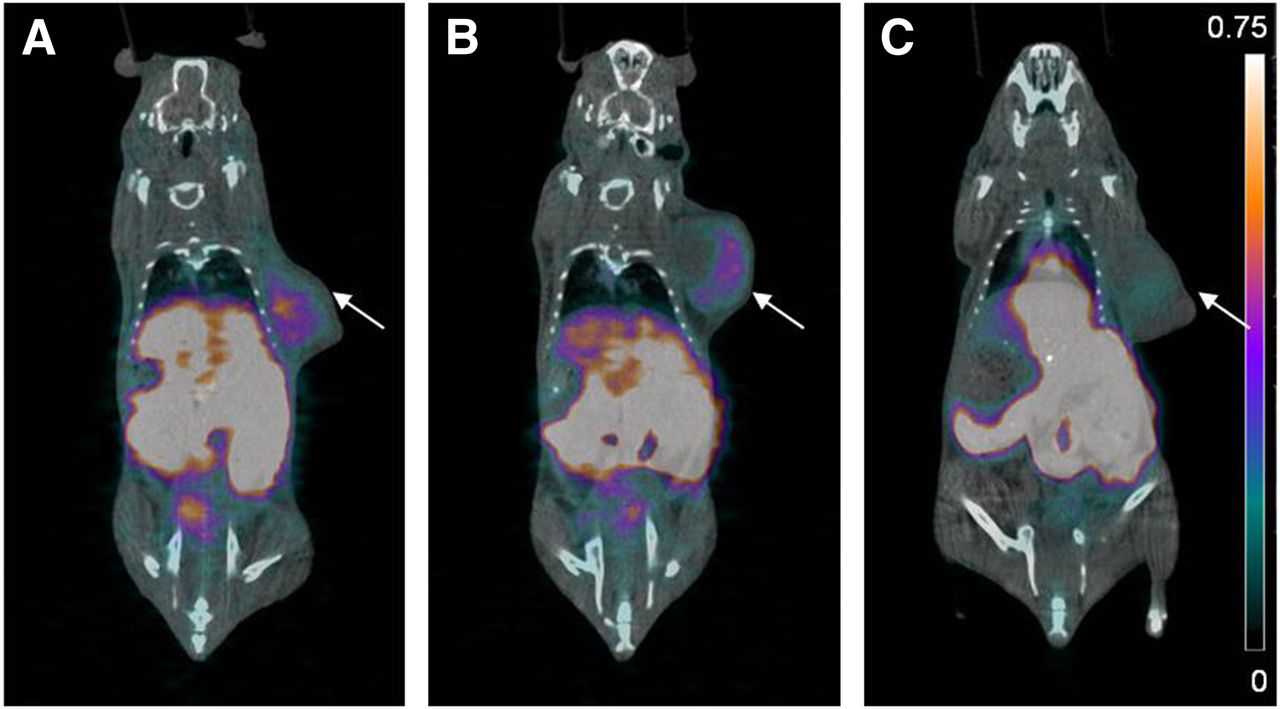
PET/CT images obtained at 1 h after injection (Figs. 4 and 5) were consistent with the biodistribution data. Higher uptake was observed in the kidneys, liver, and gastrointestinal tract. HT-29 tumor xenografts were clearly visualized after injecting 18F-AmBF3-AEBS (Fig. 4A), 18F-AmBF3-ABS (Fig. 4B), 18F-AmBF3-(AEBS)3 (Fig. 5A), or 18F-AmBF3-(ABS)3 (Fig. 5B). Blocking studies were performed for 18F-AmBF3-(ABS)3 that generated highest tumor-to-background contrast. Preinjection of acetazolamide effectively blocked uptake of 18F-AmBF3-(ABS)3 in the tumor and reduced tumor-to-background contrast (Fig. 5C).
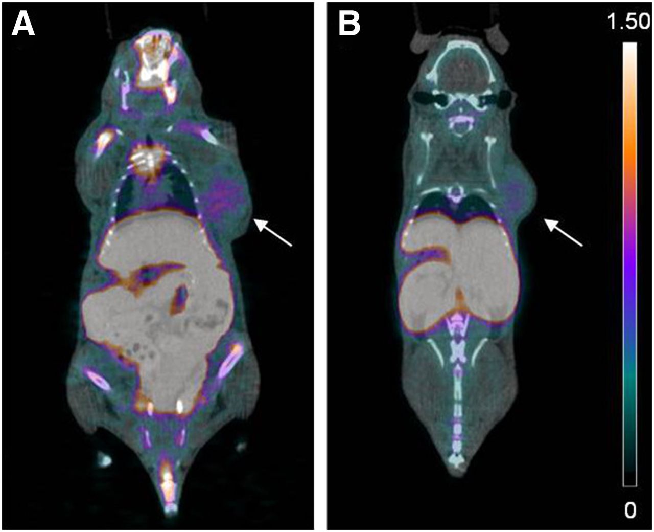
PET/CT images of monomeric 18F-sulfonamides at 1 h after injection of 18F-AmBF3-AEBS (A) and 18F-AmBF3-ABS (B). Tumors are indicated by arrows. Scale bar unit is %ID/g.

PET/CT images of trimeric 18F-sulfonamides at 1 h after injection of 18F-AmBF3-(AEBS)3 (A), 18F-AmBF3-(ABS)3 (B), and 18F-AmBF3-(ABS)3 (C) preblocking with acetazolamide. Tumors are indicated by arrows. Scale bar unit is %ID/g.
DISCUSSION
The highly conserved catalytic domain of CA isoforms represents a major challenge for the design of CA-IX–selective imaging agents (6). Off-target binding to intracellular CAs can significantly reduce tumor binding and image contrast (25). Strategies to confer CA-IX selectivity for small-molecule inhibitors have focused on limiting transport across the plasma membrane. These include incorporating bulky entities such as fluorescein isothiocyanate, albumin binders, sugar derivatives, or charged species to CA-targeting pharmacophores (26–30). On the basis of these findings, we initiated the development of multivalent PET tracers for imaging CA-IX expression in tumors.
Previously, our group reported the synthesis of a dual-mode imaging agent by coupling rhodamine and cycloRGD (arginine-glycine-aspartic acid) to an AmBF3-conjugated alkyne 4 amenable to 18F labeling (31). Using the modular adaptability of this synthon, we coupled 3 AEBS/ABS moieties via copper-catalyzed azide alkyne cycloaddition to target CA-IX expression. To the best of our knowledge, there are few examples of such multimeric inhibitors that have been labeled for imaging, and none has been labeled with 18F-fluoride nor used to visualize tumor-associated CA-IX activity. For comparison and as controls, we coupled AEBS/ABS to AmBF3-conjugated alkyne 3 to synthesize monovalent tracers to determine whether trimerization is essential to selectively target CA-IX in vivo.
Whereas more specific inhibitors can be designed to enhance selectivity to CA-IX, we sought to test the use of a trivalent scaffold with simple and rather nonspecific inhibitors to demonstrate that in vivo selectivity can be generated simply by altering cell permeability of the tracer to reduce uptake in the blood. AEBS and ABS are known to inhibit CA-IX; however, they by themselves lack isoform selectivity. According to the literature, the Ki values of AEBS and ABS for CA-IX are 33 and 294 nM, respectively (32–34). For reasons unknown, incorporation of AEBS or ABS into AmBF3-conjugated radiosynthons resulted in consistent or significantly improved binding affinities for CA-IX. AmBF3-AEBS and AmBF3-ABS exhibited binding affinities of 8.0 and 6.6 nM, whereas AmBF3-(AEBS)3 and AmBF3-(ABS)3 had binding affinities of 35.7 and 8.5 nM. Despite retaining good binding affinity to CA-I and -II, the in vivo binding of bulky 18F-AmBF3-(AEBS)3 and 18F-AmBF3-(ABS)3 to these 2 intracellular off-target CA isoforms is highly unlikely. Trimerization of AEBS and ABS to form AmBF3-(AEBS)3 and AmBF3-(ABS)3, respectively, affords cell impermeability due to the high molecular weight (>1 kDa). On the other hand, the high binding affinity of AmBF3-(AEBS)3 and AmBF3-(ABS)3 to CA-XII is advantageous as CA-XII is also found upregulated in hypoxic tumors (35).
Recently, we reported a facile 18F-19F isotopic exchange reaction on AmBF3-bioconjugates for the preparation of PET tracers (5). Features of this radiolabeling strategy include 1-step synthesis without azeotropic drying, good radiochemical yields, high purity, specific activity and in vitro/vivo stability, and relative ease of purification without high-performance liquid chromatography (5). This approach for 18F labeling has been successfully applied to RGD and somatostatin analogs for cancer imaging (31,36). The AmBF3-conjugated CA-IX–targeting tracers reported here were both obtained in good radiochemical yields (16.3%–36.8%) and specific activity (40–207 GBq/μmol) within 30 min of synthesis time. These radiosynthesis data (radiochemical yield and specific activity) are comparable with those previously reported for other AmBF3-conjugates (31,36).
For in vivo evaluations, imaging and biodistribution studies were conducted with immunodeficient mice inoculated with HT-29 human colorectal cancer cells. HT-29 cells express high levels of CA-IX under hypoxic conditions (37). Biodistribution (Table 3) and PET/CT images (Figs. 4 and 5) showed that tracers cleared rapidly through the hepatobiliary and renal pathways. At 1 h after injection, 18F-AmBF3-AEBS and 18F-AmBF3-ABS had higher tumor accumulation (0.56 ± 0.11 and 0.64 ± 0.08 %ID/g) than their trimeric counterparts (0.30 ± 0.10 and 0.33 ± 0.07 %ID/g); however, tumor-to-background ratios were substantially lower. Most importantly, tumor-to-blood ratios were 1.01 ± 0.25 and 1.24 ± 0.12 for 18F-AmBF3-AEBS and 18F-AmBF3-ABS, respectively, suggesting that both tracers may have bound intracellular CAs in blood. Although monomers may be more pertinent for traversing through aberrant tumor vasculature and binding to CA-IX, they appear to be unable to differentiate between CA isoforms. In regards to the trimers, 18F-AmBF3-(ABS)3 demonstrated superior tumor-to-muscle (9.55 ± 2.96 vs. 4.94 ± 2.76) and tumor-to-blood (3.93 ± 1.26 vs. 2.88 ± 1.81) ratios. The difference may be explained by tracer lipophilicity. With 3 additional ethylene moieties, 18F-AmBF3-(AEBS)3 proved more lipophilic and had a higher LogD7.4 value than 18F-AmBF3-(ABS)3. With the exception of the testes, stomach, kidney, and bone, uptake in nontarget tissues was higher for 18F-AmBF3-(AEBS)3. On the basis of this observation, it appears that image contrast may be improved by selecting more hydrophilic CA-IX–targeting pharmacophores (38). Because 18F-AmBF3-(ABS)3 yielded the most promising results of the evaluated tracers, additional biodistribution studies were performed at 2 h after injection to determine whether tumor uptake or contrast would improve over time. Although tumor uptake for 18F-AmBF3-(ABS)3 decreased slightly to 0.24 ± 0.05 %ID/g by 2 h after injection, tumors were readily visualized in PET images (data not shown).
Of the small-molecule inhibitors that have been radiolabeled for CA-IX imaging, only a subset has been evaluated in vivo (Table 1). Akurathi et al. reported biodistribution data for 99mTc-labeled AEBS derivatives (compounds B–F, Table 1) in HT-29 tumor xenografts (18,19). Tracers showed limited retention in tumors (≤0.5 %ID/g at 0.5–4 h after injection) with low contrast (≤1.0 tumor-to-blood ratio). Our research group evaluated 18F-labeled U-104 (compound H) (21), FEC (7-(2-fluoroethoxy)coumarin, compound N) (21), and tertiary-substituted benzenesulfonamides (compound K) (20) as CA-IX imaging agents. U-104 suffered from isoform selectivity, whereas FEC and tertiary-substituted benzenesulfonamides lacked stability in vivo. 18F-VM4-037 (compound I), an ethoxzolamide derivative developed by Siemens, is the only radiolabeled small-molecule inhibitor that advanced to phase I clinical trial (17). Biodistribution and dosimetry studies in healthy volunteers showed high uptake in the liver and kidneys, with minimal clearance. The authors noted that the pharmacokinetics of 18F-VM4-037 will preclude its use for imaging lesions in these organs and that pathologic studies correlating 18F-VM4-037 uptake and intratumoral CA-IX expression were required. Interestingly, Peeters et al. recently published preclinical biodistribution/imaging data for 18F-VM4-037, with the tracer failing to detect CA-IX expression in either glioma or colorectal cancer xenograft models (39).
Given the prognostic and therapeutic value of CA-IX, the development of CA-IX imaging agents will have a significant clinical impact. Notwithstanding somewhat low tumor uptake, both 18F-AmBF3-(AEBS)3 and 18F-AmBF3-(ABS)3 enabled clear visualization of CA-IX–expressing HT-29 tumor xenografts to provide some of the highest tumor-to-blood and tumor-to-muscle ratios ever reported. Preinjection of acetazolamide significantly blocked tumor uptake to near-background levels for 18F-AmBF3-(ABS)3, demonstrating target specificity. These data represent a great advancement over previous attempts and demonstrate the effectiveness of increasing cell impermeability to achieve CA-IX selectivity. Further synthetic modifications to reduce gastrointestinal uptake and the use of more specific inhibitors to increase tumor uptake are ongoing. In the meantime, we suggest that this method has great potential to enhance in vivo imaging of extracellular enzymatic activity through the simple application of a multivalent approach that mitigates generalized uptake.
CONCLUSION
We synthesized and evaluated four 18F-labeled sulfonamides as PET imaging agents targeting CA-IX. Trimerization of sulfonamide derivatives successfully conferred CA isoform selectivity in vivo. 18F-AmBF3-(ABS)3 showed specific and selective uptake in CA-IX–expressing tumor xenografts with good contrast. The use of multivalent enzyme inhibitors represents a viable strategy to selectively image extracellular enzyme activity with PET.
DISCLOSURE
The costs of publication of this article were defrayed in part by the payment of page charges. Therefore, and solely to indicate this fact, this article is hereby marked “advertisement” in accordance with 18 USC section 1734. This work was supported by the Canadian Cancer Society, the Canadian Institutes of Health Research, and the Leading Edge Endowment Fund, including a Rix Family scholarship. No other potential conflict of interest relevant to this article was reported.
Acknowledgments
We thank Navjit Hundal-Jabal, Nadine Colpo, Milan Vuckovic, Wade English, and Julius Klug for technical assistance.
Footnotes
↵* Contributed equally to this work.
Published online Jul. 23, 2015.
- © 2015 by the Society of Nuclear Medicine and Molecular Imaging, Inc.
REFERENCES
- Received for publication January 6, 2015.
- Accepted for publication July 9, 2015.


