


Abstract
The purpose of this study was to observe the effect of fasting and feeding on 18F-FDG uptake in a mouse model of human non–small cell lung cancer. Methods: In in vivo studies, 18F-FDG small-animal PET scans were acquired in 5 mice bearing non–small cell lung cancer A549 xenografts on each flank with continuous feeding and after overnight fasting to observe the changes in intratumoral distribution of 18F-FDG and tumor 18F-FDG standardized uptake value (SUV). In ex vivo studies, intratumoral spatial 18F-FDG distribution assessed by autoradiography was compared with the tumor microenvironment (including hypoxia by pimonidazole and stroma by hematoxylin and eosin stain). Five overnight-fasted mice and 5 fed mice with A549 tumors were observed. Results: Small-animal PET scans were obtained in fed animals on day 1 and in the same animals after overnight fasting; the lapse was approximately 14 h. Blood glucose concentration after overnight fasting was not different from fed mice (P = 0.42), but body weight loss was significant after overnight fasting (P = 0.001). Intratumoral distribution of 18F-FDG was highly heterogeneous in all tumors examined, and change in spatial intratumoral distribution of 18F-FDG between 2 sets of PET images from the same mouse was remarkably different in all mice. Tumor 18F-FDG mean SUV and maximum SUV were not significantly different between fed and fasted animals (all P > 0.05, n = 10). Only tumor mean SUV weakly correlated with blood glucose concentration (R2 = 0.17, P = 0.03). In ex vivo studies, in fasted mice, there was spatial colocalization between high levels of 18F-FDG uptake and pimonidazole-binding hypoxic cancer cells; in contrast, pimonidazole-negative normoxic cancer cells and noncancerous stroma were associated with low 18F-FDG uptake. However, high 18F-FDG uptake was frequently observed in noncancerous stroma of tumors but rarely in viable cancer cells of the tumors in fed animals. Conclusion: Host dietary status may play a key role in intratumoral distribution of 18F-FDG. In the fed animals, 18F-FDG accumulated predominantly in noncancerous stroma in the tumors, that is, reverse Warburg effect. In contrast, in fasted status, 18F-FDG uptake was found in hypoxic cancer cells component (Pasteur effect). Our findings may provide a better understanding of competing cancer glucose metabolism hypotheses: the Warburg effect, reverse Warburg effect, and Pasteur effect.
The glucose analog 18F-FDG combined with PET has emerged as an important clinical tool for cancer detection, staging, and monitoring of response and is routinely used in the clinical management of several cancer types. However, the mechanisms involved in 18F-FDG uptake in cancer are not fully understood, and the current literature is mixed and contradictory.
The German chemist and physician Otto Warburg found that Ehrlich ascites cancer cells had increased glucose demand. He observed that even in the presence of ample oxygen, cancer cells prefer to metabolize glucose by aerobic glycolysis due to mitochondrial dysfunction, the so-called Warburg effect (1). Increased glucose demand has been considered as one of the fundamental features of cancer (2), and this has been exploited clinically for cancer detection by 18F-FDG PET. In operational terms, the Warburg effect increases the relative uptake of 18F-FDG in tumors said to be unrelated to the microdistribution of hypoxia. However, growing evidence indicates that 18F-FDG uptake is significantly higher in hypoxic cancer cells than normoxic cancer cells (3–11), and 18F-FDG uptake in the latter is typically low and not statistically different from stromal or necrotic regions (3). These findings raise the question of whether the Warburg effect actually applies to normoxic cancer cells (4).
We and others have recently documented that hypoxic cancer cells have significantly higher 18F-FDG uptake than aerobic ones in vivo animal studies (3–7) as well as in in vitro cell culture studies (8–11) in various cancer cell lines. Therefore, in the absence of oxygen, cancer cells seem to have increased demand for glucose or 18F-FDG uptake, and this may be more logically explained by anaerobic glycolysis or the Pasteur effect (3,4,7).
On the other hand, Lisanti et al. recently proposed the reverse Warburg effect in a human breast cancer model (12). They demonstrated that aerobic glycolysis actually takes place in tumor-associated fibroblasts and not in cancer cells (12–16), which challenges the Warburg effect. This reverse Warburg effect hypothesis has not yet been explored in any 18F-FDG study.
Blood glucose levels of tumor-bearing subjects have been considered as a key factor affecting 18F-FDG uptake in PET studies (17–21). Therefore, fasting is a routine procedure in clinical and preclinical studies. Variable effects of fasting on blood glucose levels as well as 18F-FDG uptake have been observed (17–21). The effect of changes in blood glucose levels on intratumoral spatial distribution of 18F-FDG is unknown.
In the present study, we used small-animal PET to observe 18F-FDG uptake in the same animals in both fed and fasting states, and 18F-FDG uptake was related to blood glucose levels. We also tested the effect of fasting or feeding on intratumoral spatial 18F-FDG distribution using digital autoradiography and immunohistochemical staining.
MATERIALS AND METHODS
Cancer Cell Line, Nude Mice, and Cancer Models
The human non–small cell lung cancer A549 cell line, purchased from American Type Culture Collection, was used in our experiments. A549 cells were maintained in F-12K medium (American Type Culture Collection) supplemented with 10% fetal bovine serum (Gemini), 1% glutamine, and 1% antibiotic mixture (Cellgro). Cells were grown in a humidified incubator at 37°C in an air atmosphere containing 5% carbon dioxide. Exponentially growing cells were harvested with a 0.25% (w/v) trypsin–0.53 mM ethylenediaminetetraacetic acid solution.
All experiments were performed using 6-wk-old female athymic NCr-nu/nu mice purchased from NCI-Frederick Cancer Research Institute. Nude mice were maintained and used according to institutional guidelines. The experimental protocol was approved by the Institutional Animal Care and Use Committee of University of Louisville. Animals were housed 5 per cage and kept in the institutional small-animal facility at a constant temperature and humidity. Food pellets and water were provided ad libitum.
Subcutaneous tumors were initiated by injecting 5 × 10⁶ tumor cells in 0.1 mL of phosphate-buffered saline into each flank of the mouse, and xenografts grew to approximately 1 cm in diameter 3–4 wk later.
Experimental Procedures
Mouse Blood Glucose Measurement
Blood glucose level was measured using an ACCU-CHEK nano glucose meter and ACCU-CHEK SmartView strips (Roche Diagnostics) immediately before each 18F-FDG injection; 2 glucose measurements were taken and the average value was used. Glucose strips were validated using d-glucose water solution (50 and 100 mg/dL), and 5 strips for each concentration were tested; in addition, in a pilot study, 5 measurements were performed in blood from a nude mouse (21). The readings by the glucose meter were validated to be accurate.
In Vivo Small-Animal PET Imaging
18F-FDG was purchased from the P.E.T. NET Pharmaceuticals Inc. facility at University of Louisville Hospital. All animals were imaged prone using the dedicated 3-dimensional small-animal R4 MicroPET (Concorde Microsystems) system. The R4 MicroPET scanner has a transaxial field of view of 10 cm and an axial field of view of 7.8 cm. The resulting list-mode data were sorted into 2-dimensional histograms by Fourier rebinning and the images reconstructed by an iterative reconstruction algorithm into a 128 × 128 × 63 (0.72 × 0.72 × 1.3 mm) matrix. All animals were fed on day 1, and animals were fasted overnight for the day-2 PET study. Mice were injected without anesthesia via the tail vein with 18F-FDG (9.7 ± 0.3 MBq) and allowed freedom of movement for approximately 60 min. Animals were then anesthetized by inhalation of an isoflurane (1.5%)–air mixture, and a 10-min PET scan was obtained. We included 5 mice with each flank bearing an A549 xenograft (i.e., 2 tumors per mouse) in the small-animal PET study.
PET Image Analysis
Inveon Research Workplace software (version 3.0; Siemens) was used for small-animal PET image analysis. All image sets for each animal were visually examined using a rotating (cine) 3-dimensional display. The window and level settings were adjusted for the best intratumoral distribution visibility. Three-dimensional regions of interest were manually drawn around the edge of the tumor xenograft activity by visual inspection using Inveon Research Workplace for each scan, and the mean and maximal activities were recorded from entire ROIs. The standardized uptake value (SUV) was used to represent tissue 18F-FDG uptake as follows: SUV = region-of-interest activity concentration multiplied by mouse weight divided by injected activity (21).
Ex Vivo Autoradiography and Histologic Assay
In a separate group of mice, 5 fasted mice and 5 fed mice (total 10 mice) were included. A mixture of 18F-FDG (7.4 MBq) and pimonidazole (2 mg) was injected via the tail vein 1 h before animal euthanasia (total injection volume, 0.2 mL) as described previously (3,4). Blood glucose levels were measured.
As we described previously (3,4,7,22–24), immediately after animal sacrifice, tumor tissues were removed for subsequent processing. Subcutaneous xenografts were frozen and embedded in optimal-cutting-temperature medium (Sakura Finetek). Immediately thereafter, 5 contiguous 7-μm-thick tissue sections were cut using a 3050S cryostat microtome (Leica, Inc.) and adhered to poly-l-lysine–coated glass microscope slides (Fisher Scientific, Inc.).
Autoradiograms were obtained by placing the tumor sections in a film cassette against an imaging plate as described previously (3,4,22). The same plate was used throughout the experiments; the plate was exposed for approximately 20 h and read by a Cyclone Plus imaging system (PerkinElmer, Inc.). For digital autoradiography, regions of interest (100 × 100 μm) were drawn over hypoxic cancer cell, oxic cancer cell, and stromal areas, with reference to the immunohistochemical and hematoxylin and eosin stain images obtained from the same or an adjacent section. Digital autoradiography imaging was quantified by OptiQuant software (PerkinElmer Inc.). 18F-FDG uptake was initially expressed as digital light unit per square millimeter and was subsequently converted to MBq/g based on the known section thickness and system calibration factor of 35.4 digital light units per Becquerel (3). Finally, 18F-FDG uptake in each microenvironment component was expressed as percentage injected dose per gram of tumor tissue.
To minimize issues associated with section alignment and registration, the same tumor sections used for autoradiography or contiguous adjacent sections were used for all images. Microscopic images of the distributions of pimonidazole and hematoxylin and eosin were obtained from the same or adjacent section as described after completion of autoradiography exposures as reported previously (3,4,22,24). Briefly, slides were air-dried, fixed in cold acetone (4°C) for 20 min, and incubated with SuperBlock (Pierce Biotechnology) at room temperature for 30 min. All antibodies were also applied in SuperBlock. Sections were then incubated with fluorescein isothiocyanate–conjugated antipimonidazole monoclonal antibody (Hypoxyprobe Inc.), diluted 1:25, for 1 h at room temperature.
Statistical Analysis
Statistical significance was examined by paired t test and 2-tailed Student t test when appropriate. The paired t test was used when comparing pair data from the same animal. Correlation was analyzed by Pearson correlation using GraphPad Prism (GraphPad Software Inc.). A P value of less than 0.05 was considered statistically significant.
RESULTS
Blood glucose level was measured in 5 mice enrolled in the 2-d PET studies. Changes in blood glucose after overnight fasting were not significant (P = 0.42): 93 ± 23 for feeding and 81 ± 35 mg/dL for fasting (Fig. 1A). Body weight was significantly decreased after overnight fasting; animals weighed 23 ± 2 g in the fed status and weight decreased to 20 ± 1 g after overnight fasting (P = 0.001) (Fig. 1B). 18F-FDG uptake was expressed as SUV.
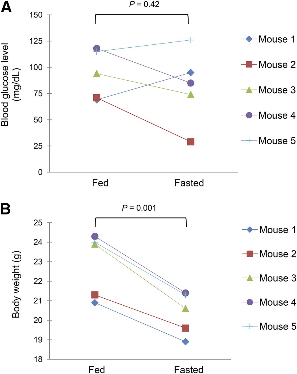
Comparison of blood glucose levels (A) and body weights (B) in fed and overnight-fasted mice bearing A549 subcutaneous xenografts (n = 5 in both cases).
PET scans were obtained in 5 mice with an A549 xenograft on each flank. On day 1, a small-animal PET scan was obtained for fed animals. On day 2, a small-animal PET scan was acquired for overnight-fasted animals. The lapse between the 2 sets of PET scans was approximately 14 h. Paired PET images are presented in Figure 2, Figure 3, and Supplemental Figure 1 (supplemental materials are available at http://jnm.snmjournals.org). The intratumoral distribution of 18F-FDG was highly heterogeneous in all tumors examined, and spatial intratumoral distribution of 18F-FDG on day-1 and day-2 PET images from the same mouse was remarkably different in all mice (Figs. 2 and 3; Supplemental Fig. 1). However, overall tumor 18F-FDG mean SUV (SUVmean) was not significantly different between fed (0.45 ± 0.13) and fasted status (0.49 ± 0.20) (P = 0.50, n = 10) (Fig. 4A), and there was no significant difference in maximum SUV (SUVmax) between fed (0.94 ± 0.37) and fasted status (1.02 ± 0.33) (P = 0.52) (Fig. 4B).

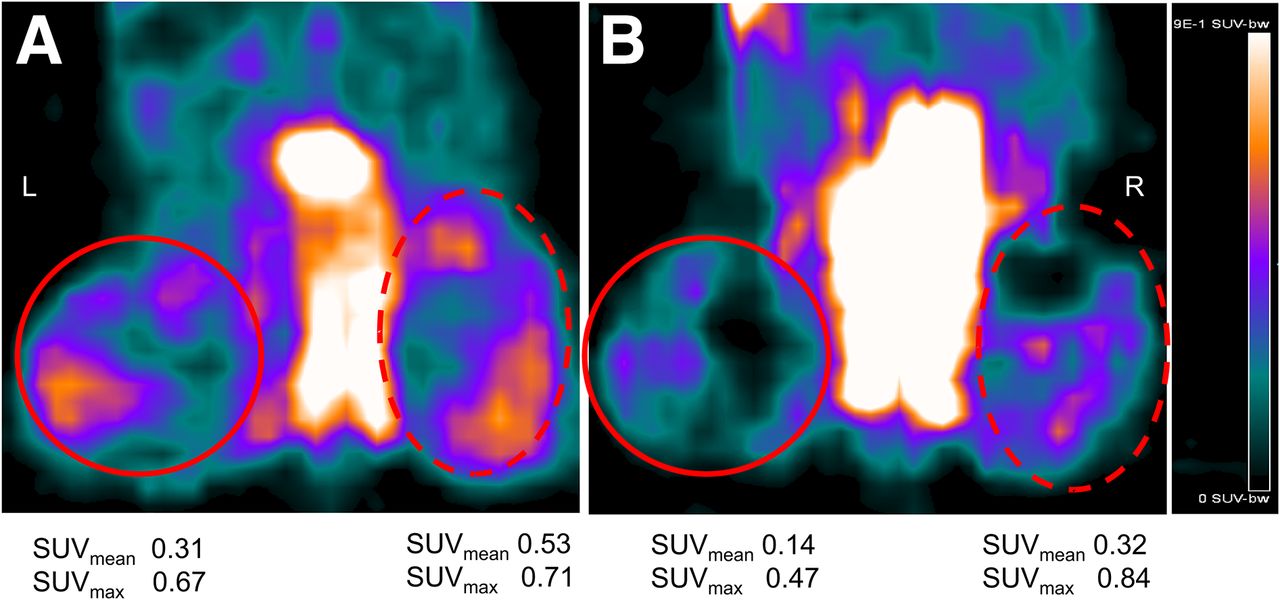
Coronal 18F-FDG PET sections of A549 xenograft–bearing mouse-1 in fed (A) and fasted (B) status. Tumors on both flanks are circled. Apparent differences in intratumoral spatial distribution of 18F-FDG was visualized. There is increase in blood glucose level after fasting from 69 mg/dL (fed) to 95 mg/dL.

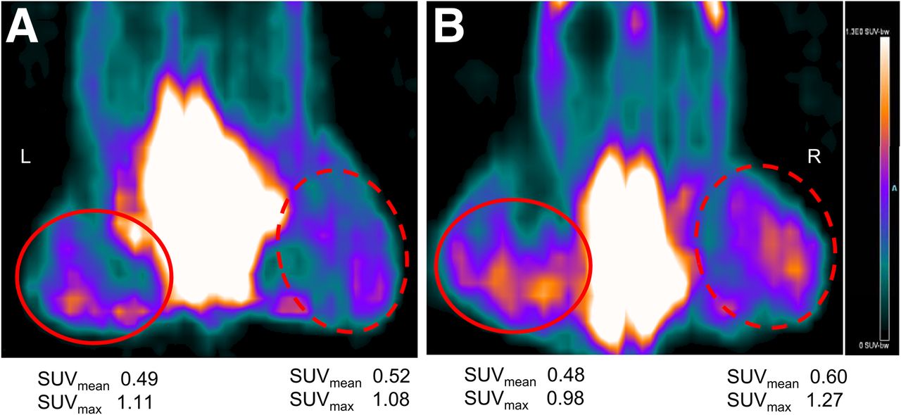
Coronal 18F-FDG PET sections of A549 xenograft–bearing mouse-3 in fed (A) and fasted (B) status. Tumors on both flanks are circled. Apparent differences in intratumoral spatial distribution of 18F-FDG were visualized. There is decrease in blood glucose level after fasting from 94 mg/dL (fed) to 74 mg/dL.

(A) Comparison of tumor SUVmean between fed and fasting status in 5 mice bearing A549 tumors. (B) Comparison of tumor SUVmax between fed and fasted status. There was no significant difference in SUVmax between fed (0.94 ± 0.37) and fasted status (1.02 ± 0.33) (P = 0.52). (C) Correlation between tumor SUVmean and blood glucose levels. (D) Correlation between tumor SUVmax and blood glucose levels.
Although there was weak correlation between tumor SUVmean and blood glucose level (R2 = 0.17, P = 0.03) (Fig. 4C), the correlation between tumor SUVmax and blood glucose level was not significant (R2 = 0.14, P = 0.052) (Fig. 4D). No significant correlation was found between changes in blood glucose concentrations and changes in SUVmax or changes in SUVmean (data not shown).
In a separate experiment, 18F-FDG autoradiography was compared with immunohistochemical/histologic staining visualization of the tumor microenvironment to observe the effect of fasted (in 5 mice) and fed (in 5 mice) status on intratumoral spatial distribution of 18F-FDG in A549 tumors. In A549 xenograft tumors after the host animal was fasted overnight, there was spatial colocalization between high levels of 18F-FDG uptake and pimonidazole binding to hypoxic cancer cells; in contrast, pimonidazole-negative aerobic cancer cells and noncancerous stromal cells showed low 18F-FDG uptake (Fig. 5A). Similar findings have been reported elsewhere (3,4,7,22). However, in the fed animals 18F-FDG accumulated predominantly in noncancerous stroma in the tumors, and there was little 18F-FDG uptake in viable cancer cells, regardless of tumor oxygenation status (Fig. 5B; Supplemental Fig. 2). 18F-FDG uptake in stroma significantly increased in fed status, but 18F-FDG accumulation in hypoxic cancer cells significantly decreased, and there was no change in 18F-FDG uptake in oxic cancer cells regardless of feeding or fasting (Fig. 5C). The blood glucose level was 95 ± 34 mg/dL (n = 5) for fed mice and 84 ± 31 mg/dL (n = 5) for fasted mice (P > 0.05). These levels were not statistically different from mice-enrolled PET studies in either fed or fasted status.

Comparison of intratumoral distribution of 18F-FDG by autoradiography with immunohistochemical–histologic staining visualization of microenvironment components in A549 tumors. (A) Fasted status. There was spatial colocalization between high levels of 18F-FDG uptake and pimonidazole binding (green) in hypoxic cancer cells (arrowhead). Noncancerous stroma was associated with low 18F-FDG uptake (arrow); blood glucose level of mouse was 79 mg/dL. (B) In fed animal with blood glucose level of 114 mg/dL, 18F-FDG accumulated predominantly in noncancerous stroma in tumor (arrow), and there was little 18F-FDG uptake in viable cancer cells whether stained positively or negatively for pimonidazole (arrowhead). (C) 18F-FDG uptake (percentage injected dose per gram [%ID/g]) in each microenvironment component. *P < 0.001, n = 10–20 measurements from 5 tumors. All scale bars = 2 mm. NS = not significant.
DISCUSSION
18F-FDG PET is routinely used in the clinical management of cancers, and its utility is usually thought to be due to enhanced aerobic glycolysis in tumors—that is, the Warburg effect. Increased glucose metabolism has been considered a fundamental feature of cancer. However, the Warburg effect hypothesis has been challenged in several ways.
First, we have documented that the Ehrlich ascites cells, which Warburg used, were severely hypoxic, and glucose demand measured by 18F-FDG uptake was high (4,7). Ascites cancer cells may use anaerobic glycolysis (Pasteur effect) to generate adenosine triphosphate. The higher uptake of 18F-FDG in hypoxic ascites cancer cells may be explained by the Pasteur effect (low oxygen tension and high glucose utilization) not aerobic glycolysis (high oxygen and high glucose utilization; the Warburg effect) (4).
Second, multiple studies have recently documented that hypoxic cancer cells have significantly higher 18F-FDG uptake than aerobic ones (Fig. 4A) (3–11). Therefore, high glucose demand may be a general feature of hypoxic cancer cells not normoxic ones.
Third, Lisanti et al. have recently proposed the reverse Warburg effect. On the basis of their observations in human breast cancers, they concluded that so-called aerobic glycolysis actually takes place in tumor-associated fibroblasts not in cancer cells (12–16). Here, we found that in fed mice, intratumoral high 18F-FDG activity was predominantly associated with noncancerous stroma and neither hypoxic nor aerobic cancer cells showed higher 18F-FDG uptake (Figs. 5B and 5C; Supplemental Fig. 2). Accordingly, we hypothesized that, under fed status, intratumoral stroma adjacent to functional blood vessels use most glucose supplied by the circulation, whereas viable cancer cells do not, which may be explained by the reverse Warburg effect. In contrast, 18F-FDG uptake was predominantly found in hypoxic cancer cells in tumor-bearing animals in the fasted condition (Figs. 5A and 5C). Similar results have been observed by us and others (3–7). Therefore, host dietary status may play a key role in intratumoral distribution of 18F-FDG.
In a group of 5 mice, we found blood glucose levels did not significantly change after overnight fasting; however, animal body weight was significantly decreased by an average of 3 g after 14-h fasting (Fig. 1). This decrease in body weight can be explained by the fact that animals self-regulate to keep glucose levels constant using stored energy sources or gluconeogenesis. It is not surprising that 18F-FDG uptake measured by small-animal PET was not changed in the tumors (Fig. 4).
In general, blood glucose level has been considered a key factor affecting 18F-FDG PET imaging. Our PET findings indicated that tumor 18F-FDG SUVmean had weak correlation with blood glucose level (R2 = 0.17, P = 0.03), but 18F-FDG SUV max did not correlate with blood glucose level (Figs. 4C and 4D). In addition, changes in tumor 18F-FDG uptake did not correlate with changes in glucose levels. Our data indicate that blood glucose level may not be the major factor affecting 18F-FDG SUV in cancer-growing mice without documented glucose metabolism abnormalities.
There is apparent discrepancy in tumor 18F-FDG uptake between patients and mice. Tumor 18F-FDG uptake in nonfasted patients generally is much reduced, compared with the fasted case; however, there were no significant changes in tumor uptake between fasted and nonfasted states in mice. The mechanism inducing the apparent discrepancy is unclear, but difference in stroma between mice and humans may play a role.
In ex vivo studies using autoradiography–immunohistochemical assays, we observed 18F-FDG uptake in each component of the tumor. Apparently higher 18F-FDG accumulation may have shifted from hypoxic cancer cells to stroma components in the fed state (Fig. 5; Supplemental Fig. 2). Therefore, the PET findings of change in spatial intratumoral distribution of 18F-FDG between fed and fasted status can be explained. SUVs reflect the entire tumor level, and spatial changes in 18F-FDG distribution among tumor microenvriomental components may not necessarily alter overall 18F-FDG uptake. Our findings presented in Figure 4 are not contradicted by autoradiography–immunohistochemical findings (Fig. 5; Supplemental Fig. 2) and PET images (Figs. 2 and 3). In clinical settings, it is important to observe intratumoral distribution of 18F-FDG in addition to SUV in serial PET/CT studies to manage cancer therapy in oncology patients.
In this study, we used SUV instead of percentage injected dose per gram to present 18F-FDG uptake. We chose the SUV parameter because animal body weight significantly changed, and the body weight factor has to be considered for accurate data presentation.
Our study has the following limitations: the lapse between the 2 sets of PET scans was approximately 14 h, slightly over 7 half-lives decay for 18F. This time was chosen to minimize the residual radioactivity effect remaining from the first injection into the second imaging. Residual 18F-FDG from the first PET study was considered completely decayed. And the overnight fasting frequently mimics patients' situations in clinical PET studies wherein they are required to fast for at least 6 h. Published data have documented that the tumor microenvironment may change over short periods of time in some cancer types (25,26). Our published results confirmed that an A549 xenograft tumor microenvironment is mostly stable during a 10- to 14-h time interval (22). Paired PET studies were also conducted in 2 fasted mice with A549 tumors within 14 h, and intratumoral distribution of 18F-FDG was highly reproducible (data not shown). Thus, demonstrated mismatch patterns of 18F-FDG PET images between fasted and fed status were valid and not the consequence of significant spatial change in the intratumoral microenvironment.
Animal body weight significantly decreased after overnight fasting, but glucose levels were not different from fed status. Apparently the prolonged fasting puts animals into a condition of metabolic stress. A drop in blood glucose leave was expected but not found; stress-triggered homeostatic responses generate more new glucose to keep blood glucose more or less constant. A short period of fasting should be observed in a future study.
Host dietary status may play a key role in intratumoral distribution of 18F-FDG. However, insulin and other hormone levels and their effects on 18F-FDG uptake were not included in the present study, and future investigations may need to explore these important issues in other cancer cell lines.
CONCLUSION
In this study, host dietary status may play a key role in intratumoral distribution of 18F-FDG. In the fed animals, 18F-FDG accumulated predominantly in noncancerous stroma in the tumors—that is, reverse Warburg effect. The effect of blood glucose level on 18F-FDG uptake in A549 tumors was minimal. Our findings may provide a better understanding of 3 competing intratumoral glucose metabolism hypotheses: the Warburg effect, reverse Warburg effect, and Pasteur effect.
DISCLOSURE
The costs of publication of this article were defrayed in part by the payment of page charges. Therefore, and solely to indicate this fact, this article is hereby marked “advertisement” in accordance with 18 USC section 1734. This study was supported by the Kentucky Lung Cancer Research program award (cycle 9), Inner Mongolia Natural Science Foundation awards (2012MS1153, 2014MS0835, and 2014MS0859), and National Natural Science Foundation of China award (81360227). No other potential conflict of interest relevant to this article was reported.
Acknowledgments
We thank Dr. John Eaton from University of Louisville for critically reading the manuscript and Dr. Haixun Guo for technical supports of PET studies.
Footnotes
↵* Contributed equally to this work.
Published online Feb. 26, 2015.
- © 2015 by the Society of Nuclear Medicine and Molecular Imaging, Inc.
REFERENCES
- Received for publication September 8, 2014.
- Accepted for publication January 26, 2015.


Jump to section
Related Articles
Cited By...
- No citing articles found.