


Abstract
P2045 is a peptide analog of somatostatin with picomolar affinity for the somatostatin receptor subtype 2 (SSTR2) upregulated in some pancreatic tumors. Studies were conducted in rat AR42J pancreatic tumor xenograft mice to determine whether 188Re-P2045 could inhibit the growth of pancreatic cancer in an animal model. Methods: 188Re-P2045 was intravenously administered every 3 d for 16 d to nude mice with AR42J tumor xenografts that were approximately 20 mm3 at study initiation. Tumor volumes were recorded throughout the dosing period. At necropsy, all tissues were assessed for levels of radioactivity and evaluated for histologic abnormalities. Clinical chemistry and hematology parameters were determined from terminal blood samples. The affinity of nonradioactive 185/187Re-P2045 for somatostatin receptors was compared in human NCI-H69 and rat AR42J tumor cell membranes expressing predominantly SSTR2. Results: In the 1.85- and 5.55-MBq groups, tumor growth was inhibited in a dose-dependent fashion. In the 11.1-MBq group, tumor growth was completely inhibited throughout the dosing period and for 12 d after the last administered dose. The radioactivity level in tumors 4 h after injection was 10 percentage injected dose per gram, which was 2-fold higher than in the kidneys. 188Re-P2045 was well tolerated in all dose groups, with no adverse clinical, histologic, or hematologic findings. The nonradioactive 185/187Re-P2045 bound more avidly (0.2 nM) to SSTR2 in human than rat tumor membranes, suggesting that these studies are relevant to human studies. Conclusion: 188Re-P2045 is a promising therapeutic candidate for patients with somatostatin receptor–positive cancer.
Somatostatin (SST) is a neuropeptide and endocrine cell product that negatively regulates the hormonal secretions including somatotropin, glucagon, insulin, thyrotropin, and gastrin (1). Secretion of these hormones from neuroendocrine tumors can lead to a life-threatening hormonal imbalance that can be inhibited by SST. However, SST is rapidly inactivated by peptidases in vivo, with a circulating half-life of less than 2 min that precludes its development as a therapeutic agent. Thus, SST analogs (SSTAs) with enhanced biologic stability have been synthesized and approved for the neuroendocrine tumor therapy (2).
The potent inhibitory actions of SSTA are initiated by their high-affinity binding to the G-protein–coupled SST receptor (SSTR). SSTR subtype 2 (SSTR2), 1 of 5 cloned receptor subtypes, is the most commonly overexpressed subtype in pancreatic tumors (2,3). The expression of SSTR2 has also been reported in tumor neovasculature but not in normal vasculature (4). Tumor expression of SSTR provides the basis for detection and staging of disease by the administration of SSTA coupled to γ-emitting radioisotopes followed by scintigraphy (5). For example, pentreotide ([diethylenetriamine pentaacetic acid] octreotide) labeled with 111In is approved for the detection of neuroendocrine tumors (6), and depreotide labeled with 99mTc was previously approved for the detection of lung cancer (7).
Cytotoxic moieties have been coupled to peptides and antibodies and thereby targeted to upregulated receptors on tumors (1,8). The peptide approach has some advantages. Peptides are smaller and can diffuse more readily into tumor tissue. Furthermore, peptides are less likely to produce an immune response and can be transported into tumor cells by receptor-mediated endocytosis. They are also cleared rapidly from the circulation, resulting in decreased whole-body exposure. As such, SSTA labeled with cytotoxic isotopes have been used to deliver radiotherapeutics to tumors overexpressing SSTR (2,9). For example, clinical trials have been conducted with the SSTA 90Y-DOTATOC and 177Lu-DOTATATE for the treatment of neuroendocrine tumors (10). Another SSTA, P2045 labeled with 188Re, was evaluated in a clinical trial for lung cancer (9) in which there was a good correlation between the visualization of lung tumors with 99mTc-P2045 and 188Re-P2045 uptake (11).
Neuroendocrine tumors of the pancreas, gut, and lung overexpress SSTR (12,13), and for this reason 188Re-P2045 may be an effective therapy for this class of tumors. The studies herein describe proof of concept for 188Re-P2045 therapy in the AR42J rat pancreatic tumor xenograft nude mouse.
MATERIALS AND METHODS
188Re-P2045 and Control Preparation
A lyophilized mix of P2045 peptide trifluoroacetate (53 μg of free peptide), sodium α-d-glucoheptonate dihydrate (25 mg), edetate disodium hydrate (100 μg), and SnCl2 (850 μg) was reconstituted with 1.1 GBq of Sodium Perrhenate 188Re from a generator (Oak Ridge National Laboratories) and boiled for 15 min. Lyophilized gentisic acid (20 mg) and ascorbic acid (10 mg) were reconstituted with saline and added to the cooled 188Re-P2045 solution. Peptide control saline was added in place of 188Re. Vehicle was decayed generator eluate.
Quality Analysis of 188Re-P2045
188Re-P2045 was determined using instant thin-layer chromatography silica gel (Gelman) developed in saturated saline, 1:1 methanol:1.0 M ammonium acetate, and methyl ethyl ketone.
For high-performance liquid chromatography (HPLC), 188Re-P2045 was eluted on a linear gradient 50%–54% A to B over 32 min on a Zorbax 300SB reversed-phase column (880995-902; Rockland Technologies). Mobile phase A was 0.1% trifluoroacetic acid in water. Mobile phase B was 0.1% trifluoroacetic acid in 90/10 methanol/water.
88Y-DOTATOC Preparation
88YCl3 (555 MBq; NEN) in 12 μL of 0.05 M HCl was added to 10 μg of DOTATOC (Diatide) in 1.0 mL of saline with 0.5 μL of 1 mM YCl3. The vial was boiled for 25 min, cooled to room temperature, and passed through a QMA Light Sep-Pak column (Waters). The column was rinsed with 1.0 mL of ammonium acetate and 2.0 mL of phosphate-buffered saline and eluted with a solution of 50% ethanol. Solvent was removed by vacuum, and 88Y-DOTATOC was reconstituted with saline.
185/187Re-P2045 Preparation
Nonradioactive 185/187Re-P2045 was prepared by reacting peptide with a 1.5-fold molar excess of Bu4NReOCl4 in anhydrous dimethylformamide at room temperature (13). Products were isolated by reversed-phase HPLC.
Binding Studies
NCI-H69 or AR42J cell membranes were incubated with 125I-(Tyr11)-SST14 (NEN) at 37°C (pH 7.4) in the presence of test peptides as described previously (14). Membrane-bound 125I-SST was isolated by filtration. Nonspecific binding was determined in the presence of excess SST14 (10 μM; Sigma). The inhibition of specific binding was calculated at 6 concentrations of test peptide (0.01–100 nM) and plotted using a Stineman function (Kaleidagraph 4.5; Synergy Software) from which the IC50 values were calculated. (IC50 value is defined as the concentration of agent required to inhibit the specific binding of 125I-SST14 by 50%.)
Animal Model
University of Alabama at Birmingham’s institutional animal care and use committee approved all animal studies. Washed cells were mixed with Matrigel (Collaborative Biochemical Products) and injected subcutaneously into outbred athymic nude mice Crl:NU/NU-nuBR (Charles River). Resulting tumor xenografts were minced, combined with a 50:50 mix of Matrigel and medium, and injected into the right flank of study mice. After 3 wk, mice with uniform tumor volumes (≈20 mm3) were selected for study.
Study Design
The study design is outlined in Table 1. Tumor-bearing mice were divided into 10 groups. There were 6 multiple-dose therapeutic study groups (1–6) and 4 single-dose biodistribution study groups (7–10). Groups 1–5 (n = 10) received an intravenous injection (100 μL) on study days 0, 3, 6, 9, 12, and 16: decayed 188W/188Re generator eluate (group 1), 530 ng of nonradiolabeled P2045 (group 2), 1.85 MBq of 188Re-P2045 containing 88 ng of peptide (group 3), 5.5 MBq of 188Re-P2045 containing 265 ng of peptide (group 4), or 11.1 MBq of 188Re-P2045 containing 530 ng of P2045 peptide (group 5). A single dose of 88Y-DOTATOC was administered to groups 6 (22.2 MBq, intraperitoneally), 9 (11.1 MBq, intravenously), and 10 (11.1 MBq, intravenously). Groups 7 and 8 received a single dose of 188Re-P2045 (9.25 MBq). The dose administered to each animal was quantified in syringes before and after injection using a dose calibrator (Atomlab 100; Biodex Medical Systems). Therapeutic treatment groups 1–4 were sacrificed on study day 16. Group 5 (11.1 MBq of 188Re-P2045) was observed for an additional 12 d without dosing and sacrificed on study day 28. A single technician measured tumor length, width, and height using Veirner calipers (Fisher Scientific) in a masked fashion.
Design of Repeated-Dose Study in AR42J Tumor Mice
Biodistribution
The biodistribution of radioactivity was determined in groups 3–5 and 7–10. On the last study day, mice in groups 3, 4, and 5 received an intravenous dose of 7.4 MBq of 188Re-P2045 and were sacrificed 4 h after injection. Mice in groups 7 and 8 received an intravenous dose of 9.25 MBq of 188Re-P2045 and were sacrificed 4 and 24 h after injection. Mice in groups 9 and 10 received a dose of 7.4 MBq of 88Y-DOTATOC and were sacrificed 4 and 24 h after injection. Sacrifice was by cardiac puncture exsanguination under isoflurane anesthesia. Tissues were fixed in 10% formalin and counted for radioactivity (Minaxiγ Auto-Gamma 5000 series γ counter; Packard). All counts per minute data were corrected for radioactive decay using calibrated standards.
Imaging Studies
Three mice from group 8 were imaged with an Anger Sigma 410 Radioisotope Gamma Camera (Ohio Nuclear) equipped with a pinhole collimator (model PHW) at 4 and 24 h after injection.
Clinical Observations, Necropsy, Hematology, and Histopathology
Blood was collected in ethylenediaminetetraacetic acid. Serum was collected without anticoagulant. All tissues were examined during necropsy for gross abnormalities. Tissues were fixed in 10% formalin, embedded in paraffin, sectioned at 5 μm, and stained with hematoxylin and eosin. A single tumor section from each animal was used to determine the area of necrotic tissue with Image-Pro Plus software (version 3.0) with a Hitachi single-chip camera mounted on an Olympus AH-1 microscope. Calibration was performed using neutral-buffered saline–traceable stage micrometers. A board-certified pathologist graded all tissue samples in a masked fashion.
Statistical Analysis
Tumor volumes were compared using ANOVA, followed by Dunett test to compare each treatment group with the vehicle control group (Graphpad Instat, version 3; GraphPad Software).
RESULTS
Radiochemical Purity of 188Re-P2045
The radiochemical purity of the 188Re-P2045 determined from instant thin-layer chromatography and HPLC was greater than 89% for all 188Re-P2045 solutions used in the study (Supplemental Table 1; supplemental materials are available at http://jnm.snmjournals.org).
Purity of 185/187Re-P2045
The purity of 185/187Re-P2045 was greater than 95% by HPLC.
Binding Studies
P2045 and its nonradioactive oxo-rhenium complex, 185/187Re-P2045, bound with picomolar affinity to SSTR in the rat AR42J and human NCI-H69 tumor cell membranes (Table 2). The IC50 in AR42J and NCI-H69 membranes was 0.43 and 0.2 nM, respectively. The IC50 of the nonradioactive rhenium complex was 0.50 nM in AR42J and 0.23 nM in NCI-H69 cell membranes. The standard errors were less than 40% of the mean for all IC50 values. The IC50 values for P2045 peptide and 185/187Re-P2045 are similar to that of native SST (0.2 nM) in NCI-H69 human small cell lung cancer membranes.
IC50 Values for Inhibition of 125I-SST–Specific Binding to AR42J Rat Pancreatic and NCI-H69 Human Lung Cancer Cell Membranes
Inhibition of Tumor Growth
The mean tumor volumes are presented in Supplemental Table 2 and Figure 1. The tumor growth in the P2045 peptide control mice was not significantly different from the decayed eluate controls. However, there was a dose-related delay in tumor growth in the 188Re-P2045–treated mice relative to the generator eluate controls. Statistically significant differences in tumor volumes were observed 6 d after the first injection when the tumors in the high-dose 188Re-P2045 group were smaller than those in the decayed eluate control group (P < 0.05). Nine days after the first injection, all 188Re-P2045 treatment groups showed a significant decrease in tumor growth (P < 0.05, 0.05, and 0.01 in the low-, medium-, and high-dose groups, respectively). Thirteen days after the first injection, the tumor volumes in the low-, medium-, and high-dose groups were 91, 44, and 13 mm3 (P < 0.01), respectively, whereas the tumor volume in the eluate control group was 318 mm3. In the high-dose group, there was slight regression in mean tumor volume observed over the dosing period, although this was not statistically significant. Tumor growth in the 88Y-DOTATOC–treated mice was also inhibited.
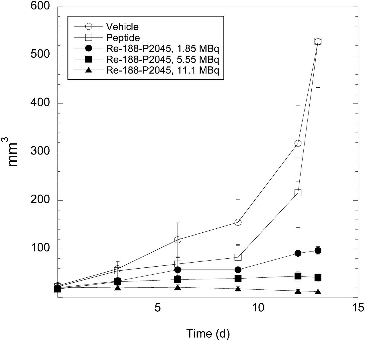
AR42J tumor volume (mm3) with 188Re-P2045, compared with P2045 (peptide) and vehicle (mock eluate) control treatment.
All mice were sacrificed on study day 16 or earlier except for the high-dose 188Re group. The mice in this group were followed for an additional 12 d with no dosing to determine the duration of the therapeutic effect. The mean tumor volume did not increase significantly until 12 d after the last dose (study day 28).
Biodistribution
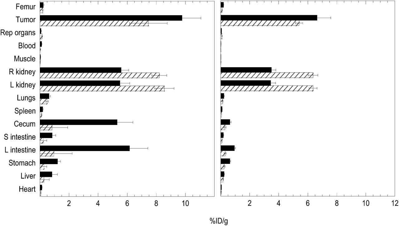
The biodistribution of 188Re-P2045 and 88Y-DOTATOC is shown in Figure 2. At 4 h after injection, the levels of 188Re-P2045 in the tumor were 9.8 %ID/g. At 24 h, the tumor retained roughly two thirds of the radioactivity (6.6 %ID/g) observed at 4 h. In contrast, 188Re-P2045 cleared rapidly from the blood such that the tumor-to-blood ratios increased from 83 to 289 at 4 and 24 h, respectively (Supplemental Table 3). The highest radioactivity in the 188Re-P2045 groups was observed in the tumor. There was roughly twice as much radioactivity in the tumors as in the kidneys at 4 h after injection. In contrast, the level of 88Y-DOTATOC in the kidneys was higher than the level in the tumors.

Biodistribution of 188Re-P2045 and 88Y-DOTATOC in AR42J tumor xenograft mice. Mice were injected with 9.25 MBq of 188Re-P2045 (black bars) or 22.2 MBq of 88Y-DOTATOC intraperitoneally (striped bars). Mice were sacrificed at 4 (left) and 24 (right) hours after injection, and tissues were collected and counted for radioactivity. Data are mean ± SEM. L = large; S = small.
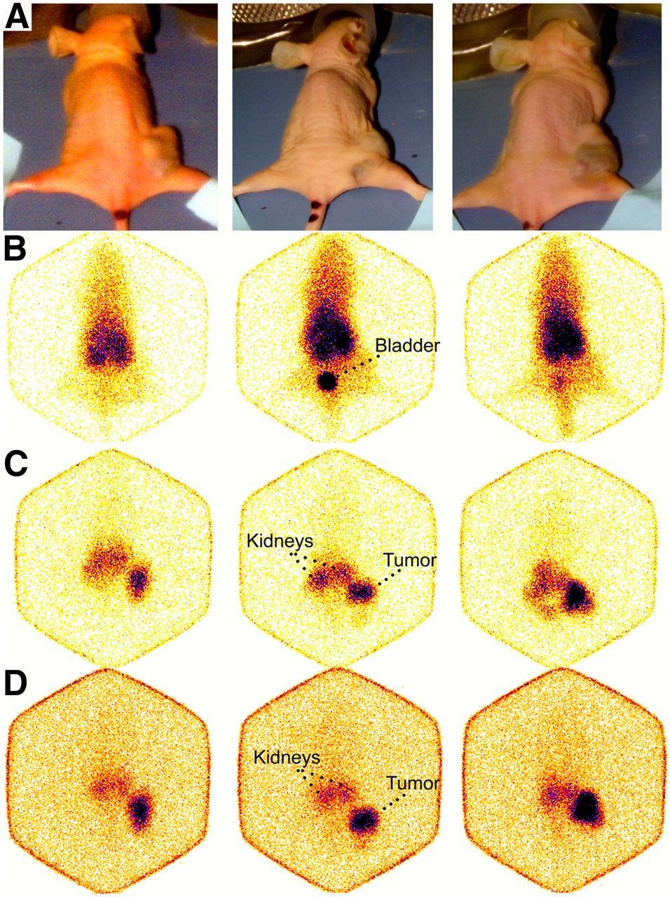
Scintigraphy of 3 representative AR42J mice after an intravenous injection of 188Re-P2045 (9.25 MBq of 188Re-P2045) is shown in Figure 3. Three minutes after injection, the bladder was visible along with the core blood volume. Eight hours after injection, the tumor and kidneys were visible and by 24 h after injection the radioactivity was predominantly located in the tumor.

Scintigraphy of AR42J tumor xenograft mice injected with 9.25 MBq of 188Re-P2045. Three individual mice bearing AR42J tumor xenografts on their right flank (A) and corresponding scintigraphic images at 3 min (B), 8 h (C), and 24 h (D) after injection.
The biodistribution of 188Re-P2045 in the tumor, blood, kidneys, and gut after multiple doses of 188Re-P2045 in the therapeutic arm of the study is shown in Table 3. The radioactivity in the tumor and tissues of mice receiving multiple doses of 188Re-P2045 was not different from mice that received a single dose. The tumor uptake of 188Re-P2045 in study groups receiving 6 doses of 188Re-P2045 over 16 d was 10.2, 10.3, and 10.4 %ID/g in the low-, medium-, and high-dose groups, respectively. In all 188Re-P2045 groups, the radioactivity in the tumor was roughly 2 times higher than that in the kidneys and there were relatively small amounts retained in the gut.
Biodistribution of 188Re-P2045 in AR42J Rat Pancreatic Tumor Xenograft Nude Mice After 6 Doses of 188Re-P2045 (Groups 3, 4, and 5)
Clinical Observations, Necropsy, Hematology, and Histopathology
188Re-P2045 was well tolerated, and adverse clinical findings were not observed in any of the 188Re-P2045 study groups. There were no significant differences in mean animal weights, and the organ–to–body weight ratios remained in the range of historic data for healthy untreated mice (15). No macroscopic abnormalities were observed during necropsy. No abnormal histopathology was noted in any tissues. As expected, undifferentiated tumor cells typical of the AR42J tumor were observed in all tumor sections, and there was no significant difference in the percentage of necrosis in any of the groups tested.
The clinical chemistry and hematology results are shown in Supplemental Table 3. The percentage of neutrophils in the 188Re-P2045 treatment groups were elevated severalfold, compared with the vehicle control group (P < 0.01). All other hematologic parameters were unchanged in the 188Re-P2045 treatment groups. In addition, the clinical chemistry biomarkers of renal function, blood urea nitrogen, and creatinine were unchanged in the 188Re-P2045 groups, compared with vehicle controls.
DISCUSSION
The current study demonstrates that 188Re-P2045 inhibits tumor growth in a model of pancreatic cancer. Two controls were used, 1 with free peptide and 1 with decayed generator eluate. There was a 22-fold increase in tumor growth by study day 16 in the mock eluate control group, and free peptide did not inhibit tumor growth. In comparison, the intravenous administration of 188Re-P2045 once every 3 d for 16 d resulted in a dose-related delay in tumor growth in the low- and mid-dose groups of 2- and 5-fold, respectively, compared with the first day of dosing. In the high-dose group, tumor growth was completely inhibited by 188Re-P2045 treatment. In fact, the mean tumor volume decreased steadily in the high-dose group over the course of treatment, although this decrease was not statistically significant. After the last dose of 188Re-P2045 was administered, tumor volumes in the high-dose group were measured over time with no additional dosing to assess the duration of the therapeutic effect. An increase in mean tumor volume was not observed until 12 d after the last dose was given, suggesting a long-lived therapeutic affect.
188Re-P2045 was safe and well tolerated in all dose groups. No mortality or abnormal behavior, body weights, organ weights, or histology were observed in the 188Re-P2045 groups. Clinical chemistries were not different between 188Re-P2045–treated and vehicle-treated mice. A small but statistically significant increase in the percentage of circulating neutrophils was observed in all 188Re-P2045 treatment groups, possibly due to the activation of neutrophils in response to tumor cell apoptosis induced by ionizing radiation (16).
188Re-P2045 cleared rapidly from the blood and nontumor tissues and paralleled that of the blood clearance. In contrast, 188Re-P2045 cleared slowly from the tumor. The tumor-to-blood ratios were 83 and 289 at 4 and 24 h after a single dose. Gamma camera images and biodistribution studies demonstrated that most of the radioactivity was localized within the tumor xenograft. This result was confirmed in biodistribution studies. It is likely that receptor-ligand internalization is responsible for the delayed clearance of 188Re-P2045 from tumors (17,18). Although difficult to demonstrate in vivo, 188Re-P2045 is internalized after binding to human SSTR2 transfected into HEK-293 cells in vitro (17,19). It follows that P2045 is likely an SSTR agonist because only agonists are internalized (20).
G-protein–coupled receptors desensitize after prolonged exposure to agonists (21). Downregulation of SSTR has been observed in tumors exposed to SSTA (22). Receptor desensitization results in a decreased number of receptors on the cell surface and may cause a decrease in 188Re-P2045 in tumors after repeated doses. To test this possibility, the radioactivity in the tumors of animals receiving a single dose of 188Re-P2045 was compared with tumors in the study animals that had received multiple doses of 188Re-P2045. The radioactivity in all tumors was roughly the same, suggesting that either desensitization did not occur at the doses administered or that the SSTR had resensitized during the 3-d dosing interval (23).
The exposure of kidneys to peptide radiotherapeutic agents is a dose-limiting factor in clinical studies (24). Radiolabeled peptides are mainly excreted via the kidneys and are partly reabsorbed in the renal proximal tubules leading to kidney retention. High radiation dose to the kidneys can lead to kidney failure; therefore, it is important to minimize the kidney retention of radiotherapeutic peptides (25). Structural modifications to the chelating portion of depreotide were made to produce a library of peptides with low kidney and high tumor retention. P2045 was chosen from this library for its favorable biodistribution characteristics in SSTR2-expressing murine xenograft models. The murine kidney retention of radiolabeled P2045 was roughly 25-fold lower and the tumor uptake 4-fold higher than that of depreotide (14). Importantly, markers of renal function, including blood urea nitrogen and creatinine, were normal in the 188Re-P2045 treatment groups. Furthermore, kidney histopathology was normal in the 188Re-P2045 treatment groups. The high tumor retention coupled with low kidney exposure makes 188Re-P2045 a promising cancer therapeutic candidate.
The AR42J tumor xenograft model was chosen for its SSTR density (14). However, the AR42J tumor originates from rat pancreatic tissue. To determine whether this model was predictive of 188Re-P2045 binding to human SSTR, the binding affinity of 185/187Re-P2045 for SSTR was compared in membranes prepared from rat AR42J and human NCI-H69 tumor cells expressing predominantly SSTR2. Because the IC50 values were similar (3), the AR42J model was considered representative of human SSTR2-expressing tumors.
177Lu-DOTATATE is considered the current gold standard for the treatment of SSTR2-expressing tumors (26). Although we conducted no 177Lu-DOTATATE studies in our laboratory, the important comparators are published (26,27). The binding affinity of 177Lu-DOTATATE for human SSTR2-transfected cell membranes was reported to be 1.5 ± 0.4 SEM nM (27), and the binding affinity of 177Lu-DOTATATE for rat SSTR2 expressed in AR42J membranes was 0.21–0.23 nM (28). In contrast, 185/187Re-P2045 bound more avidly to human H69 SSTR2 (0.23 ± 0.1 nM, Table 2) and less avidly to rat SSTR2 expressed in AR42J tumor membranes (0.43 ± 0.19, Table 2). These studies suggest that 188Re-P2045 binds with approximately 7-fold-higher avidity to human SSTR2 and with 2-fold-lower avidity to rat SSTR than 177Lu-DOTATATE. It follows that the AR42J tumor model might favor higher tumor levels of 177Lu-DOTATATE but clinical studies might favor tumor uptake of 188Re-P2045. Levels of radioactivity in the AR42J tumor were similar for both agents (Fig. 2) (26).
The physical characteristics of 188Re are superior to those of 177Lu. 188Re is inexpensively produced from an on-site 188W/188Re generator (29,30). The generator is shipped to hospitals where its shelf-life is several months, and the specific activity of the eluted 188Re is greater than 25.9 MBq/mL (30). Additionally, 188Re has a decay half-life of 17 h that is ideal for radiotherapy because its radioactive decay parallels the biologic clearance of 188Re-P2045. Furthermore, the same 188Re chelator can be used to complex 99mTc to P2045 for imaging (5), and both P2045-chelated products distribute similarly in the body (14). Because different SSTAs have different signature affinities for each of the SSTR subtypes (2), the ideal scenario is to use the same peptide for diagnosis as for radiotherapy. In contrast, 177Lu must be shipped on-demand from a facility with a cyclotron and has a half-life of 6.65 d (26).
CONCLUSION
This study demonstrates that 188Re-P2045 inhibited tumor growth in a model of SSTR2-expressing pancreatic cancer in a dose-related fashion. The agent was well tolerated in all treatment groups and cleared rapidly from the blood and nontarget tissues. Levels of 188Re-P2045 were low in the kidneys, and parameters of kidney function were normal. Levels of 188Re-P2045 were high in the tumor, and levels were not diminished by multiple doses. Collectively, these data indicate that 188Re-P2045 is a promising therapeutic candidate for the treatment of cancers expressing SSTR2. 188Re-P2045, also known as Tozaride (Andarix Pharmaceuticals), is currently in active development and has recently received orphan drug designation in the United States for small cell lung and pancreatic cancer.
DISCLOSURE
The costs of publication of this article were defrayed in part by the payment of page charges. Therefore, and solely to indicate this fact, this article is hereby marked “advertisement” in accordance with 18 USC section 1734. Christopher P. Adams owns and Carol A. Nelson and Christopher P. Adams own stock options in Andarix Pharmaceuticals, a company developing 188Re-P2045 for clinical use. No other potential conflict of interest relevant to this article was reported.
Acknowledgments
This publication is dedicated to Dr. Michael T. Azure who died in 2013. He is greatly missed by his friends and colleagues. We thank Dr. Trenton Schoeb for the histopathology evaluations.
Footnotes
Published online Oct. 30, 2014.
- © 2014 by the Society of Nuclear Medicine and Molecular Imaging, Inc.
REFERENCES
- Received for publication April 13, 2014.
- Accepted for publication September 11, 2014.


Jump to section
Related Articles
Cited By...
- No citing articles found.