


Abstract
Non–small cell lung cancer (NSCLC) is a highly morbid and mortal cancer type that is difficult to eradicate using conventional chemotherapy and radiotherapy. Little is known about whether radionuclide-based pharmaceuticals can be used for treating NSCLC. Here we embedded the therapeutic radionuclide 188Re in PEGylated (PEG is polyethylene glycol) liposomes and investigated the biodistribution, pharmacokinetics, and therapeutic efficacy of this nanoradiopharmaceutical on NSCLC using a xenograft lung tumor model and the reporter gene imaging techniques. Methods: Human NSCLC NCI-H292 cells expressing multiple reporter genes were used in this study. 188Re was conjugated to N,N-bis(2-mercaptoethyl)-N′,N′-diethylethylenediamine (BMEDA) and loaded into the PEGylated liposome to form a 188Re-liposome. The tumor growth rates and localizations were confirmed using bioluminescent imaging and SPECT/CT after the 188Re-BMEDA or 188Re-liposome was intravenously injected. The accumulation of the nanodrug in various organs was determined by the biodistribution analysis and the nano-SPECT/CT system. The pharmacokinetic and dosimetric analyses were further determined using WinNonlin and OLINDA/EXM, respectively. Results: The biodistribution and nano-SPECT/CT imaging showed that PEGylated 188Re-liposome could efficiently accumulate in xenograft tumors formed by NCI-H292 cells that were subcutaneously implanted in nude mice. Pharmacokinetic analysis also showed that the retention of 188Re-liposome was longer than that of 188Re-BMEDA. In an orthotopic tumor model, ex vivo γ counting revealed that the uptake of 188Re-liposome was detected in tumor lesions but not in surrounding normal lung tissues. Moreover, we evaluated the therapeutic efficacy using bioluminescent imaging and showed that the lung tumor growth was suppressed but not eradicated by 188Re-liposome. The life span of 188Re-liposome–treated mice was 2-fold longer than that of untreated control mice. Conclusion: The results of biodistribution, pharmacokinetics, estimated dosimetry, nano-SPECT/CT, and bioluminescent imaging suggest that the PEGylated liposome–embedded 188Re could be used for the treatment of human lung cancers.
According to a 2008 World Cancer Report published by the World Health Organization, lung cancer is the leading cause of death among various types of cancers. More than 80% of human lung cancers are categorized as non–small cell lung cancer (NSCLC). Lobectomy is the major therapeutic strategy to treat NSCLC, followed by adjuvant chemotherapy or external-beam radiotherapy (1–3). However, advanced NSCLC exhibits a high frequency of resistance to chemotherapy and radiotherapy, suggesting that alternative approaches need to be developed for NSCLC treatment (4–6).
Radiopharmaceuticals are radionuclides that can be used for diagnosis or therapy of internal diseases. Different decay modes of radionuclides lead to emission of high-energy photons or charged particles for medical applications. 99mTc pharmaceuticals can emit γ rays (140 keV) that are detectable using the γ camera or a SPECT scanner for clinical diagnosis. This radionuclide can be used for imaging various organs, depending on the types of biochemical compounds that chelate to 99mTc (7,8). However, whether radiopharmaceuticals can provide both therapeutic and diagnostic value for human lung cancers remains to be addressed.
188Re is a noteworthy therapeutic radionuclide with potential value in diagnosis because it emits both γ rays (155 keV) and an extremely high energy of β particles (2.12 MeV) during decay. Its atomic radius is similar to technetium, and both radionuclides share common chemistry (9,10). Moreover, the tissue penetration of emitted β particles is only 5 mm, but it is sufficient to suppress the proliferation of human cancer cells with little damage to surrounding normal tissues (11). However, 188Re is non–tissue-specific, so that it has to be conjugated to biochemical compounds that can target tumor tissues for radionuclide-based therapy. For instance, it has been reported that conjugation of 188Re to monoclonal antibodies and hydroxyethylidene diphosphonate can suppress cancer growth and relieve the pain caused by bone metastasis, respectively (12–14). Recently, 188Re has been embedded in nanoscale liposomes via N,N-bis(2-mercaptoethyl)-N′,N′-diethylethylenediamine (BMEDA) chelator and proved to be ideal for targeting in the local and lung metastatic colorectal cancer animal model (15). Additionally, conjugation of 188Re or 99mTc to the somatostatin receptor–binding peptide analog P2045 has been demonstrated to be a potent radiopharmaceutical for lung cancer therapy and diagnosis, and it has entered phase I clinical trials (16–18). Whether 188Re conjugated to nanoscale liposomes is also beneficial for the treatment of human NSCLC would be of interest to investigate further.
In this study, we embedded 188Re-BMEDA in PEGylated (PEG is polyethylene glycol) liposomal particles for the treatment of human NSCLC in the xenograft tumor model. The pharmacokinetics and the absorbed dose estimation of 188Re-liposome administration were revealed by SPECT/CT imaging and biodistribution analysis. The current results demonstrated that the 188Re-liposome exhibited longer retention and better tumorous accumulation than 188Re-BMEDA in vivo. The results of bioluminescent imaging (BLI) showed that tumor growth was suppressed, but not eradicated, by 188Re-liposome, and the survival rate of treated tumor-bearing mice was higher than that of mock-treated control mice. Taken together, the current data suggest that use of 188Re-liposome for lung cancer treatment would be a potent clinical application.
MATERIALS AND METHODS
Cell Lines
Human NSCLC NCI-H292 cells (Bioresource Collection and Research Center) were maintained in RPMI-1640 (Invitrogen Inc.) with a supplement of 10% fetal bovine serum (ThermoFisher Scientific Inc.). To produce lentiviral particles, 293T cells were cultured in Dulbecco modified Eagle medium with the 10% fetal bovine serum supplied. Cells were incubated at 37°C in a humidified incubator containing 5% CO2.
Polycistronic Plasmid and Lentiviral Infection of Reporter Genes
The LT-3R polycistronic lentiviral plasmid, which has constitutive expression of green fluorescent protein, firefly luciferase, and herpes simplex virus type I thymidine kinase (HSV1-tk), was constructed and produced as previously described (19). The infected cells were subjected to fluorescence-activated cell sorting (FACSAria; BD Biosciences) for isolation of stably expressing cells.
Establishment of Tumor-Bearing Animal Model
The stable cell line with multiple reporters, named H292-GLT, was inoculated on nude mice with subcutaneous or orthotopic injection. For subcutaneous implantation, 5 × 10⁶ cells were suspended in OPTI-MEM (Invitrogen Inc.) and mixed with Matrigel Matrix High Concentration (#354248; BD Biosciences) and then injected into the left hind limb of nude mice. For orthotopic inoculation, a 1-cm incision was made on the left chest wall of nude mice to precisely locate the insertion of the needle, and 106 H292-GLT cells were injected through a 29-gauge syringe in a mixture with Matrigel. The size of the subcutaneous tumor (mm3) was calculated as  (20). All the experimental animals were housed and cared for by following the standard protocols laid out by the National Laboratory Animal Center and approved by the Institutional Animal Care and Use Committee (IACUC) of National Yang-Ming University.
Preparation of 188Re-BMEDA and 188Re-Liposome
The procedure to prepare the radioactive material–embedded liposome was previously described (21,22) and is illustrated in Figure 1A. Briefly, 188Re was milked from a 188W/188Re generator (Institut National des Radioelements, Fleurus, Belgium) (23). The eluted sodium perrhenate (Na188ReO4) was conjugated with BMEDA (ABX GmbH) by an hour of incubation in an 80°C water bath. The PEGylated liposome (phospholipids [13.16 μmol/mL]) (NanoX; Taiwan Liposome Co. Ltd.) was added into the 188Re-BMEDA solution for another 30-min incubation in a 60°C water bath. Labeling of 188Re-BMEDA was tested by instant thin-layer chromatography (iTLC) using a radio–thin-layer chromatography scanner (Bioscan AR2000; Bioscan, TriFoil Imaging Inc.). The embedded liposome was isolated from free 188Re-BMEDA by PD-10 chromatographic column (GE Healthcare) elution. The liposomal loading efficiency (%) of 188Re-liposome was calculated as (Eq. 1)

Procedures for production and quality validation of PEGylated 188Re-liposome. (A) Flowchart for 188Re-liposome production. (B) iTLC analysis for assessing embedding efficiency.
Biodistribution and Pharmacokinetic Analysis
Forty lung cancer–bearing mice (5 mice per group for each time point) were intravenously injected with 188Re-BMEDA or 188Re-liposome (1.85 MBq/100 μL). After certain times (1, 4, 24, and 48 h), the mice were sacrificed by CO2 asphyxiation for the collection of samples. Each collected organ was weighed and counted by a γ counter (Cobra II Auto-Gamma Counter; PerkinElmer Inc.), and the results were presented as percentage injected dose per gram. For pharmacokinetic analysis, the tumor-bearing mice were injected with 188Re-BMEDA or 188Re-liposome (1.85 MBq/100 μL). The blood samples were collected by tail vein puncture with microliter capillary tubes at different times, including 0.083, 0.25, 0.5, 1, 2, 4, 8, 16, 24, and 48 h. The samples were counted and calculated by Pharsight WinNonlin 5.2 software (Certara L.P.).
Dosimetric Evaluation of 188Re-Liposome Absorbed Radiation Dose In Vivo
To estimate the internal absorbed radiation dose in humans, we extrapolated the calculated mean value of percentage injected dose per weighed unit (g) from mice to humans. The dissected organs were weighed and counted from different time points, and these data were input into OLINDA/EXM software following the guidelines of MIRD pamphlets and previous studies (24,25) to calculate dose estimates.
In Vivo Radionuclide-Based Imaging and BLI
To track tumor growth with multiple imaging probes, 123I-1-(2-deoxy-2-fluoro-1-d-arabinofuranosyl)-5-iodouracil (123I-FIAU) was prepared as previously described (26). Tumor-bearing mice were fed with 0.5% v/v Lugol iodine drinking water 1 h before the 123I-FIAU administration. Ten hours after intravenous 123I-FIAU injection, the mice were imaged using the nano-SPECT/CT scanner (Mediso Ltd.). To monitor and quantify tumor growth in the orthotopic tumor–bearing model, mice were injected with d-luciferin (150 mg/kg) (VivoGlo Luciferin; Promega Corp.) intraperitoneally. The In Vivo Imaging System 50 (Perkin Elmer Inc.) was used for BLI. All imaged mice were anesthetized by 1% isoflurane inhalation and warmed until they awakened.
Statistical Analysis
The results demonstrated in this study were analyzed by the Student t test, with a P value of less than 0.05 indicating a significant difference. The Kaplan–Meier plot was generated using the MedCalc (MedCalc Software) integrated program.
RESULTS
Manufacturing Procedures and Quality Validation of 188Re-BMEDA and 188Re-Liposome
Conjugation of BMEDA to rhenium perrhenate (Na188ReO4) is required for the production of 188Re-liposome, and the labeling efficiency was greater than 99% (n = 5). The manufacturing procedures of the 188Re-liposome are illustrated in Figure 1A. The iTLC distinguished the free and BMEDA-labeled 188Re from molecular weight shifting (Fig. 1B). The mean loading efficiency of 188Re into PEGylated liposome was 71.1%, which is consistent with a previous report (22). Additionally, the size and surface charge of 188Re-liposome were 84.6 ± 4.12 nm and 1.1 ± 1.9 mV, respectively, as measured by dynamic light scattering analysis.
Analysis of 188Re-Liposomal Biodistribution in Xenograft Lung Cancer Model
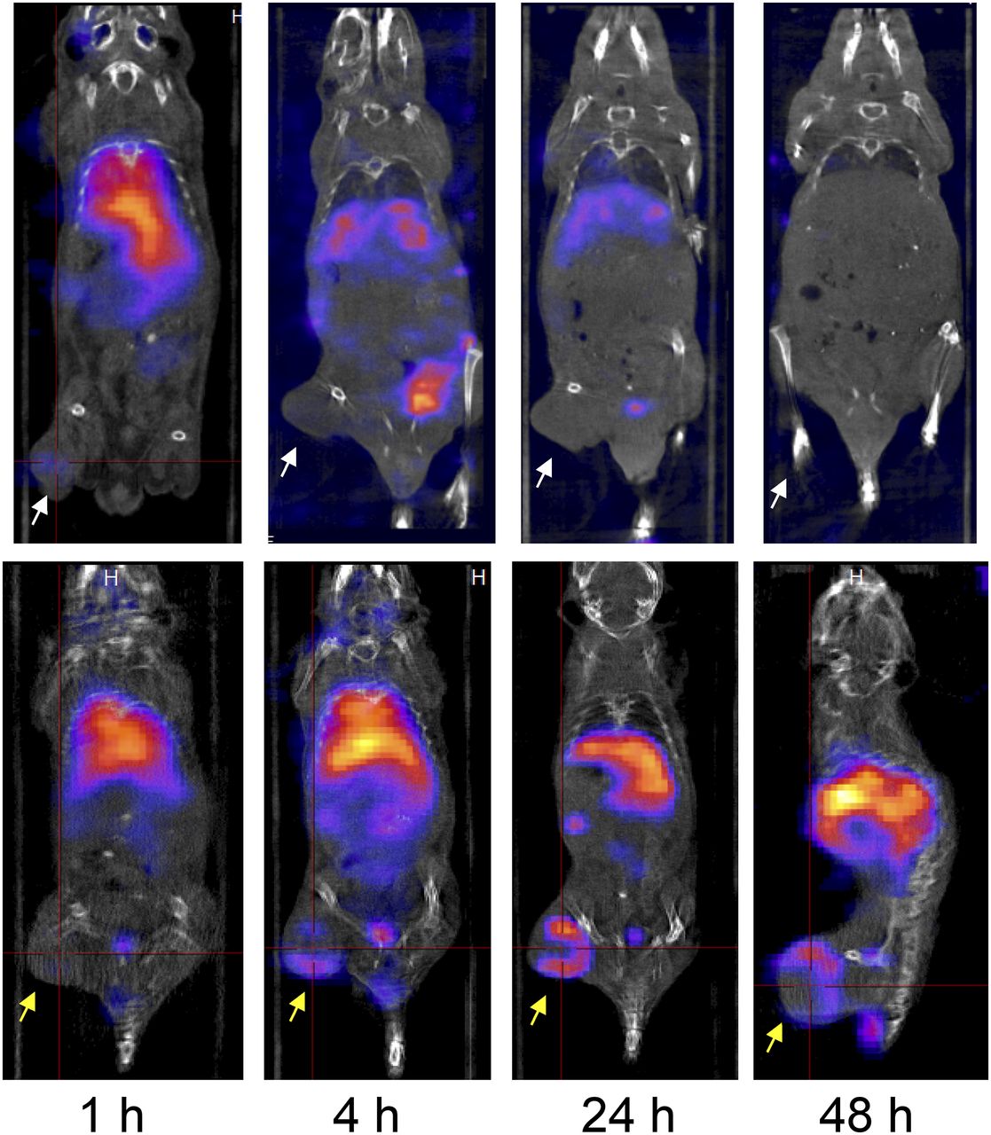
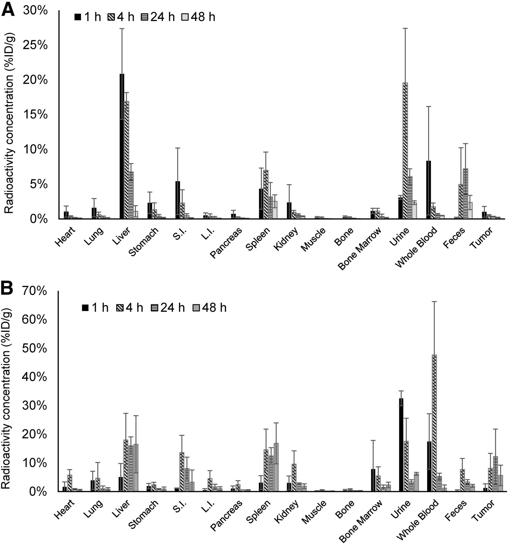
We next investigated whether 188Re-liposome could be used for detecting the tumor lesions in vivo. Human NSCLC H292-GLT cells were subcutaneously inoculated into the left hind limbs of nude mice. After the tumors reached 300 mm3, the tumor-bearing mice were separated to 2 groups and intravenously injected with either 188Re-BMEDA or PEGylated 188Re-liposome. The mice were subsequently subjected to nano-SPECT/CT at different times up to 48 h. The results showed that both 188Re-BMEDA and 188Re-liposome were rapidly detected (at 1 h) after they were injected into mice. However, 188Re-BMEDA was quickly washed out in tumor-bearing mice, whereas accumulation of PEGylated 188Re-liposome at the tumor site remained detectable up to 48 h (Fig. 2). Therefore, these results demonstrated that 188Re-liposome exhibited high retention and tumor targeting in vivo. Additionally, the quantitative biodistribution analysis of 188Re-BMEDA and 188Re-liposome showed that the distribution of these 2 radiopharmaceuticals at different times was distinct in several organs, including the liver, whole blood, spleen, and tumors. Rapid washout was observed in 188Re-BMEDA (Fig. 3).

Nano-SPECT/CT imaging of 188Re-BMEDA and 188Re-liposome distribution in NCI-H292 tumor–bearing mice. Tumors were implanted into left hind limb of each nude mouse (n = 3). After intravenous injection of 188Re-BMEDA (upper) and 188Re-liposome (lower), nano-SPECT/CT imaging was acquired at 1, 4, 24, and 48 h. Arrows indicate positions of subcutaneous tumors with or without radioactive signals.

Biodistribution analysis of 188Re-BMEDA and 188Re-liposome in tumor-bearing mice. (A) Results of 188Re-BMEDA. (B) Results of 188Re-liposome. Radioactivity in each organ is demonstrated by bar charts (n = 5). L.I. = large intestine; S.I. = small intestine.
Pharmacokinetic Analysis of PEGylated 188Re-Liposome
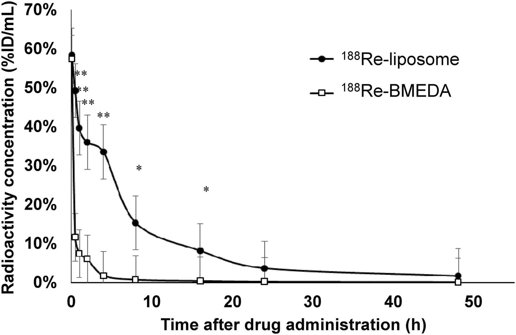
We further analyzed the pharmacokinetic curves of 188Re-BMEDA and PEGylated 188Re-liposome in vivo. Compared with 188Re-BMEDA, 188Re-liposome exhibited a long mean residence time, larger maximal concentration, and a slow pace of clearance (Table 1; Fig. 4). The area under the curve calculated from the time–activity curve showed that the liposomal encapsulation avoided rapid excretion and extended the mean residence time, suggesting that the enhanced permeability and retention effect was effective in this PEGylated radiopharmaceutical application. Moreover, we estimated the radiation dose in humans according to the results of murine biodistribution and determined that the absorbed dose ratio of tumor to lung could be up to 380.7-fold (within a 100-g tumor mass) (Table 2).
Pharmacokinetic Analysis in Human NSCLC–Bearing Mice

Pharmacokinetic time–activity curves in systemic circulation from tumor-bearing mice injected with either 188Re-BMEDA or 188Re-liposome at 0.083, 0.25, 0.5, 1, 2, 4, 8, 16, 24, and 48 h (n = 6). *P < 0.05. **P < 0.01).
Tumor-to-Nontumor Ratio of Absorbed Dose Estimation
Detection of 188Re-Liposome Accumulation in Established Orthotopic Lung Cancer Model
To evaluate the therapeutic efficacy of 188Re-liposome, a multiple-reporter-gene–integrated orthotopic tumor-bearing animal model was established. Tumor growth can be noninvasively monitored using BLI or radionuclide-based imaging in a small-animal model (Supplemental Fig. 1; supplemental materials are available at http://jnm.snmjournals.org). First, the location of the implanted tumor in the pulmonary parenchyma was confirmed using 123I-FIAU, which can be trapped by expressed HSV1-tk (Fig. 5A). The lungs dissected from the tumor-bearing mice also showed apparent bioluminescent signals that were detected in the left but not in the right lung (Fig. 5B). Using this model, we intravenously injected the PEGylated 188Re-liposome to detect the accumulation of 123I-FIAU in tumor-bearing mice. However, no apparent photon signals could be detected in the orthotopic lung tumor using nano-SPECT/CT (Supplemental Fig. 2). Therefore, we performed a biodistribution analysis in the orthotopic tumor model including the dissection of lung tissues from tumor-bearing mice and compared the data with normal mice. Using the γ counter, we found 56% increased uptake of 188Re-liposome in the tumor-bearing lungs, compared with tumor-free lungs (Fig. 5C; Supplemental Fig. 3). Additionally, fewer CD31-expressing vasculatures were observed in the orthotopic tumor model than in subcutaneous models (Fig. 5D; Supplemental Fig. 4). Therefore, these results implied that PEGylated 188Re-liposome could still accumulate in the orthotopic lung tumor.
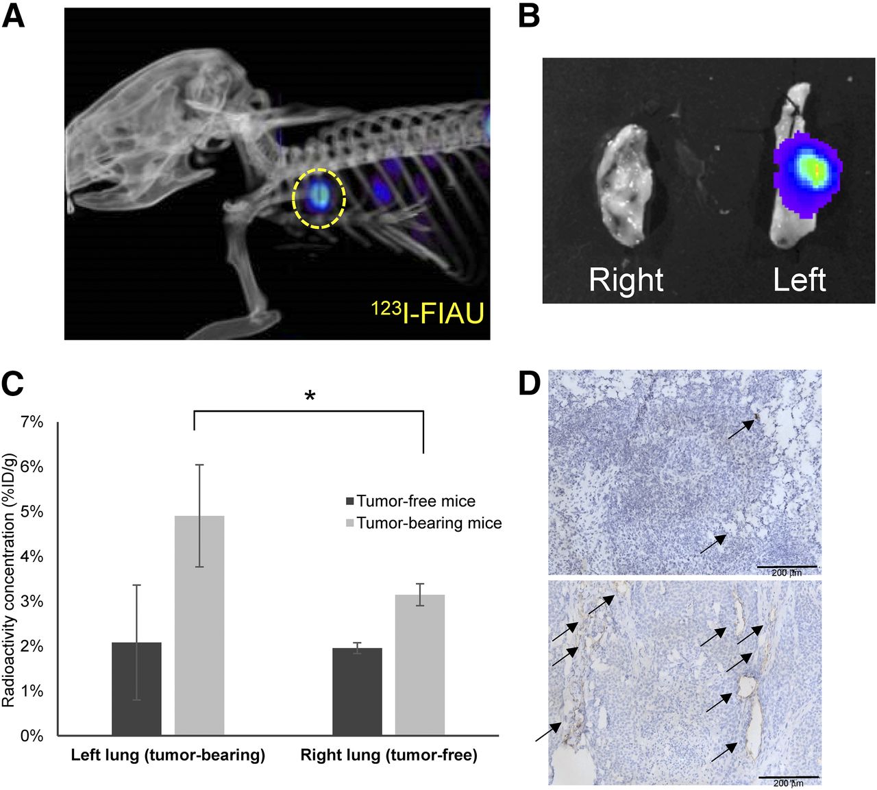
Detection of 188Re-liposome distribution in orthotopic model using HSV1-tk and luciferase dual reporter genes. (A) H292-GLT cells forming orthotopic tumor were localized by administration of 123I-FIAU and monitored by nano-SPECT/CT. Circle indicates location of tumor. (B) BLI was used to confirm formation of tumor in left lung of nude mouse. (C) Ex vivo tissues were subjected to γ counter for quantification. Significant difference between tumor-inoculated and non–tumor-inoculated mice was revealed. (D) Platelet endothelial cell adhesion molecule (PECAM-1) immunohistochemistry pathologic section showed that plenty of vasculature surrounded by PECAM-1 expression was observed in subcutaneous (lower) compared with orthotopic (upper) tumor. Arrows indicate PECAM-1–positive area. *P < 0.05.
Comparison of Therapeutic Efficacy of 188Re-BMEDA and 188Re-Liposome Using Orthotopic Lung Cancer Model
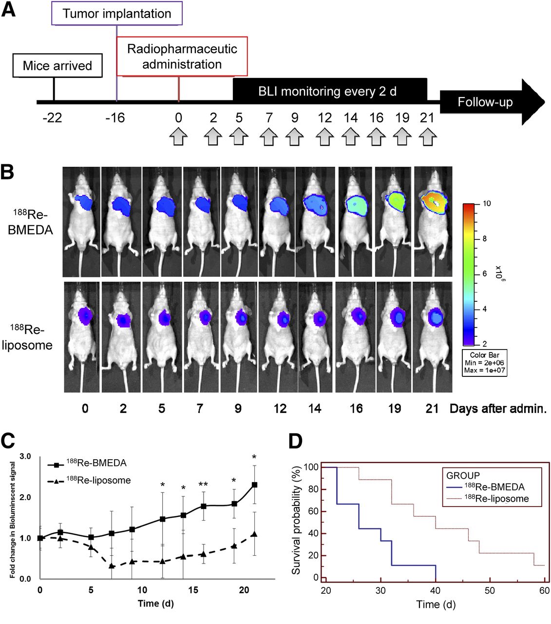
The 188Re-BMEDA and 188Re-liposome were separately injected intravenously into tumor-bearing mice with 23.68 MBq (640 μCi), the 80% maximum tolerated dose, within a single dose. BLI was used to monitor tumor growth in the thoracic cavity through the detection of luciferase activity at a fixed time interval (Fig. 6A). Bioluminescent signal correlates to tumor volume (Supplemental Fig. 1). After injection, the tumor growth was monitored up to 21 d, and the results showed that 188Re-liposome but not 188Re-BMEDA could suppress tumor growth (Fig. 6B). Quantification of the photon flux of orthotopic lung tumors further demonstrated that tumor viability was suppressed by 188Re-liposome but not by 188Re-BMEDA (Fig. 6C). However, it appeared that 188Re-liposome could not eradicate tumors but prevent their proliferation. Even so, the median survival time was longer in 188Re-liposome–treated tumor-bearing mice than in 188Re-BMEDA–treated ones (Table 3), and the Kaplan–Meier survival curves exhibited a significant difference between these 2 groups (Fig. 6D). Therefore, PEGylated 188Re-liposome could suppress lung tumor growth and extend life span in the orthotopic lung tumor model.

Comparison of therapeutic efficacy between 188Re-BMEDA and 188Re-liposome. (A) Therapeutic strategy of 188Re-BMEDA or 188Re-liposome. BLI was acquired at 10 time points up to 21 d. All animals were housed during this period until they reached endpoint (deceased or loss of 30% weight). (B) Bioluminescent images of orthotopic tumor growth. (C) Regions of interest quantified using LivingImage software (PerkinElmer Inc.). (D) Kaplan–Meier survival curve, with statistical analysis performed by log-rank test. *P < 0.05. **P < 0.01. admin. = administration; max = maximum; min = minimum.
Kaplan–Meier Survival Curve in Orthotopic Human NSCLC–Bearing Mice
DISCUSSION
Little is known about whether 188Re-embedded liposomal drugs can be used for treatment of tumor types above the diaphragm. Here we used PEGylated 188Re-liposomes to investigate their therapeutic effects on human lung cancer and showed that 188Re-based radiopharmaceuticals could be applied to this cancer type.
Good targeting of cancers is one of the most important considerations in using radiopharmaceuticals for cancer treatment. However, most of the metallic radioisotopes require chemical chelators for further encapsulation into physiologically compatible vehicles, such as liposomes. In this study, the labeling efficiency of BMEDA (>99%) to 188Re and the resultant radiochemical purity (>99%) were both similar to previous results (22), but the loading efficiency of 188Re to liposome was slightly lower (71.1% ± 7.61%) than that of the previous report (82.3% ± 4.5%) (22).
Although the circulation period of PEGylated 188Re-liposome is extended in vivo for better therapeutic efficacy, accumulation of 188Re in the liver and spleen is also increased. This phenomenon may cause potent side effects (15,22,27). Indeed, our biodistribution study revealed that accumulation of 188Re-liposome in the liver and spleen could remain up to 48 h, but 188Re-BMEDA washed out after 24 h of administration. Besides, the pharmacokinetic analysis also showed that the area under the curve of 188Re-liposome was 8.33-fold higher than that of 188Re-BMEDA, which is comparable to another report (28). The maximal absorbed dose to the liver and spleen was estimated at 4.07 and 17.4 Sv, respectively, after PEGylated 188Re-liposome treatment (see the “Materials and Methods” section). According to previous reports, the pathogenic dose to the liver and spleen is approximately 30 Sv (29). Thus, it seems that accumulation of 188Re-liposome in the liver and spleen would not result in severe radiation damage. Additionally, the estimated absorbed dose was 69.86 Gy in the subcutaneous tumor (300 mm3). Compared with the conventional clinical radiotherapeutic modality, the total tumor-absorbed dose of fractionation radiotherapy is from 60 to 80 Gy (30), which is comparable to the calculated radiation dose in the 188Re-liposome–treated tumors. However, further investigation is necessary to determine the safety of 188Re-liposome for clinical application.
One of the primary difficulties of this study was to establish an orthotopic xenograft model to mimic NSCLC in the thoracic cavity and to detect accumulation of PEGylated 188Re-liposome in the tumor lesion. Because subcutaneously implanted H292-GLT tumors exhibited significant uptake of 188Re-liposome, the orthotopic model is expected to have a similar result. Nevertheless, 188Re-liposome in the orthotopic tumor was barely detected using nano-SPECT/CT. The maximum tumor volume implanted in the pulmonary parenchyma was around 50 mm3, far smaller than the size of subcutaneously implanted tumors. Additionally, the emitted γ rays from 188Re are only 15% of overall radioactive decay, so that the partial-volume effect resulting from the instrumental limitation may also hamper tumor detection in the orthotopic model. Using the γ-counter measurement, however, we did find that the tumor-bearing lung exhibited higher radioactivity than the normal lung in mice treated with 188Re-liposome, suggesting that this radiopharmaceutical compound might be taken up by the orthotopic NSCLC model.
It has been reported that the administration of 188Re in murine colorectal cancer and human prostate cancer models could lead to significant tumor-killing effects and extend the survival of the animals through the emitted β particles (11,15,31). Although we have demonstrated that the life span of tumor-bearing mice was extended by PEGylated 188Re-liposome, the BLI data showed that the orthotopic lung tumors were not eradicated but their growth was suppressed. This observation may be consistent with the finding that the moderate accumulation of 188Re-liposome in orthotopic lung cancer cells was not sufficient to cause tumor death but did suppress tumor growth. Several lines of evidence have shown that the combination of 188Re and chemotherapy or multiple dosages of 188Re may increase the cytotoxic response in various cancers (21,28,32). It would be advantageous to design different regimes to evaluate whether 188Re-liposome treatment can eradicate NSCLC and reach maximum therapeutic efficacy.
CONCLUSION
The current results demonstrate that PEGylated 188Re-liposome can effectively suppress the growth of human NSCLC and extend the average life span of tumor-bearing mice. This radiopharmaceutical was taken up by NSCLC in vivo, but direct imaging of its accumulation in orthotopic tumor lesions may be largely dependent on tumor size. Moreover, the pharmacokinetics and dosimetry suggest that the use of PEGylated 188Re-liposome on lung cancer should be safe and deserves further investigation in larger animals. To the best of our knowledge, this is the first study investigating the efficacy of PEGylated 188Re-liposome on human lung cancer. It is anticipated that this novel radiopharmaceutical will be used to design strategies for lung cancer radiotherapy.
DISCLOSURE
The costs of publication of this article were defrayed in part by the payment of page charges. Therefore, and solely to indicate this fact, this article is hereby marked “advertisement” in accordance with 18 USC section 1734. This study was supported by grants NSC100-NU-E-010-002-NU and NSC101-2623-E-010-002-NU from the Institute of Nuclear Energy Research; grant NSC102-2628-B-010-012-MY3 from the National Science Council; grant NSC 102-2325-B-001-042 from the Taiwan Mouse Clinic, which is funded by the National Research Program for Biopharmaceuticals (NRPB) at the National Science Council of Taiwan; and an “Aim for the Top University Plan” grant from the Ministry of Education, National Yang-Ming University. No other potential conflict of interest relevant to this article was reported.
Acknowledgments
We thank Dr. Chia-Che Tsai and Wei-Hsin Hsu for help with 188Re-liposome preparation, production, and quality assurance and the Taiwan Mouse Clinic for technical support.
Footnotes
Published online Oct. 27, 2014.
- © 2014 by the Society of Nuclear Medicine and Molecular Imaging, Inc.
REFERENCES
- Received for publication March 19, 2014.
- Accepted for publication September 12, 2014.


Jump to section
Related Articles
Cited By...
- No citing articles found.