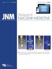
Since its original characterization in breast cancer nearly 30 y ago, the human epidermal growth factor receptor 2 (HER2) has become a key target for cancer therapeutics (1,2). HER2 is overexpressed in approximately 20% of breast cancers and is an adverse prognostic factor as it promotes cell proliferation and tumor metastasis; however, monoclonal antibody therapy targeting HER2 with agents such as trastuzumab combined with chemotherapy has produced dramatic improvements in recurrence rates and overall survival in patients with HER2-overexpressing tumors. In this issue of The Journal of Nuclear Medicine, Gebhart et al. have conducted one of theSee page 1862
largest multicenter studies to date to evaluate the role of 18F-FDG PET imaging for response assessment to neoadjuvant treatment in HER2-positive breast cancer in an imaging substudy of the Neo-ALTTO clinical trial (3).
The window-treatment design of the study was novel, as it allowed the metabolic response to HER2- and epidermal growth factor receptor–targeted therapies to be measured before the addition of weekly paclitaxel chemotherapy. Trastuzumab is a humanized monoclonal antibody that targets HER2 and is now routinely used in HER2-overexpressing breast cancer in the neoadjuvant, adjuvant, and metastatic settings. Lapatinib has a dual mode of action and also targets epidermal growth factor receptor as well as HER2. Patients were treated with lapatinib, trastuzumab, or the combination of lapatinib and trastuzumab for 6 wk followed by a 12-wk period during which the selected therapy was given in combination with paclitaxel before surgery. The main study, published earlier this year by Baselga et al., involved 455 patients and found a higher rate of pathologic complete response with the lapatinib–trastuzumab combination arm than with lapatinib or trastuzumab alone (51.3% vs. 29.5%) (4).
So the question here is can early 18F-FDG PET response predict subsequent pathologic complete response in HER2-positive breast cancer? Eighty-six patients enrolled to undergo 18F-FDG PET imaging; 66 of the 86 were able to complete all of the planned scans at baseline, after 2 wk, and after 6 wk of therapy. Imaging response was assessed within 30 centers in 14 countries. The degree of metabolic response was determined using maximum standardized uptake value (SUVmax), and the imaging was validated by a central core laboratory. The metabolic response at 2 wk correlated with that seen at 6 wk, and there was a significantly larger decrease in SUVmax at both time points in patients who subsequently had a pathologic complete response compared with those who did not (P = 0.02). The results from this study suggest that 18F-FDG SUVmax at 2 wk after initiation of therapy does correlate with ultimate pathologic response and that 18F-FDG response was twice as likely to be seen in clinical responders, compared with nonresponders. The definition of 18F-FDG response used by the investigators was based on the European Organization for Research and Treatment of Cancer (EORTC) criteria, which differ from the PET Response Criteria in Solid Tumors (PERCIST). The EORTC criteria use thresholds for partial metabolic response that vary depending on the time of assessment after treatment (a minimum of 15% decrease in standardized uptake value [SUV] after one cycle and greater than 25% after more than one cycle, whereas PERCIST has a constant threshold of 30% or greater reduction in lean body mass–normalized SUV [SUL] peak (5,6)).
The authors are to be commended for their study, which shows that multicenter studies using PET can be achieved; this will hopefully move us further along the path from Response Evaluation Criteria in Solid Tumors (RECIST) to a place where an appropriate biologic endpoint can be accepted as evidence of response to biologic therapy. Previous neoadjuvant studies in HER2-positive patients have had conflicting results. Humbert showed that the change in 18F-FDG SUVmax after 1 course of treatment may predict clinical response for patients treated with docetaxel, trastuzumab, and carboplatin (n = 37). In contrast, Koolen et al. suggested that changes in 18F-FDG PET uptake in 25 patients with HER2-positive breast cancer after 6–8 wk of paclitaxel, trastuzumab, and carboplatin were less useful, but the limited numbers of patients in both studies prevents definitive conclusions (7,8).
Tumor response assessment in the neoadjuvant setting is becoming even more important now because the Food and Drug Administration is considering approval of pathologic complete response in high-risk early stage breast cancer as a valid marker of efficacy for novel agents (9). Historically in breast cancer, new drugs are approved if they have been shown to have a survival benefit in the metastatic setting in phase III studies. Such phase III studies take a long time to report, and new approaches are needed to accelerate novel drug development. The suggested new mechanism is likely to lead to a new expedited model for drug development, preventing the current high rates of attrition of novel compounds where patients with heavily pretreated advanced metastatic disease are treated with a single agent to which they are unlikely to respond due to innate tumor resistance. Pathologic complete response is an important outcome in neoadjuvant breast cancer treatment, which has been shown to translate into future benefits in overall and disease-free survival. The ability of early 18F-FDG PET scanning to serve as a useful surrogate will need further validation in trials such as Neo-ALTTO with well-defined subgroups of patients.
Recently, it has been recognized that there may be at least 10 distinct molecular subtypes of breast cancer distinguishable by genomic sequencing. Tumor glucose utilization measured by 18F-FDG can vary with molecular subtype; several investigators have shown that significantly higher uptake levels of 18F-FDG are seen in triple-negative and HER2-positive phenotypes. Increased glycolysis in HER2-positive cancers is thought to arise by signaling via the PI3K/AKT/MTOR cascade because activated AKT causes increased transport and metabolism of glucose (10). Treatment regimes are evolving and becoming more personalized, and as we divide breast cancer into more subtypes it is likely that multicenter studies will be needed to recruit a significant number of patients to prove treatment efficacy. Multicenter studies are commonplace for treatment response assessment; however, relatively few of these studies contain a molecular imaging component because they continue to rely on RECIST.
This study shows us that the change in 18F-FDG uptake after treatment may help predict clinical response; however, it does not show that baseline 18F-FDG is a useful predictive biomarker for HER2-targeted therapy. Many patients do have intrinsic resistance to HER2-targeted therapy, and it would be useful if a predictive biomarker could identify this group up front so that alternative treatments could be used. HER2 status is most commonly evaluated on a tissue biopsy specimen with immunohistochemistry or by fluorescent in situ hybridization or silver in situ hybridization. Tumor heterogeneity is being recognized as an important feature in resistance to targeted therapies that may not be captured by a biopsy from a single region, and this may be where molecular imaging probes under development—such as 111In- and 68Ga-radiolabeled Affibody molecules (Affibody), which bind to HER2 with high affinity—could be used in the future to detect this tumor heterogeneity noninvasively (11). The safety and predictive utility of these new molecular imaging probes in a clinical setting need to be firmly established, especially now with the advent of new HER2-directed therapies and combinations that are likely to increase treatment costs. Another option could use 3′-deoxy-3′-18F-fluorothymidine, which was thought to be more useful than 18F-FDG in assessing response in HER2-overexpressing tumors in a preclinical model (12). Further efforts should concentrate on the standardization of PET response characteristics globally and integration of 18F-FDG and other novel PET biomarkers with other molecular biomarkers so that true phenomic profiling can be achieved to direct the optimum treatment strategy for individual patients.
DISCLOSURE
The author's work is supported by an NIHR Clinician Scientist Fellowship, NIHR/CS/009/009. No other potential conflict of interest relevant to this article was reported.
Footnotes
Published online Sep. 26, 2013.
- © 2013 by the Society of Nuclear Medicine and Molecular Imaging, Inc.
REFERENCES
- Received for publication June 13, 2013.
- Accepted for publication July 9, 2013.


