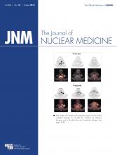
REPLY: The work that we have recently described tested the feasibility of the use of Affibody molecules (Affibody AB) and PET to predict tumor response to ErbB2-targeted therapy. It is clear that additional studies will be needed to dissect the mechanistic events underlying the observed changes. Differences among tumor cell lines could affect responses as well.
Clinical studies show that the assessment of ErbB2 level by immunohistochemistry produces variable results among laboratories. This variation may be due to differences in immunohistochemistry staining techniques and scoring criteria (1). For antigen-retrieval processes, the solution used (e.g., citrate buffer or ethylenediaminetetraacetic acid and their pH), the duration of heating, and antigen retrieval may all affect detection of the ErbB2 antigen by immunohistochemistry (2). Different anti-ErbB2 antibodies used for immunohistochemistry staining have also been shown to produce different degrees of ErbB2 staining in tumors, even in the presence of gene amplification (3), although applying calibration may help in minimizing those differences (4). The HercepTest (Dako) using Dako antibody was proposed as the standardized immunohistochemistry method to overcome the problem of interlaboratory variations. The scoring system uses the intensity of ErbB2 staining as its basis, and an ErbB2-positive tumor is defined as a tumor with greater than 10% of cells stained 3+ (5). Despite use of the HercepTest, there still was a high discrepancy between local and central ErbB2 testing in the N9831 Intergroup Adjuvant Trial, with a concordance of only 81.6% for a diagnostic test for the presence of the ErbB2 protein (6). The American Society of Clinical Oncology and College of American Pathologists recommended an algorithm defining positive, equivocal, and negative values for both ErbB2 protein expression and gene amplification. A positive ErbB2 result from immunohistochemistry staining is defined by uniform, intense membrane staining of more than 30% of invasive tumor cells instead of the original 10% (5). However, despite this new algorithm and definition, not all laboratories have adopted this new guideline and there still are variable results in ErbB2 testing among laboratories. Furthermore, both the old and the new “ErbB2 counting” definitions have problems in detecting subtle ErbB2 changes induced by the treatment. For example, if trastuzumab decreases ErbB2 staining from 100% of the cells to 40%, both the old and the new ErbB2 definition will score pre- and post-treatment samples as “positive” and may fail to detect ErbB2 changes after trastuzumab treatment.
After trastuzumab treatment, we found in human breast carcinoma BT474 xenografts a significant reduction of tracer uptake related to ErbB2 decrease rather than tumor size reduction (7). The observable reduction in PET signals could be due to partial-volume effect, but this possibility is rather unlikely since the images were acquired with high contrast and in the absence of background activity. When large enough regions are drawn around the tumor, the partial-volume effect does not cause any loss of signal, and the signal that is measured indicates the actual activity distribution. Moreover, for PET quantification, we deliberately chose the value related to maximum counts per pixel within the tumor that is least affected by partial-volume effect. Importantly, we have also shown that tumor ErbB2 membrane staining and PET changes correlated with tumor volume after 5 doses of trastuzumab treatment (7). There was a correlation between PET and immunohistochemistry, and the radionuclide concentrations measured with PET agreed with the radioactivity concentrations obtained by γ-counting (data were not presented). Although there was a large overlap in ErbB2 staining between the trastuzumab-treated and control groups, we found a significant reduction of ErbB2 downregulation after 5 doses of trastuzumab. This finding is consistent with several cell line experiments from different groups finding that trastuzumab downregulates ErbB2 receptors (8–10). After a single dose of trastuzumab, we could clearly see the differences in ErbB2 membrane staining between control and trastuzumab-treated samples, but the intensity percentage scoring failed to detect these changes (7).
The dose and duration of trastuzumab will clearly affect the amount of ErbB2 downregulation and the detection of ErbB2 changes by immunohistochemistry. In our paper, the dose of trastuzumab was deliberately high (50 mg/kg loading dose followed by 4 more doses of 25 mg/kg each) to ensure that changes in receptor expression ErbB2 would be possible (7). Reddy et al. (11) treated BT474 xenografts with a lower dose of 10 mg/kg for only 6 d. They found a decrease in PET tracer using C6.5 diabody but could not detect any ErbB2 changes by immunohistochemistry. They concluded that “The exact mechanism by which trastuzumab treatment inhibits C6.5db binding is not yet understood.” On the other hand, McLarty et al. (12) reported that trastuzumab reduced ErbB2 membrane staining in SKBR3 cells and in MDAMB361 and MDAMB361 xenograft models. In this case, mice were treated only with 4 mg of trastuzumab per kilogram for 3 d or 3 wk. At 3 d, the authors did not see ErbB2 membrane changes, but 3 wk later immunohistochemistry analysis of tumor tissues indicated significant ErbB2 downregulation, associated with almost complete eradication of viable tumor cells. This finding is consistent with our study as we did not see a difference in intensity percentage scoring after a single dose of trastuzumab (7); we observed differences in ErbB2 membrane staining after 5 doses of the drug (7). We believe that the differences seen in ErbB2 staining between Reddy et al. (11), McLarty et al. (12), and our study (7) may be related to the dose and duration of trastuzumab used. However, the differences may also be related to the ErbB2 testing methods and the scoring criteria, which could not detect subtle ErbB2 changes after trastuzumab treatment.
Trastuzumab was used as monotherapy before surgery in patients with primary operable ErbB2-positive breast tumors in a pilot study by Gennari et al. (13). They observed no change in ErbB2-positive staining using monoclonal antibody CB11 in the trastuzumab-treated samples. However, they provided figures from only 1 patient, as shown in their Figures 2A and 2B (13). Although these figures suggest some changes between pre- and posttreatment samples, the authors found no variations in the ErbB2 status (13). Furthermore, the use of a different anti-ErbB2 antibody and different scoring criteria may also have contributed to failure to detect ErbB2 changes between pre- and posttreatment samples.
Tagliabue et al. state in their letter that no changes in ErbB2 receptor status evaluated by immunohistochemistry were found in most patients with operable (14) or locally advanced (15) ErbB2-positive breast cancers after neoadjuvant exposure to trastuzumab in combination with chemotherapy. In the neoadjuvant study by Harris et al. (14), the ErbB2 status was based on the HercepTest, and 4 of 18 patients had lower immunohistochemistry scores. It is important to emphasize that HerceptTest criteria (i.e., 10% of cells positive) are not sensitive in detecting subtle ErbB2 changes induced by treatment. Therefore, it is possible that the HerceptTest could pick up ErbB2 changes in only a few patients because of the scoring criteria used. Mohsin et al. (15) used the Allred scoring system for ErbB2 changes between baseline and after 1 or 3 wk of treatment and did not see a difference. However, this is not the standard method for ErbB2 testing and may not be able to differentiate between a weak ErbB2 intensity present in the whole tumor mass and a high ErbB2 intensity in just certain parts of the tumor, further underscoring the problems of using immunohistochemistry as the only screening test.
Mittendorf et al. (10) reported ErbB2 gene amplification loss in 1 of 3 patients, although the protein level measured by immunohistochemistry was not shown. It could be interesting to correlate gene level with protein expression. Tagliabue and colleagues argued that this loss of ErbB2 amplification is due to selection of ErbB2-negative cells rather than trastuzumab-induced ErbB2 downmodulation. Although we agree that selection of ErbB2-negative cells is a possibility, it is also possible that trastuzumab downregulates ErbB2 receptors resulting in tumor shrinkage but there is clonal expansion of the other ErbB2-negative cells.
We did not find a correlation between the impaired Affibody localization in xenografts and decreased vascularization. In fact, we saw the highest vessel count in those tumors with greater ErbB2 loss as assessed by PET although an elevated number of vessels was found only in the group of animals showing a dramatic decrease in 18F-FBEM-HER2:342-Affibody uptake (PET [%ID/g] ≤ 0.55). We confirmed that the tumor size was not related to the average vessel count per field, and thus, we did not simply select tumors that responded to trastuzumab because of a better vascularization.
Regarding the comment by Tagliabue et al. citing their recent report that “tumor shrinkage induced by trastuzumab-containing therapy can sometimes be followed by an inflammatory reaction that masks any decrease in neoplastic mass” (16), that report was a retrospective study assessing the use of trastuzumab beyond progression, and therapy included a variety of additional chemotherapy agents. ErbB2 levels were not evaluated in progressing tumors.
In summary, we agree that there have been various controversial reports on the effect of trastuzumab on ErbB2 receptor downregulation in cell lines, xenograft models, and human studies. Modeling the therapeutic activity of any antibody in human xenografts is challenging. Their vascular system is derived from the host. Subcutaneous transplants do not recapitulate the systemic and metabolic effects of spontaneous cancers, and they fail to capture the contribution of the immune system, which is saved in syngeneic systems. Therefore, these models may not completely mimic the response in human patients.
The particular animal model, the dose of trastuzumab, and the duration of trastuzumab could all affect the amount of ErbB2 downregulation by trastuzumab observed in preclinical studies. Furthermore, differences in immunohistochemistry staining techniques, antibodies used, and scoring criteria could account for differing results in the assessment of ErbB2 status by immunohistochemistry in both animal and human studies.
Footnotes
Published online Sep. 4, 2012.
- © 2012 by the Society of Nuclear Medicine and Molecular Imaging, Inc.


