


Abstract
We have developed a practice procedure for prostate lymphoscintigraphy using SPECT/CT and filtered 99mTc-sulfur nanocolloid, as an alternative to the proprietary product 99mTc-Nanocoll, which is not approved in the United States. Methods: Ten patients were enrolled for this study, and all received radiotracer prepared using a 100-nm membrane filter at a commercial radiopharmacy. Whole-body scans and SPECT/CT studies were performed within 1.5–3 h after the radiotracer had been administered directly into 6 locations of the prostate gland under transrectal ultrasound guidance. The radiation dose was estimated from the first 3 patients. Lymphatic drainage mapping was performed, and lymph nodes were identified. Results: The estimated radiation dose ranged from 3.9 to 5.2 mSv/MBq. The locations of lymph nodes draining the prostate gland were similar to those found using the proprietary product. Conclusion: When the proprietary radiolabeled nanocolloid indicated for lymphoscintigraphy is not available, prostate lymph node mapping and identification are still feasible using filtered 99mTc-sulfur nanocolloid.
It is estimated that 50% or more patients with high-risk prostate cancer will experience a relapse after definitive treatment (1). There are substantial data suggesting that many of these relapses may be due to microscopic metastasis in the pelvic lymph nodes (2–4). Recent surgical data have indicated that the incidence of positive nodes is higher than once thought (2).
Nomogram predictors (5) help identify a potential high risk of involved pelvic nodes. To confirm the microscopically involved nodes, lymphadenectomy combined with pathologic analysis is necessary. Radical prostatectomy combined with an extended pelvic lymph node dissection (6) is one of the standard treatments for localized prostate cancer. However, in some cases, this treatment represents an overly aggressive and invasive approach.
External-beam radiotherapy or brachytherapy is presented with the same challenges. However, there is no method to assess nodal involvement unless anatomic imaging shows enlarged (>1 cm in diameter) lymph nodes. Recent studies suggest therapeutic value to treating potentially involved nodes by radiation (1). For example, the phase III trial by the Radiation Therapy Oncology Group (RTOG-9413) demonstrated that prophylactic pelvic lymph node irradiation improves progression-free survival for high-risk patients, suggesting that treatment of the primary tumor and local lymph nodes can be curative (7).
Pelvic irradiation can increase the probability of treatment side effects. The exact volume of nodes to include in the radiation field is therefore critical and has been much debated (8). Currently, most whole-pelvis radiotherapy planning is based on assumptions about standardized anatomic lymphatic drainage patterns (9). However, as shown by the results of radioguided surgical lymph node dissection, the patterns of each patient's lymphatic drainage from the prostate are highly variable (9,10).
Whole-pelvis irradiation of patient-specific lymphatic drainage with a highly conformal radiotherapy technique such as intensity-modulated radiotherapy may improve long-term tumor control outcomes (11). 99mTc-nanocolloids (colloidal particles < 100 nm) can be used to achieve personalized whole-pelvis radiation planning (9,12).
The most popular 99mTc-nanocolloid is a commercial product called 99mTc-Nanocoll (GE Healthcare), a colloid of human serum albumin (13). Extensive studies using this nanocolloid have been performed to map the sentinel lymph nodes of the prostate (14–16). However, this product has not yet received clearance from the U.S. Food and Drug Administration. In the United States, 99mTc-sulfur colloid is used in breast lymphoscintigraphy as well as for other applications (17). The 99mTc-sulfur colloid can be formed into a nanocolloid through the use of a 100-nm polycarbonate membrane filter, resulting in a range of particle sizes similar to that of 99mTc-Nanocoll (13). The important factor contributing to the kinetic properties of a radiocolloid is the distribution of particle sizes, and that factor has yet to be studied for filtered 99mTc-sulfur nanocolloid for prostate lymphoscintigraphy. We report our feasibility study of a SPECT/CT practice procedure we developed for prostate lymphoscintigraphy using filtered 99mTc-sulfur nanocolloid.
MATERIALS AND METHODS
Subject Recruitment and Adverse Event Monitoring
Subjects were recruited following a protocol approved by the Institutional Review Board. The filtered 99mTc-sulfur nanocolloid, as a Food and Drug Administration–approved agent, was used for this open-label indication of lymph node scintigraphy. To be included, patients had to have a definitive diagnosis of prostate cancer, be clinically eligible to receive intensity-modulated radiotherapy with pelvic lymph nodal irradiation, and be scheduled for such treatment. Ten patients (age range, 56–81 y; prostate-specific antigen level, 3.55–70 ng/mL) were enrolled. Reportable adverse events were to be communicated 1 wk after radiotracer administration, but no adverse events occurred.
Administration of Agent
An arrangement was made with a local commercial radiopharmacy (GE Healthcare) that all orders for this study were to be prepared using a 100-nm membrane filter. After 99mTc-sulfur colloid is passed through a 100-nm polycarbonate membrane filter, most colloidal particles (∼90%) are between 30 and 80 nm, with a peak at 53.9 nm (18). The agent was administered in the Urology Clinic at the University of California, San Francisco, Medical Center by one urologist with transrectal ultrasound guidance.
Filtered 99mTc-sulfur nanocolloid (40.7–111 MBq) was divided equally into 6 fractions of 0.5 mL each and was administered into 3 locations (apex, mid portion, and base) for each lobe (left and right) of the prostate gland. Although the administration was supposed to be evenly distributed to all 6 locations, we noted that some patients received uneven doses resulting in uneven flow patterns of nodal uptake.
Whole-Body and SPECT/CT Parameters
Our studies used a SPECT/CT system with a low-amperage CT scanner (Infinia Hawkeye 4; GE Healthcare) at the University of California, San Francisco, China Basin Imaging Center. All SPECT images were reconstructed using ordered-subsets expectation maximization involving CT-based attenuation correction. The SPECT projections were acquired with a 128 × 128 matrix over 60 angles covering 360° at 15 s per stop. The reconstructed images were 64 × 64 × 64 matrices postfiltered using a Butterworth filter. The anteroposterior whole-body scans were obtained before the SPECT/CT scans to ensure a sufficient distribution of filtered 99mTc-sulfur nanocolloid from the gland. SPECT/CT covering the pelvis and thorax was performed 1.5–3 h after administration. For the matching SPECT field of view, abdominopelvic CT was performed without a contrast agent. A tube voltage of 140 kVp and tube current of 2.5 mA were used, and the data were acquired using a 256 × 256 matrix and a 5-mm slice thickness. The filtered backprojection algorithm provided by the manufacturer was used for CT reconstruction.
Radiation Dose Estimation Using Simple Biokinetic Data
For the first 3 patients, we performed whole-body scans after SPECT/CT to obtain additional datasets for radiation dose estimates. For logistic convenience, we applied a simple biokinetic analysis based on 3 time points of radiotracer distribution. The first time point was not from imaging but was based on the assumption that initially the radiotracer is localized only to the administration site—the prostate gland. The 2 additional time points were before and after SPECT/CT.
OLINDA/EXM software (version 1.1) (19) was used to estimate radiation dose. We calculated residence times in the prostate, liver, and spleen from the whole-body scans using a conjugate-view method and phantom measurements that corrected for image-pixel differences in radioactivity in anterior and posterior views. For phantom measurements, we used a 6-mL syringe with a clear acrylic attenuator of known activity, calibrated using a dose calibrator.
Lymph Node Identification and Drainage Mapping
The lymphoscintigraphy results were interpreted by attending nuclear medicine physicians and then again by one of the study investigators. We used SPECT/CT images for identifying lymph nodes and drainage patterns. The identification of sentinel lymph nodes and of all secondary nodes identifiable from the imaging studies was noted in the reports.
RESULTS
Radiation Dose Estimates
The reported radiation dose estimate for 99mTc-Nanocoll is an effective dose of 7.6 mSv/MBq (20). Data from the 3 patients showed an effective dose of 3.9–5.2 mSv/MBq for the filtered 99mTc-sulfur nanocolloid, slightly lower than the effective dose from 99mTc-Nanocoll (Table 1). Even with injected doses that were lower (40.7–111 MBq) than the typical dose of 99mTc-Nanocoll (200 MBq), we were able to visualize uptake in lymph nodes draining the injection site within 1.5–3 h after administration. From the radiation dose estimation data, we also found that hepatic clearance of filtered 99mTc-sulfur nanocolloid is slightly faster than that of 99mTc-Nanocoll.
Comparison of Absorbed Dose Estimates (mSv/MBq) for 99mTc-Nanocoll and Filtered 99mTc-Sulfur Nanocolloid
Lymph Node Identification
One criterion for the success of this procedure was to be able to image lymph node uptake outside the prostate within 1–3 h, and our results were consistent with that expectation.
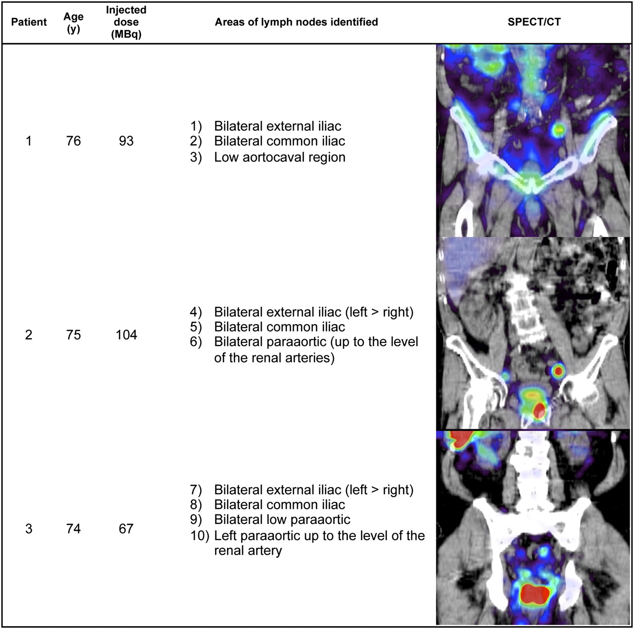
Also as expected, lymph nodes were sometimes identified outside the pelvic area, a common feature of the prostatic lymphatic drainage pattern. Figures 1–3 show the demographics, injected dose, and lymphatic uptake locations for each patient. We also show a coronal SPECT/CT view displaying at least one lymph node that took up filtered 99mTc-sulfur nanocolloid. A descriptive list of lymph nodes identified as draining the prostate gland is also included in Figures 1–3.

Age distribution, radiotracer administration, identified lymph node locations, and representative coronal view of SPECT/CT for study patients 1–3.

Age distribution, radiotracer administration, identified lymph node locations, and representative coronal view of SPECT/CT for study patients 4–6.

Age distribution, radiotracer administration, identified lymph node locations, and representative coronal view of SPECT/CT for study patients 7–10.
DISCUSSION
In this report, we have shown that the nonproprietary filtered 99mTc-sulfur nanocolloid can be used to perform prostate lymphoscintigraphy within 1.5–3 h after injection. Proper preparation of the radiotracer is important because the required size of the colloid, less than 100 nm, is critical to ensure fast drainage from the prostate gland, as has been shown for the proprietary product. We recommend that others who use our practice procedure work with their local radiopharmacy to obtain sulfur colloid that has been prefiltered with a 100-nm membrane filter.
A limitation of our study was that only patients with an intact and previously untreated prostate were included. We chose not to include postprostatectomy patients because of uncertainty as to the appropriate places to inject the radiotracer in this setting and the known altered drainage patterns after previous therapies (15). Another limitation of our study was the absence of definitive proof as to exactly how the information obtained through this lymphoscintigraphy technique may have altered our treatment fields. Such a detailed analysis is under way. On the basis of the work of others, we do expect to find evidence supporting the routine use of sentinel node imaging (9,16).
Unlike common use of lymphoscintigraphy for surgical management of cancer, our prostate lymphoscintigraphy was to use SPECT/CT to provide tomographic information on lymphatic drainage through sentinel and secondary nodes, allowing radiation treatment for these patients to be planned using individualized irradiation fields. Clearly, in at least 2 cases the sentinel node images resulted in a substantial altering of the fields. In these 2 cases, the primary drainage included common iliac nodes above the level of L5–S1 (our standard superior border). In addition, in all cases we applied slightly wider margins and delivered a slightly higher dose to sentinel nodal areas. We also used tighter margins and consequently lower doses to nonsentinel nodal drainage areas. This strategy, at least in theory, should result in enhanced nodal control and reduced toxicity due to smaller irradiated volumes to uninvolved normal tissues, such as the small bowel.
Finally, another interesting methodologic approach that could enhance our practice procedure for SPECT/CT prostate lymphoscintigraphy is to use high-amperage CT, commonly applied in diagnostic multidetector CT scanners, instead of the low-amperage CT that was used in our studies, when combined with SPECT data. The high-amperage CT can reveal small lymph nodes in greater detail; thus, it will be easier to identify the anatomic locations of lymph nodes on SPECT images.
CONCLUSION
We successfully performed a 10-patient study to establish a SPECT/CT practice procedure for prostate lymphoscintigraphy, with a goal of using the information obtained for planning radiation treatment. The use of filtered 99mTc-sulfur nanocolloid seems appropriate for this indication, and the 1.5- to 3-h imaging window is favorable in clinical practice.
DISCLOSURE STATEMENT
The costs of publication of this article were defrayed in part by the payment of page charges. Therefore, and solely to indicate this fact, this article is hereby marked “advertisement” in accordance with 18 USC section 1734.
Acknowledgments
We thank Marilyn Robinson at the University of California, San Francisco, for her help with coordinating the clinical study. We also thank Chang-Lae Lee, who helped with radiation dose calculations using biokinetic data. This study was partially funded by National Cancer Institute grant K25 CA114254. No other potential conflict of interest relevant to this article was reported.
- © 2011 by Society of Nuclear Medicine
REFERENCES
- Received for publication December 9, 2010.
- Accepted for publication February 28, 2011.


Jump to section
Related Articles
Cited By...
- No citing articles found.