


Abstract
The aim of this study was to determine the uptake of L-[methyl-11C]-methionine (11C-MET) in the normal brain of patients younger than 20 y, to facilitate more accurate diagnoses in young patients. Methods: Eighty-two patients were categorized into 4 groups according to their age. They underwent 11C-MET PET, and a standardized uptake value (SUV) was determined for different brain regions including the frontal lobe, parietal lobe, cerebellum, and brain stem. Results: Compared with all other parts of the brain, the cerebellum had the highest SUV. A tendency for a positive relationship between SUV and age was found in all regions, and a significant relationship with SUV was found in the frontal lobe and cerebellum. Conclusion: The character of SUV in the normal brains of children is different from that of adults, and these normal SUV data will play an important role as a critical reference value.
The tracer L-[methyl-11C]-methionine (11C-MET) is useful for PET in neurooncology ( 1, 2), making it possible to assess the characteristics of lesions that could not be diagnosed by other means ( 1, 3– 7). 11C-MET uptake is also informative about the malignancy of lesions, as is 18F-FDG PET ( 6, 7). For younger patients, it is critical to get accurate information about lesions, such as the degree of malignancy and extent of lesions, as early as possible. Therefore, it is essential to have an accurate understanding of 11C-MET PET data. To date, 11C-MET PET has been interpreted mainly using the lesion-to-normal (L/N) ratio—comparison between 11C-MET uptake in the lesion and that in the corresponding normal region in the contralateral hemisphere ( 8– 10). In cases of a highly malignant lesion, it would be relatively easy to point out where the lesion is, but when the disease has low malignancy, it may be difficult because interpretation of the result is semiquantitative. Thus, an absolute index is desirable. Uda et al. determined the normal 11C-MET uptake and extent of variation ( 11), but there is no report about normal uptake among children. In the current study, we evaluated normal 11C-MET uptake in persons younger than 20 y.
MATERIALS AND METHODS
Patients
Between February 1994 and May 2008, 1,228 patients underwent 11C-MET PET at Osaka City University Hospital. Of these patients, 136 were 20 y or younger. Exclusion criteria included the following: infiltrative grade II–IV neoplasm according to the World Health Organization classification ( 12), extramedullary or midline tumors greater than 20 mm in diameter (3 patients), edematous changes in the brain (3 patients), and history of radiation therapy or chemotherapy (48 patients). The remaining 82 patients (40 male and 42 female; mean age ± SD, 12.4 ± 6.1 y; range, 0–20 y), who were not taking any drugs that could influence brain metabolism, were divided into 4 groups according to their age: group 1, 0–5 y; group 2, 6–10 y; group 3, 11–15 y; and group 4, 16–20 y. The standardized uptake value (SUV) of normal brains was examined according to age and each region, including the frontal lobe, parietal lobe, cerebellum, and brain stem.
The Ethics Committee of Osaka City University Graduate School of Medicine approved this PET study. Informed consent was obtained from all patients or their parents.
PET
PET was performed with an Eminence B PET scanner (Shimadzu); the spatial resolution was 4.5 mm (in full width at half maximum), and slice thickness was 5.6 mm. Scans were obtained parallel to the orbitomeatal line of the patients. After 4 h of fasting, 11C-MET (6 MBq/kg) was injected intravenously over 30 s. After a transmission scan was obtained, a static scan of 10 min was begun 20 min after injection.
Imaging Analysis
Two experienced nuclear medicine radiologists interpreted the scans. The PET images were reconstructed by measured attenuation correction. In both analyses, the region of interest (ROI) was placed manually in the axial plane within the frontal cortex, parietal cortex, cerebellum, and brain stem ( Fig. 1). The ROIs of these regions, except for the brain stem, were demarcated manually within the lateral side of the gray matter by reference to the MRI data. In the brain stem, the ROI was placed over the whole brain stem at the middle pons level. Mean pixel counts in the ROIs were normalized to SUV using the subject's body weight with the following equation: SUV = ([mean pixel count/pixel volume]/[injected radioisotope activity/body weight]) × calibration factor.
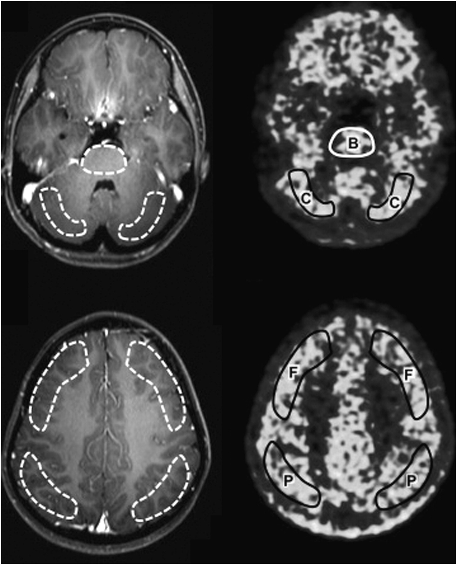
Sample of 11C-MET PET images. ROIs were taken manually in axial plane. Coregistration between MR images and PET scans is adequate. B = brain stem; C = cerebellum; F = frontal cortex; P = parietal cortex.
Statistical Analysis
The differences between each part of the brain were analyzed in every group. The sample consisted of 54 patients for whom we had all SUV data, including the right frontal lobe, right parietal lobe, right cerebellum, and brain stem. Statistical analysis was performed by non–repeated-measures ANOVA, with post hoc tests corrected using Bonferroni and Student–Newman–Keuls analysis.
The relationship between SUV and age was analyzed in every part of the brain with linear regression and Spearman correlation tests. The samples used in these analyses were 158 frontal hemispheres (79 right hemispheres and 79 left hemispheres), 118 parietal hemispheres (59 right hemispheres and 59 left hemispheres), 156 cerebellar hemispheres (77 right hemispheres and 79 left hemispheres), and 62 brain stems. In all statistical analyses, significance was defined as a P value less than 0.05.
RESULTS
Frontal Lobe
SUVs (mean ± SD) in the frontal lobe were 1.133 ± 0.295 and 1.088 ± 0.379 in group 1, 1.067 ± 0.264 in group 2, 1.101 ± 0.268 in group 3, and 1.199 ± 0.258 in group 4 ( Table 1). A significant linear regression between SUV and age (P = 0.0282) was found ( Fig. 2).
SUV in Each Part of Brain in Every Group
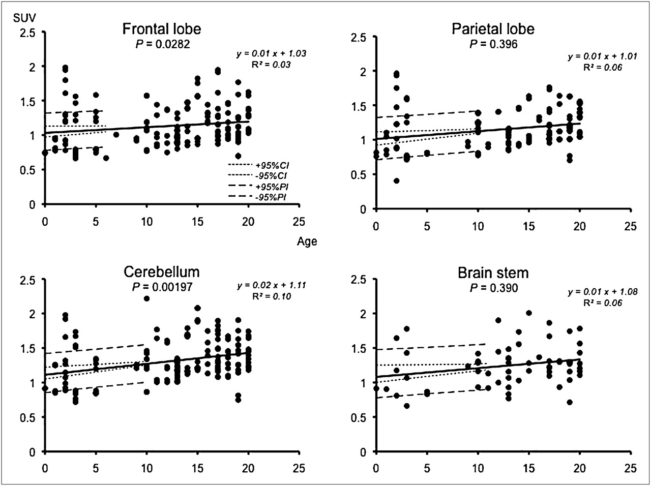
Regression and scatterplots for analysis of age-associated SUV change in frontal lobe, parietal lobe, cerebellum, and brain stem. In all parts of brain, positive linear regression was found, but P value of regression coefficient was statistically significant only in frontal lobe and cerebellum.
Parietal Lobe
SUVs (mean ± SD) in the frontal lobe were 1.149 ± 0.290 and 1.052 ± 0.399 in group 1, 1.114 ± 0.209 in group 2, 1.122 ± 0.220 in group 3, and 1.235 ± 0.243 in group 4 ( Table 1). Significant differences between groups 1 and 4 (P < 0.01) were found, but no significant linear regression between SUV and age (P = 0.396) was found ( Fig. 2).
Cerebellum
SUVs (mean ± SD) in the frontal lobe were 1.312 ± 0.309 and 1.145 ± 0.347 in group 1, 1.319 ± 0.345 in group 2, 1.327 ± 0.301 in group 3, and 1.394 ± 0.251 in group 4 ( Table 1). Significant differences between groups 1 and 2 (P < 0.05), groups 1 and 3 (P < 0.01), and groups 1 and 4 (P < 0.01) were found. A significant linear regression between SUV and age (P = 0.00197) was also found ( Fig. 2).
Brain Stem
SUVs (mean ± SD) in the frontal lobe were 1.245 ± 0.306 and 1.096 ± 0.367 in group 1, 1.222 ± 0.165 in group 2, 1.268 ± 0.3422 in group 3, and 1.298 ± 0.265 in group 4 ( Table 1). No statistically significant difference between SUV and age was found ( Fig. 2).
Comparison Between Each Part of Brain
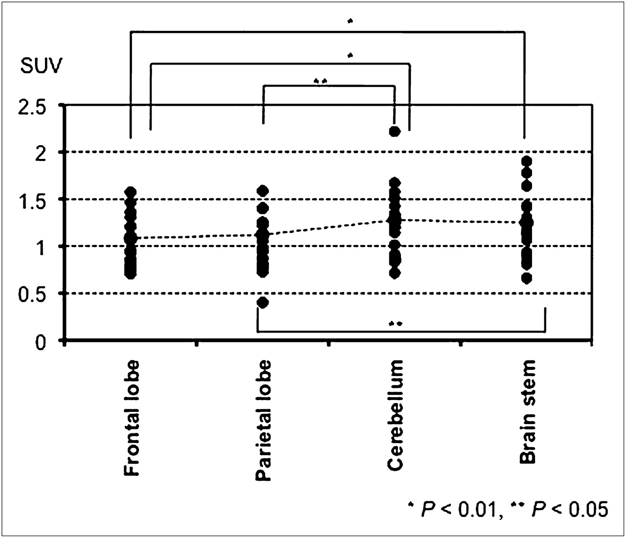
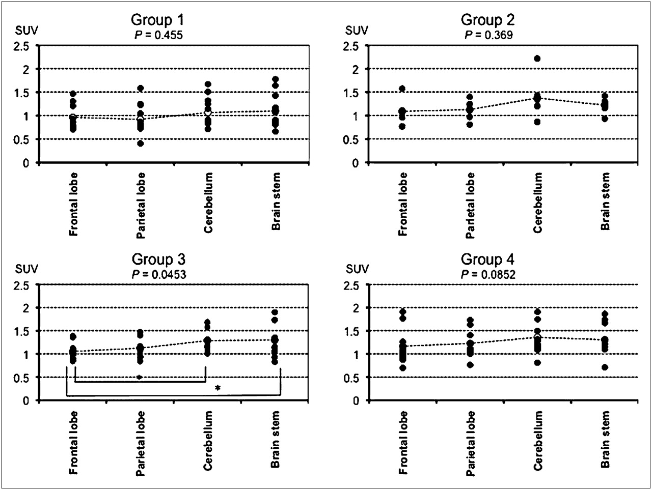
Fifty-four of the 82 patients were analyzed, and significant differences between the frontal lobe and cerebellum (P < 0.01), frontal lobe and brain stem (P < 0.01), parietal lobe and cerebellum (P < 0.05), and parietal lobe and brain stem (P < 0.05) were found ( Fig. 3). In group 3, significant differences between the frontal lobe and cerebellum (P < 0.01) and frontal lobe and brain stem (P < 0.01) were found. But in groups 1, 2, and 4, there was no significant difference between each part of the brain ( Fig. 4).

SUV in different parts of brain among patients aged 0–20 y. Significant differences between frontal lobe and cerebellum, frontal lobe and brain stem, parietal lobe and cerebellum, and parietal lobe and brain stem were observed.

SUV in different brain parts in every group. In group 1, SUV was highest in brain stem, followed by cerebellum, but significant difference was not observed. In group 2, SUV was highest in cerebellum but also was not statistically significantly different from other regions. In group 3, significant differences between frontal lobe and cerebellum and frontal lobe and brain stem were found. In group 4, although SUV was highest in cerebellum, difference was not statistically significant. *P < 0.01.
DISCUSSION
11C-MET PET has been used for the description of lesions and for less invasive evaluation of malignancy. Many papers show that PET is more sensitive and specific than MRI in detecting intracranial tumors ( 6, 9, 10), because PET can detect amino acid metabolism in various cells directly. In general, amino acid metabolism is low in normal brain tissue and high in tumor tissue. According to this difference in 11C-MET accumulation, 11C-MET PET can display clear contrast images for lesions. Therefore, 11C-MET PET would be an informative modality for detecting the boundary between an active lesion and normal brain tissue ( 6). Some reports have also described 18F-FDG PET as being helpful in assessing the degree of malignancy ( 3– 6), and 11C-MET PET is also associated with malignancy and may provide valuable information on clinical tumor aggressiveness and prognosis ( 6, 7, 13).
In general, 11C-MET PET images have been evaluated mainly by using an L/N ratio that compares 11C-MET uptake in the lesion with that in the corresponding normal region in the contralateral hemisphere ( 8– 10). Because the reference normal tissue will influence the result of the L/N ratio as a denominator, it is important that an appropriate location be chosen for the ROI used to calculate the normal reference value ( 14).
There are variations in normal 11C-MET accumulation for each part of the brain and variation between different ages ( 11). The reliability of the L/N ratio will, then, decline when uptake in the lesion is not so high. In addition, in the case of tumors near the midline or brain stem, it will be difficult to set normal ROIs in the contralateral region. Therefore, it is important to know the absolute amount of SUVs in the normal brain.
In a previous study, Uda et al. reported the SUV in the normal adult brain ( 11), and such data are an important indicator of amino acid metabolism. To date, there has been no report about normal uptake of 11C-MET in brains throughout development, including infancy, childhood, and young adulthood. If standard amino acid metabolism varies by age, the SUV may be a more suitable indicator than the L/N ratio in 11C-MET PET.
In previous reports, O'Tuama et al. showed a significant age-dependent decline of 11C-MET uptake in maturing adults ( 15), and Uda et al. also reported a slightly negative linear regression, although no statistically significant difference was observed ( 11). The reason for this age-associated decline in 11C-MET uptake was explained as a developmental decline in the activity of the neutral amino acid transporter of the blood–brain barrier ( 15). In the present study, we found that uptake of 11C-MET in all parts of the brain we studied gradually increased with age until 20 y, possibly reflecting high activity of the neutral amino acid transporter and brain protein synthesis to meet the needs of brain metabolism. These results suggest that the brains of younger persons are immature and still developing.
On the other hand, 18F-FDG PET, the local cerebral metabolic rate for glucose, is higher in infants at 3–5 mo old ( 16, 17). This tendency is different from our result in 11C-MET PET because the metabolism of glucose represents not only the activity of neurocytes but also the activity of the neural network, including structures around synapses, whereas amino acid metabolism indicates the extent of protein synthesis. 11C-MET uptake also increases linearly as the brain matures during young adulthood. After brain weight reaches a plateau, the uptake begins to decrease, reflecting developmental decline, as described in a previous report ( 15).
In each part of the brain, SUV was highest in the cerebellum in this population, similar to results reported for adult humans investigated by Uda et al. ( 11). The plasticity of synapses is important for motor learning ( 18), which is critically governed by the cerebellum. Activity of the neural network will increase amino acid metabolism in neurocytes, and this higher motor learning will continue for a lifetime. In addition, there is higher cell density in the cerebellum, especially in the granular cell layer, than is found in other brain regions. Therefore, total amino acid metabolism will increase. These are possible reasons for high accumulation in the cerebellum.
We obtained more reliable information about 11C-MET SUV in normal brains, including age-associated and regional changes. This study provides useful information for clinical determinations such as operative indications, which are affected by the malignancy of the lesion.
When continuous long-term follow-up is necessary in some children and younger patients, information about normal metabolism variation associated with aging is useful in making judgments regarding the effectiveness of treatment, and thus it is essential to make accurate evaluations according to a patient's age and brain region.
CONCLUSION
The present study evaluated the accumulation of 11C-MET and SUVs in the normal brain among children and young adults and found significant age-associated differences in some regions. To make more accurate evaluations in 11C-MET PET, age-associated criteria will be necessary in children and young adults.
- © 2011 by Society of Nuclear Medicine
REFERENCES
- Received for publication August 31, 2010.
- Accepted for publication October 21, 2010.


