


Abstract
Data collection in preclinical small-animal PET studies has been hindered by the small number of recordings typically obtained for a single radiosynthesis. Therefore, we tested procedures for obtaining 8 simultaneous small-animal PET recordings from the brains of 8 mice using an acrylic anesthesia distributor (the Octamouse), with the dopamine D2/3 ligand 18F-fallypride serving as a test substance for brain receptor imaging. Methods: The effect of scatter correction on the small-animal PET recordings was first evaluated in phantom studies in which sources of different radioactivity concentration were placed within the chambers of the Octamouse. Next, potential effects of mass on the 18F-fallypride binding potential (BPND) in the striatum were tested in groups of mice receiving 18F-fallypride at 2 different specific activities (140 and 50 GBq/μmol), with and without scatter correction. Finally, the relationship between BPND and injected dose of 18F-fallypride (3.5–17 MBq/mouse) was tested. Results: Scatter correction improved the contrast between sources and air space within the Octamouse phantom. The magnitude of 18F-fallypride BPND in mouse striatum was invariant across the tested range of specific activities, and scatter correction increased BPND by a mean of 6%; covariances of the inter- and intraoperator variability of BPND were 10%. There was a positive correlation between radiochemical dose and BPND with (R2 = 0.53) and without (R2 = 0.63) scatter correction, which was driven by increasing area under the percentage injected dose curve in the striatum. Conclusion: The quantitation of emission sources placed within the Octamouse is linear over a wide range of source activities. In the striatum of living mice, the magnitude of 18F-fallypride BPND was highly reproducible between operators and was constant over a 3-fold range of specific activities, indicating a lack of significant occupancy. Scatter correction improved quantitation but did not entirely correct for the dependence of BPND on injected dose, which was deemed to arise because of effects propagating from detector dead time when the total radiochemical dose in the field of view exceeded 50 MBq. Given this consideration, we were still able to quantify 18F-fallypride BPND in 16 mice from a single radiosynthesis, an economy that should be generalizable to brain studies of diverse radioligands.
Preclinical small-animal PET studies with short-lived radiotracers can entail an inefficient use of scanning resources when only a few acquisitions are obtained from a single radiosynthesis. This inefficiency is especially the case for radiotracers requiring prolonged acquisitions, such as the dopamine D2/3 receptor ligand 18F-fallypride, for which 120-min emission recordings enable a stable estimation of the binding potential (BPND) in mice. Thus, we have previously conducted small-animal PET studies of the pharmacologic modulation of 18F-fallypride BPND in mouse striatum (1,2), obtaining from each radiosynthesis recordings from a mean of 5 animals. In our experience, statistical variability of the endpoint is such that a minimum group size of approximately 8 animals is necessary for detecting 20% changes in the magnitude of 18F-fallypride BPND and likewise for the binding of the serotonin 5-hydroxytryptamine1A antagonist 18F-MPPF (3). Indeed, other researchers have used head-to-head small-animal PET recordings in pairs of rats (4) or as many as 4 simultaneous recordings for mice (5). For efficiency and to reduce the variability of the binding estimates, we consequently developed an anesthesia distributor equipped with 8 acrylic chambers (Octamouse), with external dimensions suitable for placement within the field of view of the Inveon DPET (Preclinical Solutions, Siemens Healthcare Molecular Imaging).
In the present study, we validated the use of the Octamouse for efficiently obtaining brain emission recordings from up to 16 mice from a single 18F-fallypride radiosynthesis. Because counting artifacts arising from acrylic components surrounding the mice may influence the quantitation of brain signals, and thus interfere with the reliable estimation of BPND, we first conducted phantom studies in which the detector response was tested for a wide range of source radioactivities placed within the chambers of the Octamouse. We then tested the effects of scatter correction on quantitation. Next, we tested the effects of scatter correction on the estimation of 18F-fallypride BPND in the striatum of living mice. In addition, we also tested for potential mass effects due to afternoon use of 18F-fallypride obtained from a morning synthesis, during which time the specific activity decreased 3-fold. The inter- and intraoperator variabilities of our BPND estimates were assessed. Finally, we tested the dependence of BPND on the 18F-fallypride radiochemical dose in the range of 3.5–17 MBq/mouse.
MATERIALS AND METHODS
Octamouse
The Octamouse is constructed from acrylic, with groups of 4 cylindric chambers on either side of a central distributor for anesthesia gas; each cylinder has an internal diameter of 2.5 cm and accommodates mice weighing up to 22 g. The entire apparatus weighs 320 g and measures 16 × 7 × 7 cm (Fig. 1A), thus fitting within the field of view of an Inveon DPET scanner.
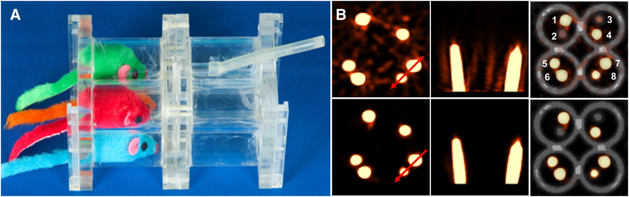
Octamouse (A) and reconstructed images (B) from phantom study without (upper row) and with (lower row) scatter-correction algorithm. Red arrows indicate position of line profiles shown in Figure 2A. From left to right are depicted coronal plane through syringes, horizontal plane, and fused PET–transmission image of source syringes within Octamouse. White numerals from 1 to 8 indicate chambers containing the following 18F concentrations: 1, 2.1 MBq/mL; 2, 0.014 MBq/mL; 3, 0.05 MBq/mL; 4, 2.1 MBq/mL; 5, 0.6 MBq/mL; 6, 2.1 MBq/mL; 7, 2.1 MBq/mL; and 8, 0.18 MBq/mL.
Phantom Studies
For an initial validation of the Octamouse, we performed a phantom study using 1-mL plastic syringes (internal diameter, 4.8 mm) filled with aqueous solutions containing 18F at concentrations of 2.1, 0.6, 0.18, 0.05, and 0.014 MBq/mL. Pairs of tubes of differing radioactivity were placed in each of the 8 chambers of the Octamouse, which was then positioned in the center of the field of view of the tomograph. To construct a transmission image of the Octamouse, we obtained a 30-min single-frame emission scan, followed by a 7-min transmission scan with a rotating 57Co point source for attenuation correction, and finally a 30-min transmission scan.
Animal Studies
All experiments with animals were performed in compliance with the Law on Animal Experimentation of Germany. Thirty-two female BALB/c mice (mean weight ± SD, 20.2 ± 1.6 g) purchased from Charles River Laboratories were housed in a temperature- and humidity-controlled room with a 12-h light–dark cycle, with free access to food and water.
Radiochemistry
18F-fallypride was synthesized using a SynChrom R&D automatic synthesis module (Raytest Isotopenmessgeräte), as previously described (2,6).
Small-Animal PET Mouse Recordings
Anesthesia was induced with 2.5% isoflurane administered in an acrylic container and was maintained throughout the small-animal PET recordings with 1.5% isoflurane, delivered at 1.5 L/min via the central distributor of the Octamouse. After cannulation in a tail vein, groups of 8 mice were positioned within the Octamouse, which was then placed within the aperture of the Siemens Inveon DPET. Dynamic emissions lasting 120 min were initiated on serial intravenous slow-bolus injections of 18F-fallypride (3.5–17 MBq) in 200 μL of saline, followed by a transmission scan using a rotating 57Co point source, as previously described (2). In practice, we performed nearly simultaneous injections of 4 mice by holding 2 syringes in each hand. After a pause of 30 s (to gain access to the other side of small-animal PET scanner), we repeated the procedure and deleted the first 30-s frame in the subsequent analysis of the second group of 4 mice.
Effect of Specific Activity
To elucidate a potential effect of 18F-fallypride specific activity on our BPND results, we made morning and afternoon small-animal PET recordings from a single radiosynthesis.
Inter- and Intraoperator Variability
To test the interobserver reliability of the BPND estimates, the data analysis procedure was repeated in a group of 14 animals (6 high and 8 low specific activity) by a second operator. Intraoperator variability was assessed through a reanalysis by the first operator at 2 wk after the initial analysis. Inter- and intraobserver variability was calculated for each individual mouse as follows:
Effect of Injected Activity
To optimize the radiochemical dose for 18F-fallypride BPND estimates, we performed multiple experiments using doses ranging from 3.5 to 17 MBq of 18F-fallypride, always in 200 μL of saline.
Image Reconstruction and Analysis
Phantom.
Emission and transmission data were rebinned to sinograms, and scatter sinograms were produced using Siemens software, based on a standard approach (7). Sinograms were reconstructed using a combined reconstruction algorithm with two 3-dimensional ordered-subset expectation maximization iterations (Siemens Medical Solutions), followed by eighteen 3-dimensional maximum a posteriori iterations with a zoom factor of 1.0 and attenuation correction with and without scatter correction. The final result was a 128 × 128 × 159 matrix, with voxel dimensions of 0.86 × 0.86 × 0.80 mm.
Cylindric volumes of interest (VOIs) were positioned within the bores of the syringes. Decay- and attenuation-corrected mean radioactivity concentrations were measured within the VOIs and calculated relative to the true radioactivity concentrations of the solutions, which were measured in a calibrated γ-counter (Packard Cobra). Finally, line profiles were measured transecting the apparatus (Fig. 1B) and passing through adjacent pairs of syringes (Fig. 2A), with and without scatter correction.

(A) Line profiles of syringe sources with and without scatter-corrected reconstructions. Inset shows magnification of one of the voids between syringes, indicating that scatter correction reduced measurements toward true zero background. (B) Corresponding linear relationship between source radioactivity concentrations measured in calibrated γ-counter and measured by small-animal PET.
Mouse Recordings
List-mode data were rebinned to sinograms, consisting of frames increasing systematically from 30 s to 10 min, for a total of 20 frames, and scatter sinograms were produced using Siemens software, based on a standard approach (7). Sinograms were reconstructed as described for the phantom study. In the specific activity study, a zoom factor of 1.3 was also used, but without scatter correction, resulting in voxel dimensions of 0.66 × 0.66 × 0.80 mm. The small-animal PET files were converted to NIfTI data format via PMOD software (PMOD Technologies Ltd.). The summation images were calculated for each dynamic 18F-fallypride recording, converted to Montreal Neurological Institute format (MNI), and manually coregistered to a digital MRI volume atlas of the mouse brain (8) using Register-1.3.6 and Minc-2.0.9 software (MNI–BIC Software). Registration was accomplished using rigid-body transformation with 9° of freedom, as described previously (2). Then, the 120-min dynamic 18F-fallypride images were resampled to the common space; the absence of substantial head movement was verified by frame-by-frame examination of the resampled dynamic images. After transformation back to NIfTI format, images were processed further using the PMOD software. Voxelwise parametric maps of the binding potential (BPND ≈ k3/k4) were calculated using linear graphical analysis (9), with a reference tissue input derived from a 0.8-mm-diameter spheric VOI placed within the cerebellum. Mean BPND maps were calculated by group for each condition and were resampled into the common stereotactic space. Mean magnitudes of the voxelwise estimates of BPND were calculated for the left and right striatum, using spheric VOIs as described previously (2). An asymmetry index ([left – right]/[left + right]) was calculated for the striatum, as described in humans (10). Because no asymmetry (1.4%, not significant) was observed in the group of 32 mice, the left and right striatal VOIs were combined. In the specific activity experiment (n = 14), we also placed a spheric VOI on the petrosal bone.
Statistical Analysis
Results were reported as the mean value and SD. Unpaired t tests were used in the comparisons of the different groups.
RESULTS
Phantom Results
Figure 1B shows emission recordings with syringes containing 0.014–2.1 MBq of 18F-fluoride superimposed on a transmission scan of the Octamouse. Line profiles passing through several syringes of different radioactivity concentrations show that, without scatter correction, the trough activities in the air spaces are overestimated (Fig. 2A).
Figure 2B shows that small-animal PET concentration measurements recorded from cylindric sources with a 4.8-mm internal diameter (i.e., twice the full width at half maximum for the reconstructed images) had a nearly perfect correlation between PET and γ-counter measurements over the entire concentration range. The efficiency of small-animal PET was 82% and nearly identical with and without scatter correction.
Mouse Results
Issues of Specific Activity, Scatter Correction, Observer Variability, and Zoom Factor.
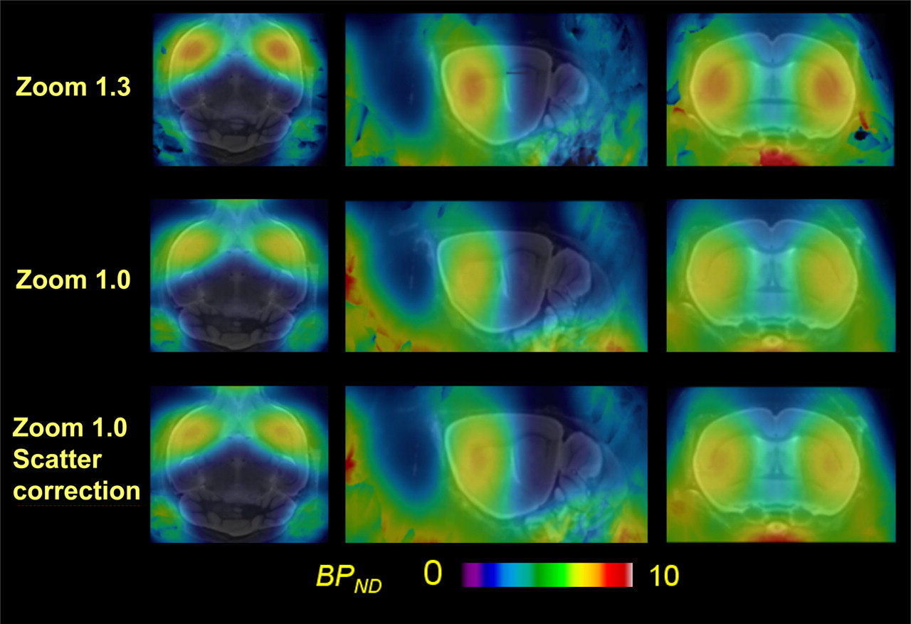
In the specific activity study, morning mice received a mean dose of 6.2 ± 0.8 MBq of specific activity (140 GBq/μmol), whereas afternoon mice received a mean dose of 5.8 ± 0.7 MBq (not significant) of specific activity (50 GBq/μmol). The mean striatal BPND was invariant with specific activity (Table 1). Compared with a zoom factor of 1.3, a lower zoom factor of 1.0, as required if the transmission recording is to encompass the entire Octamouse, resulted in a 15% underestimation of striatal BPND. For the subgroup of 14 mice receiving 5.8–6.5 MBq of 18F-fallypride, scatter correction led to a highly significant 6% increase of BPND. For this same subgroup, interobserver variance was 11.2%, and intraobserver variance was 8.6%. Figure 3 shows the corresponding mean parametric images for zoom factors 1.3 and 1.0 without scatter correction and for zoom factor 1.0 with scatter correction in 3 planes projected on the mouse brain MRI atlas.

Mean parametric maps of 18F-fallypride BPND projected on MRI mouse atlas (gray scale) in horizontal, left sagittal, and coronal planes of mice with different zoom factors and with or without scatter correction. The mean injected dose (±SD) was 5.9 ± 0.5 MBq (n = 14).
Inter- and Intraoperator Variability and Impact of Zoom Factor and Scatter Correction
Effect of Injected Radioactivity on BPND
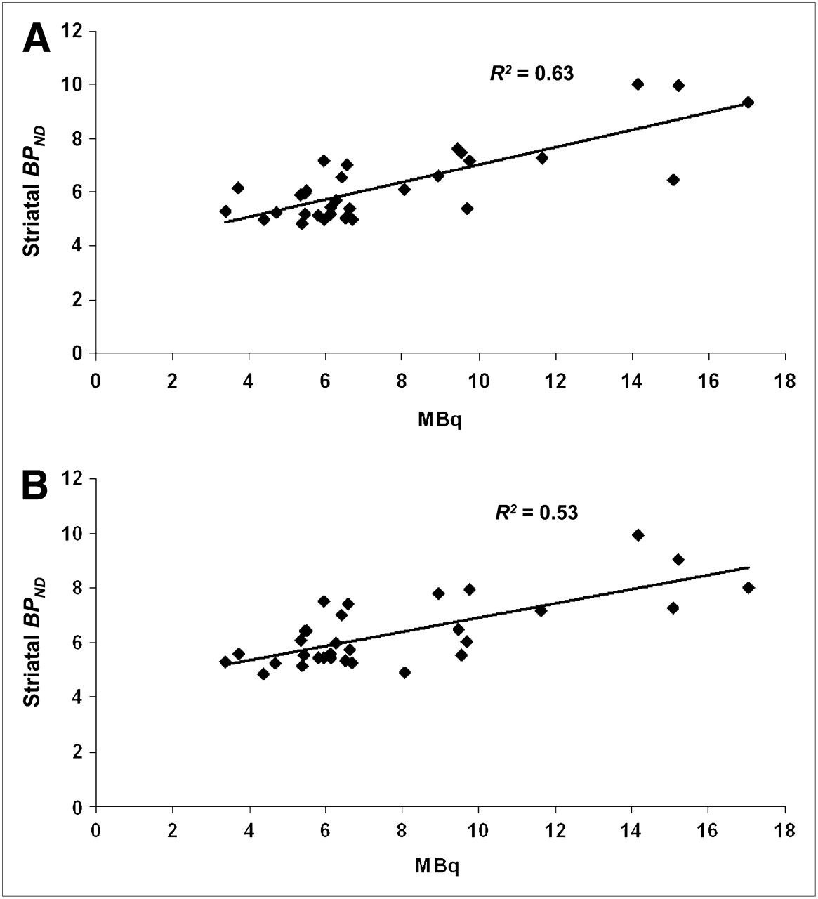
The magnitude of BPND correlated with the individual amount of injected radioactivity for all 32 mice analyzed with a zoom factor of 1.0; application of the scatter correction decreased the linear regression slope (Figs. 4A and 4B). Figure 5A shows the mean time–activity curves in the striatum, cerebellum, and petrosal bone from the 8 mice with the lowest injected radioactivity (range, 3.4–5.5 MBq; mean, 4.7 MBq). Figure 5B shows the corresponding curves from the 8 mice with the highest dose of injected radioactivity (range, 9.5−17.1 MBq; mean, 12.7 MBq); also depicted are the corresponding mean parametric maps in a representative axial slice passing through the striatum. The area under the curve (AUC) of the striatal time–activity curves were significantly lower in the group with low radiochemical dose (712 ± 107) than in the group with high dose (847 ± 131; P < 0.04), whereas the corresponding cerebellum AUCs did not differ (153 ± 40.3 vs. 144 ± 29.6; not significant), nor did the petrosal bone recording AUCs (241 ± 23.2 vs. 261 ± 58.3; not significant).

Correlation of 18F-fallypride BPND with amount of injected activity (MBq) without (A) and with (B) scatter correction (n = 32) in mouse striatum.

Mean (±SEM) time–activity curves in striatum, cerebellum, and petrosal bone of groups of 8 mice with low 18F-fallypride dose (4.7 ± 0.8 MBq) (A) and high 18F-fallypride dose (12.7 ± 3.0 MBq) (B). Inserts show corresponding mean parametric maps of 18F-fallypride BPND in axial plane passing through striatum. %ID = percentage injected dose.
DISCUSSION
The goal of this study was to establish and validate the use of the Octamouse, an acrylic insert for simultaneous small-animal PET scanning of 8 isoflurane-anesthetized mice, to optimize the yield of data acquisition from a single radiosynthesis. We first conducted phantom studies to assess scatter effects arising from the Octamouse on the quantitation of sources placed within the chambers and systematically investigated the contributions of several postprocessing steps on the quantitation of 18F-fallypride binding to dopamine D2/3 receptors in the striatum of living mice. There have hitherto been few reports describing a multichamber approach for small-animal PET recordings. One study has described an insert for simultaneous nose-to-nose scanning of 2 rats (4). Inserts have also been used for obtaining precise repositioning of animals in serial PET studies or multimodal imaging studies of rats (11) and also mice (12). Simultaneous small-animal PET recordings have been obtained in as many as 4 mice (5). However, this is the first report, to our knowledge, on the use of a multichamber holder for the scanning of 8 mice, and at least 16 mice from a single radiosynthesis. Although the use of an acrylic insert presents a clear advantage in economy, especially when large groups and long dynamic acquisitions are required, several technical issues relevant to quantitation are raised.
The phantom studies showed linear response of the small-animal PET scanner for a wide range of source radioactivities placed within the Octamouse chambers. Counting efficiency was close to 82%, as expected from the geometry of cylindric sources with diameters twice the full width at half maximum of the small-animal PET scanner. However, the phantom images revealed the presence of considerable radioactivity throughout the air spaces of the chambers, which we attributed to scatter from the relatively massive Octamouse. Indeed, application of a standard scatter-correction algorithm substantially improved the image quality (Fig. 1B). The line profiles placed across the sources indicated that scatter correction decreased the trough activities toward the true background activity, which propagated to a 6% increase in 18F-fallypride BPND. This effect was highly significant, because it occurred in almost every animal. Thus, scatter is an avoidable source of bias in the quantitation of signals arising from within the Octamouse.
We have previously characterized the binding of 18F-fallypride in the striatum of mice scanned in groups of 2–4 animals without an acrylic holder (1,2). In those studies, we used a zoom factor of 1.3 to restrict the reconstructed volume, which led to a decreased voxel size and, consequently, fewer partial-volume effects, in turn propagating a somewhat higher BPND. However, because the 1.3 zoom factor does not encompass the entire Octamouse, the attenuation and scatter correction would not properly account for mass outside the field of view. Lowering the zoom factor to 1.0 resulted in a more accurate attenuation correction throughout the field of view of the small-animal PET scanner but reduced the apparent magnitude of 18F-fallypride BPND by 14% (Table 1; Fig. 3).
Mass effects can potentially influence the outcome measure in small-animal PET studies with 18F-fallypride and other tracers. We have earlier shown by calculation that the occupancy of dopamine D2/3 receptors is less than 5% in mice receiving 10 MBq of 18F-fallypride (2). In the present study, we made a more systematic test of possible mass effects by comparing BPND estimates in groups of mice, with 18F-fallypride specific activity declining by a factor of 3; we found no associated decline in specific binding, indicating a lack of mass effects within the range of specific activity (140–50 GBq/μmol) encountered in our laboratory. Much more problematic with respect to small-animal PET quantitation is the difficulty in obtaining adequate spatial normalization for objects as small as the mouse brain. This should be remedied in the future, with the advent of hybrid small-animal PET/CT and small-animal PET/MRI but remains for the present a manual task requiring considerable skill and practice. Because BPND estimates are highly vulnerable to the misplacement of the VOIs, especially for the reference region, the manual registration is a potential source of experimental error or bias. However, our inter- and intraoperator covariances in the estimation of 18F-fallypride BPND (10%) were of the same scale as the variance with a subpopulation of 14 mice, indicating that skillful operators can obtain adequately reproducible quantitation.
In our earlier 18F-fallypride small-animal PET studies, we found a mean striatal BPND of 10 in saline-treated mice; these animals, scanned in groups of 2–4 with a zoom factor of 1.3, had all received a radiochemical dose of 10 MBq of 18F-fallypride. In the course of the present specific activity study with 14 mice, we administered only 6 MBq of 18F-fallypride and found a mean BPND of only 6. For the entire present population of 32 mice, there was a highly significant positive correlation between the injected dose and BPND. Because the magnitude of BPND is, in general, a steady-state ratio between the high binding and reference region activities, there is no a priori reason to expect dose effects, so long as receptor occupancy is minimal. Because mass effects are excluded, we had to find an alternative explanation for this unexpected phenomenon. Initial considerations focused on the potentially biasing effects of spill-in from the cranium, which can be quite significant at late 18F-fallypride circulation times (2). Defluorination of certain gaseous anesthetics in rodent liver microsomes proceeds as a zero-order process (13,14), which might have predicted mass effects on the rate of accumulation in bone of 18F-fluoride derived from the hepatic metabolism of 18F-fallypride. However, the time–activity curves from the petrosal bone did not reveal any differential labeling as a function of specific activity.
The relatively heavy acrylic holder is a source of scatter, which biased BPND estimates, as noted in the “Discussion” section. The application of a scatter-correction algorithm attenuated by 20% the slope of the relationship between individual injected dose and apparent BPND. The remaining 80% of the correlation must arise from other factors, because the scatter-corrected striatal AUC in the high-dose mice exceeded that in the low-dose mice, whereas the mean cerebellum AUCs did not significantly differ, and the petrosal uptake had only a trend toward increased signal (Fig. 5). Using a rat-sized phantom in the Inveon DPET scanner, Kemp et al. found an increasing mean relative counting rate error with increasing radioactivity (15), especially when the total radioactivity in the field of view exceeded 74 MBq for a rat-sized phantom. Given that the Octamouse is larger than the rat phantom, we can predict that the cutoff value should be lower, perhaps 50 MBq.
When total radioactivity in the field of view exceeds a certain limit, dead-time correction (which is applied globally) alters the contrast between regions of high and low radioactivity, thus plausibly accounting for the increase in apparent BPND. Indeed, our experiments with 10 MBq per mouse resulted in total activity as high as 80 MBq within the Octamouse. To avoid faulty dead-time correction, future Octamouse studies will be limited to a total of 40 MBq (8 × 5 MBq), this change having the additional advantage of reducing the maximum required injection volume to 160 μL per mouse.
We have considered some of the many possible sources of error and bias in BPND estimation by small-animal PET. In addition, it must be considered that the linear graphical Logan analysis used in the present study (9) is inherently vulnerable to noise effects, which led, in our hands, to underestimated voxelwise maps of 11C-raclopride BPND in the pig brain (16) and 18F-MPPF BPND in the rat brain (3). Quantitation of 18F-fallypride BPND in mice is vulnerable to effects with opposing biases, perhaps none so important as the effects of partial volume; the size of the mouse striatum (50 mm3) is such that small-animal PET estimates of 18F-fallypride BPND are less than half of the true value obtained by dissection of the mouse brain ex vivo (2). In the 2 earlier mouse small-animal PET studies with 18F-fallypride, the BPND estimates were 13 (17) and 3.2 (18); this difference likely reflects the high resolution of the quad-HIDAC PET (17) used by Honer et al. (<1 mm), and the relatively lower resolution of the Advanced Technology Laboratory Animal Scanner (National Institutes of Health) and Siemens Focus 120 scanner used by Skinbjerg et al. (1.6 mm) (18). Rat small-animal PET studies found 18F-fallypride BPND of 7 (19) and 8 (20), or a striatum-to-cerebellum ratio of 8 (21), whereas Tantawy et al., using a full-compartmental analysis of 2-h dynamic recordings, found a BPND in rat striatum of 13 (22). Thus, the increment in striatal volume between rats and mice does not seem to consistently result in improved quantitation of BPND, although a direct comparison of mouse and rat has not yet been made.
Whereas monkey PET studies found striatum-to-cerebellum ratios ranging from 3 to 9, depending on the time frame (6,21,23), prolonged recordings 18F-fallypride gave BPND estimates of 27 (24) and 23 (25). Thus, results in the monkey compare well with those in the human brain (26–28), as expected for the primate striatum, which is 10- to 20-fold larger than in rodents. Disregarding the possible attainment of submillimeter small-animal PET resolution, more accurate BPND estimates in mouse striatum might be obtainable through the use of a partial-volume correction (29).
CONCLUSION
The Octamouse enables the efficient use of 18F-fallypride from a single radiosynthesis, making the Octamouse a favorable device for conducting dose–response studies of pharmacologic challenges in relatively large groups of animals. The results of our study should be generalizable for small-animal PET studies with diverse neuroreceptor ligands. The next version of the Octamouse accommodates mice weighing 35 g, as will be required for longitudinal and transgenic studies.
Acknowledgments
We thank Georg Stark for technical assistance with the construction of the Octamouse and Cornelia Arszol for technical assistance. This project was supported by a grant of the Medical Faculty of the University of Munich (FöFoLe).
- © 2010 by Society of Nuclear Medicine
REFERENCES
- Received for publication April 26, 2010.
- Accepted for publication July 14, 2010.


Jump to section
Related Articles
Cited By...
- Time Courses of Cortical Glucose Metabolism and Microglial Activity Across the Life Span of Wild-Type Mice: A PET Study
- Longitudinal Assessment of Cerebral {beta}-Amyloid Deposition in Mice Overexpressing Swedish Mutant {beta}-Amyloid Precursor Protein Using 18F-Florbetaben PET
- Noninvasive Nuclear Imaging Enables the In Vivo Quantification of Striatal Dopamine Receptor Expression and Raclopride Affinity in Mice