


Abstract
Renal function is known to be a strong predictor of cardiovascular prognosis, and cardiorenal disease is increasingly investigated in medical research. In this study, we tested the hypothesis that a single combined cardiorenal scintigraphy examination with the perfusion tracer 99mTc-tetrofosmin is feasible and may allow the simultaneous investigation of cardiac and renal pathology in cardiovascular patients. Methods: Thirty patients scheduled for dual-day gated SPECT also gave their informed consent for a renal acquisition after a single injection of 370 MBq of 99mTc-tetrofosmin, at rest (30 patients) or with adenosine (21 patients), and to undergo an additional standard renal study with the glomerular tracer 99mTc-diethylenetriaminepentaacetic acid (DTPA) (74 MBq) after 1 wk (24 patients). Kidney images and renograms were obtained. Renal uptake index, expressed as the percentage ratio of kidney counts cumulated over the second minute after injection to the administered dose, was calculated as a functional renal parameter. Time to peak activity and separate kidney percentage uptake (as the percentage contribution of each kidney to total renal uptake) were also calculated. Results: Compared with 99mTc-DTPA, 99mTc-tetrofosmin provided better-quality kidney images, with a higher uptake index (13.17% ± 4.76% vs. 8.33% ± 2.45%, P < 0.001) and with comparable separate kidney percentage uptake and times to peak activity. 99mTc-tetrofosmin uptake index was significantly lower in the patients who were more compromised according to renal and cardiovascular functional parameters, and correlated with 99mTc-DTPA uptake index (r = 0.77, P < 0.001), serum creatinine (r = 0.59, P < 0.005), log brain natriuretic peptide N-terminal levels (r = −0.65, P < 0.005), myocardial and carotid intima-media wall thickness (for both, r = −0.61, P < 0.005), and the Doppler index of renal vascular resistance (r = −0.60, P < 0.005). In the 21 patients who underwent the provocative test, adenosine induced a significant decrease in renal 99mTc-tetrofosmin uptake index (from 14.12% ± 4.50% to 11.81% ± 3.33%, P < 0.005) suggesting a decrement in renal perfusion or function. Conclusion: 99mTc-tetrofosmin cardiorenal scintigraphy is feasible at low cost as a single-shot study and may allow both the evaluation of renal morphology and renograms during a cardiac study and the calculation of renal functional parameters.
The pathophysiologic relationships between heart and kidney have been known for a long time and have been ascribed to the integrated control of blood volume and arterial blood pressure by the 2 organs. Recently, the term cardiorenal syndrome has been proposed for the pathophysiologic disorder of the heart and kidneys whereby acute or chronic dysfunction in one organ may induce acute or chronic dysfunction in the other (1).
Mild renal dysfunction is known to be an independent risk factor with a strong prognostic significance in heart failure, arterial hypertension, and diabetes mellitus (2–6). On the other hand, cardiovascular disease has a high prevalence in nephropathic patients, and subclinical abnormalities of coronary reserve may be found in subjects with asymptomatic renal disease, suggesting a combined microvascular involvement of the kidney and the heart (7,8). Gated SPECT is widely used to investigate myocardial ischemia or viability and has been demonstrated to be able to detect even subclinical microcirculatory impairment in cardiopathic and nephropathic patients (9,10).
A combined cardiorenal investigation during exercise-stress gated SPECT was proposed by Hurwitz et al. in 1994 using 99mTc-sestamibi but to our knowledge has not gained clinical application (11). Compared with 99mTc-sestamibi, 99mTc-tetrofosmin is cleared more quickly from both lungs and liver, allowing earlier imaging (12–15) as is widely used in gated SPECT.
In premarketing studies, 99mTc-tetrofosmin showed a significant renal uptake and urinary clearance rate in both experimental animals and healthy human volunteers (12). These observations prompted us to test the hypothesis that a single combined cardiorenal study is possible with 99mTc-tetrofosmin and could allow simultaneous evaluation of the heart and the kidney in cardiovascular patients. To test its feasibility, we developed a cardiorenal protocol and after obtaining informed consent applied it to cardiovascular patients who were routinely scheduled for myocardial gated SPECT.
MATERIALS AND METHODS
We studied 30 consecutive patients (14 men, 16 women; mean age ± SD, 62 ± 14 y) scheduled to undergo myocardial gated SPECT to investigate myocardial ischemia or viability; coronary angiograms were available for 18 patients, and 11 of them showed coronary artery disease. Twenty-four patients were hypertensive (7 with verified renovascular disease), and 7 were diabetic; 6 patients had dilated cardiomyopathy. Vital statistics and main clinical parameters are reported in Table 1.
Main Clinical Parameters in Patients Studied
A standard dual-day scintigraphic protocol was applied for the 21 patients who underwent the stress test; adenosine was administered on the first day, without significant symptoms except for transient dyspnea in 1 patient.
The patients also gave their informed consent for a renal acquisition during the rest study (30 patients) and during adenosine infusion (21 patients) and to undergo an additional standard renal study with the glomerular tracer 99mTc-diethylenetriaminepentaacetic acid (DTPA) (74 MBq) after 1 wk (24 patients). Figure 1 outlines the main steps of the cardiorenal scintigraphic protocol.

Flow chart of scintigraphic cardiorenal acquisition protocol. G-SPECT = gated SPECT.
Although not strictly required by the study design, laboratory data and sitting blood pressure values (Vital Signs Monitor; Welch Allyn) (30 patients) were retrieved with patient consent from clinical records, along with cardiac, carotid, and renal Doppler ultrasound data, which were available in 29, 20, and 19 patients, respectively, as shown in Table 1.
Scintigraphic Acquisition Procedures
A double-head γ-camera (Millenium; GE Healthcare) equipped with a high-resolution collimator was used. After the counting linearity of the γ-camera had been checked at the doses used, the acquisition of preinjection and postinjection syringe counts were standardized by counting at a 20-cm distance from the γ-camera for 5 s and then multiplying to yield 60-s counts. After hydration (0.7 mL/kg) in the hour preceding the study, dynamic renal scintigraphy was performed after bolus injection of 370–444 MBq of 99mTc-tetrofosmin, with the patient supine.
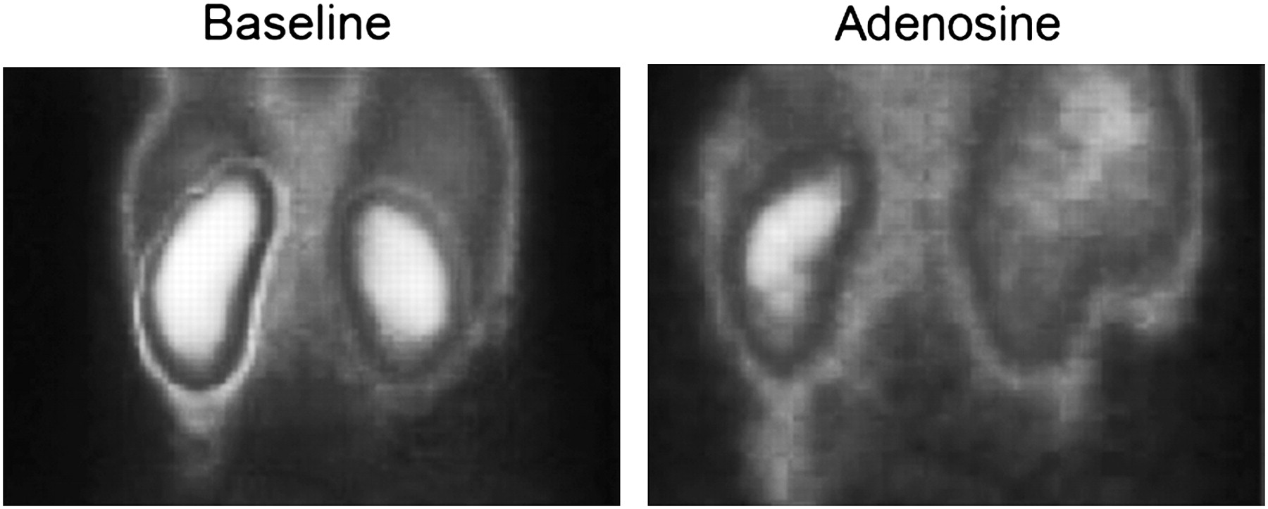
In the 21 patients who underwent the adenosine stress test (140 μg/kg/min for 6 min), 99mTc-tetrofosmin renal scintigraphy was started at the third minute of infusion. For both 99mTc-tetrofosmin and 99mTc-DTPA renal studies, a standard dynamic renal acquisition protocol was used, consisting of a first-pass phase followed by a renographic phase for a total of 30 min with a 64 × 64 matrix and 1.33 zoom (20% window centered on 140 keV). Gated SPECT was performed immediately after the renal acquisition, using 36 projections (25-s length, 16 frames per cycle), a 64 × 64 matrix, and a 15% window centered on 140 keV.
Scintigraphic Data Processing
Renal Analysis
The processing software Renal Analysis (GE Healthcare) was used to obtain renographic curves and renal images. Time to peak activity and percentage separate kidney uptake were made automatically available by the processing protocol. Kidney counts (60 s) were calculated after background subtraction over the second minute after injection and corrected for kidney depth (cm) as in the Gates formula for calculating 99mTc-DTPA uptake (16). The uptake index was used as a functional renal parameter, according to the following formula, in agreement with current practice, by integral count methods to calculate glomerular filtration rate or tubular extraction rate (16,17): percentage uptake index = background and depth-corrected kidney counts (60 s) × 100/dose counts (60 s).
Statistical Analysis
Variables were expressed as mean ± SD and range. Variables were compared by paired or unpaired 2-sided t test. Correlations were studied by linear regression analysis, reporting relationship parameters, as well as the Pearson correlation coefficient, r.
A value of P lower than 0.05 was considered statistically significant. All statistical analyses were performed using SPSS 11.0 software (SPSS Inc.).
RESULTS
99mTc-Tetrofosmin Gated SPECT
Myocardial perfusion abnormalities were found in 7 of 30 patients at baseline; 10 of 21 patients were positive for adenosine blood flow maldistribution. All these patients underwent coronary angiography, and coronary stenoses greater than 75% were found in 3.
Renal 99mTc-Tetrofosmin Scintigraphy
99mTc-Tetrofosmin and 99mTc-DTPA
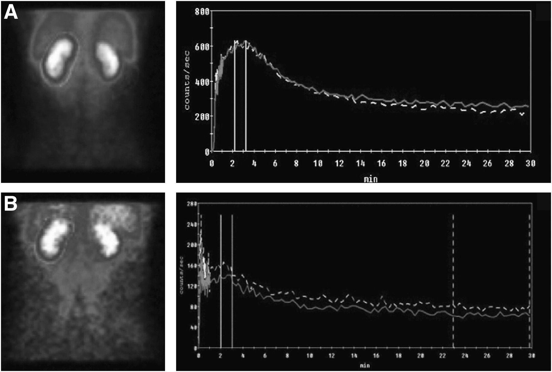
Compared with 99mTc-DTPA, 99mTc-tetrofosmin permitted better definition of kidney images and renographic curves because of a higher kidney-to-background activity ratio (Fig. 2); bilaterally or unilaterally decreased renal uptake was appreciable, as illustrated in Figure 3. In 10 patients, the amplitude of the late excretory phase of the right-kidney renogram obtained with 99mTc-tetrofosmin was increased by a superimposed biliary activity.

Examples of kidney images (over uptake interval) and renographic curves obtained with 99mTc-tetrofosmin (A), in comparison with 99mTc-DTPA (B), in patient with normal renal function.

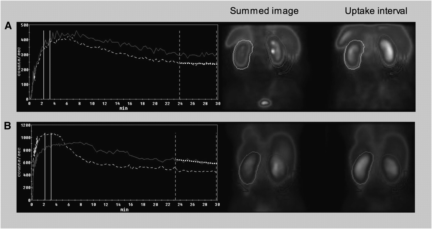
Renographic curves and kidney images obtained with 99mTc-tetrofosmin in patient with moderate reduction in renal function (creatinine clearance, 56 mL/min) (A) and patient with right renal artery stenosis (B). Continuous line = right kidney; dotted line = left kidney.
At quantitative analysis, 99mTc-tetrofosmin had a significantly higher renal uptake index than did 99mTc-DTPA, whereas no significant differences between the 2 tracers were observed for right- and left-kidney separate percentage uptake and for times to peak activities (Table 2).
Main Scintigraphic Parameters Compared Between Tetrofosmin and 99mTc-DTPA by Paired Student t Test
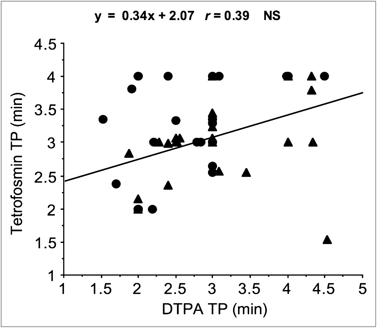
Close correlations were found between 99mTc-tetrofosmin and 99mTc-DTPA for uptake index (r = 0.77, P < 0.001) and separate kidney percentage uptake (r = 0.96, P < 0.001), with no appreciable differences in right-kidney versus left-kidney behavior (Fig. 4A). 99mTc-tetrofosmin and 99mTc-DTPA time to peak activities were weakly correlated, with no clear differences in right versus left kidney (Fig. 5).
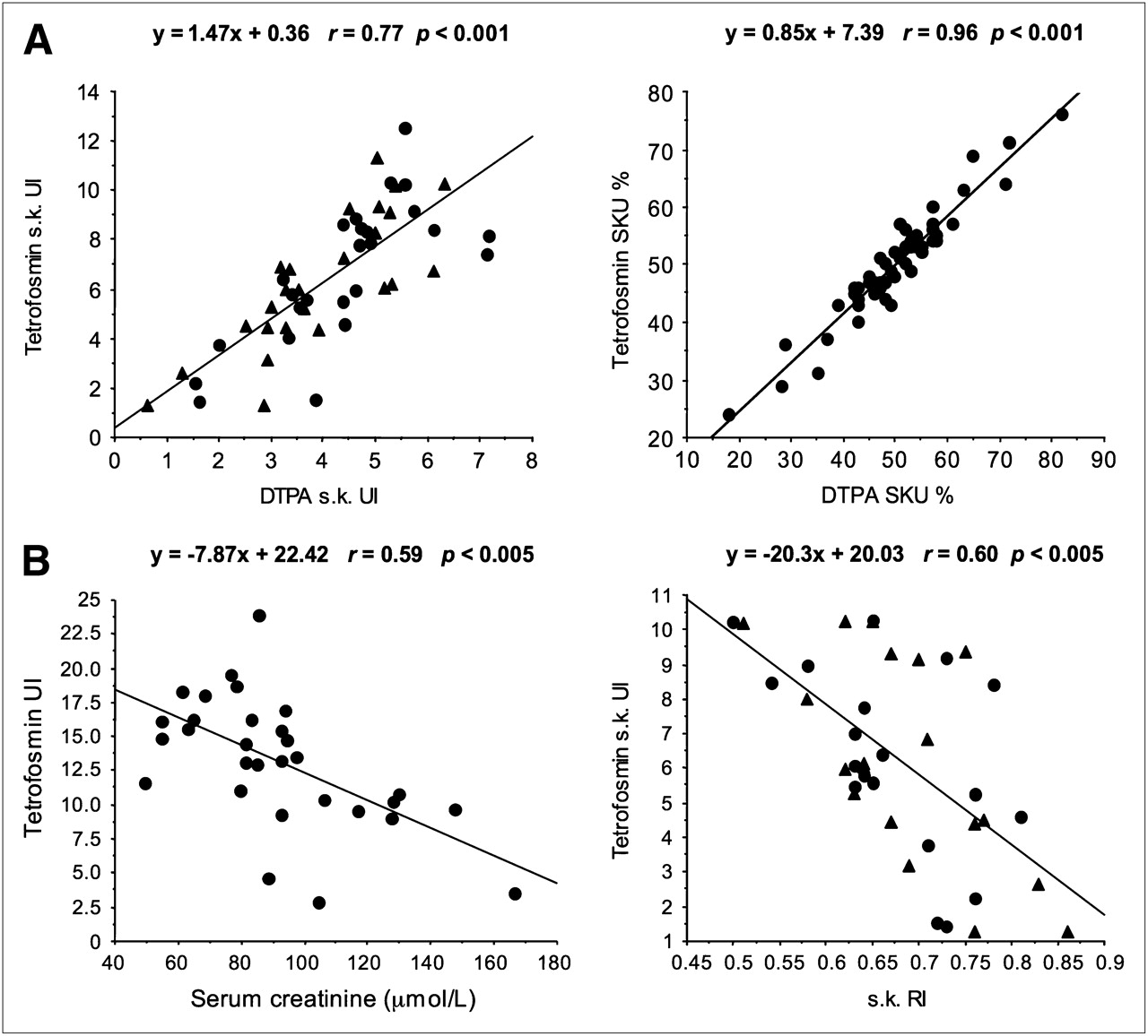
Representative correlations of 99mTc-tetrofosmin uptake with 99mTc-DTPA uptake (A) and renal functional parameters (B). • = right kidney; ▴ = left kidney; RI = Doppler ultrasound renal resistance index; s.k. = single kidney; SKU % = separate kidney uptake (percentage total-kidney uptake). Uptake index (UI) is in percentage injected dose.

Correlation between time to peak (TP) for 99mTc-DTPA and 99mTc-tetrofosmin in right and left kidneys. • = right kidney; ▴ = left kidney.
99mTc-Tetrofosmin and Renovascular Disease
In the 7 patients with angiographically proven significant renal artery stenosis, the baseline uptake index was lower in the stenotic kidney (3.67 ± 1.9 vs. 5.70 ± 2.46, P < 0.05) (Fig. 3). Because of scanty renal angiographic data in the control population (n = 3), the 99mTc-tetrofosmin uptake index was compared between renovascular and nonrenovascular patients on the basis of their renal Doppler ultrasound data. A significantly lower uptake index was observed in the affected kidneys of renovascular patients than in individuals without renal artery stenosis, considered as the average uptake of the 2 kidneys (3.29 ± 1.76 vs. 7.16 ± 2.36, P < 0.01).
Renal Scintigraphy and Gated SPECT
Comparing the group of patients with normal myocardial perfusion patterns (10 patients) and those with baseline or stress perfusion abnormalities (15 patients), we found that the resting renal uptake index was significantly lower (12.52 ± 3.98 vs. 16.09 ± 4.02; P < 0.05) in the patients with abnormal perfusion.
Comparison with Other Clinical Data
As shown in Table 3, subdividing the patients into 2 groups—those above and those below the median values of different renal and cardiovascular parameters—showed the 99mTc-tetrofosmin uptake index to be lower in those who were more compromised.
Tetrofosmin Uptake Index in Different Parameters Subdivided According to Median Values
The 99mTc-tetrofosmin uptake index was strongly related to serum creatinine and the Doppler index of renal vascular resistances (Fig. 4B). In addition, significant correlations were found with age (r = −0.76, P < 0.005), log10 brain natriuretic peptide N-terminal levels (r = −0.65, P < 0.005), and myocardial and carotid intima-media wall thickness (both, r = −0.61, P < 0.005).
Effect of Adenosine
Adenosine caused a significant decrease in renal 99mTc-tetrofosmin uptake index (from 14.12% ± 4.50% to 11.81% ± 3.33%, P < 0.005). A more pronounced decrement was observed in the 10 subjects with a baseline uptake index greater than 13.37% (median value) than in those who were below the median (n = 11) (−3.72 ± 2.66 vs. 0.75 ± 3, P < 0.005). Accordingly, a negative correlation was found between resting 99mTc-tetrofosmin uptake index and its changes after adenosine (r = −0.56, P < 0.005).
Patients with a positive adenosine test for myocardial perfusion abnormalities (n = 10) had a lower renal 99mTc-tetrofosmin uptake index at baseline than did those with a negative test (n = 11) (11.6 ± 4.5 vs. 15.23 ± 4.15, P < 0.05) and a lesser decrease in renal uptake index after adenosine (−0.92 ± 3.49 vs. −3.57 ± 2.32, P < 0.05). Of the 7 patients with renal artery stenosis, the adenosine test was performed in only 4, and no statistical analysis was performed. However, in 3 patients adenosine induced a 99mTc-tetrofosmin uptake index decrement that was clearly more pronounced on the affected side, with an increase in kidney-to-kidney asymmetry as shown in Figure 6.
Adenosine effect in patient with bilateral renal artery stenosis more severe on right side, detected by 99mTc-tetrofosmin (uptake interval).
DISCUSSION
It has been known for a long time that heart and kidney are related in cardiovascular pathophysiology. Recent studies have focused on this relationship as a link to cardiovascular risk and prognosis (1–8), and the term cardiorenal syndrome is increasingly used to define a condition of reciprocal dysfunction of the heart and kidney, with different clinical presentations depending on which organ is primarily involved (1).
The assessment of kidney function in clinical practice usually relies on estimation of the glomerular filtration rate by standard laboratory parameters such as serum creatinine or creatinine clearance; however, renal perfusion abnormalities may precede glomerular filtration changes because of the regulation of filtration pressure and may be more relevant in the early assessment of cardiorenal pathology and prognosis (20). Furthermore, clinicians often need the estimation of separate kidney function, which is noninvasively obtainable only by scintigraphic examination.
Gated SPECT with 99mTc-tetrofosmin is widely used for the study of cardiac perfusion and function and can also detect silent ischemia (9,10). The present study indicated that this tracer may also allow functional imaging of the kidneys, suggesting the application of the combined technique to the evaluation of cardiorenal disease.
We found a proportionality between 99mTc-DTPA and 99mTc-tetrofosmin renal uptake index, but the kidneys appeared to extract a greater proportion of 99mTc-tetrofosmin than of 99mTc-DTPA, with better images and renographic curves. Hepatobiliary extraction of the tracer may have disturbed the appearance of the late right-kidney image and renographic curve and explain the greater (though not significantly so) delay in average time to peak in the right kidney, as reported in Table 2, but did not appear to affect renal counting rate in the interval of functional evaluation. Taking into account the observation by Higley (12) of an approximately equal uptake of 99mTc-tetrofosmin by the kidney and liver, and the 99mTc-tetrofosmin and 99mTc-DTPA uptake index that we found, we could hypothesize for tetrofosmin a renal clearance far greater than glomerular filtration rate, suggesting a prevailing tubular extraction as for blood flow renal tracers. It may be interesting to observe that mean 99mTc-tetrofosmin renal uptake index (13%) was comparable to observations reported by Higley et al., who found a 10% fractional urinary elimination of the radioactive dose at 2 min after injection in rats and at 5 min in healthy human volunteers (12). The biodistribution study by Higley et al. (12) showed an approximately equal contribution of the kidney and liver to the elimination of the injected dose. Taking these data and our uptake index results into account, 99mTc-tetrofosmin uptake index was also proportional to renal function estimated by serum creatinine in the explored range and could also be associated with several other indicators of severity of cardiovascular disease. These findings suggest that cardiorenal scintigraphy may be useful in a wide range of clinical conditions requiring a combined evaluation of heart and kidney, such as arterial hypertension, diabetes, chronic heart or renal failure, and heart or renal transplantations (1–8,21). In addition, it may contribute valuable information to the diagnosis and functional evaluation of renovascular forms that are frequently associated with cardiovascular disease (22). Patients referred for coronarography represent an important group in which renal evaluation may distinguish healthy individuals from those at risk for the development of acute radiocontrast-induced nephropathy (23). A noninvasive study of both myocardial perfusion and renal function is also required in the preoperative assessment of patients with thoracoabdominal aortic aneurysm and peripheral vasculopathy (24,25).
The decrease in renal 99mtc-tetrofosmin uptake index after adenosine appears to agree with the statement that sustained vasoconstriction is the first response to purinergic stimulus in the kidney, where adenosine is a main effector of the tubuloglomerular feedback mechanism regulating preglomerular resistance (26,27). This effect seemed to be less pronounced in the more seriously affected kidneys, possibly suggesting a decreased vasoconstrictive purinergic renal reserve that can be scintigraphically detected. A substantial decrease in renal 99mTc-tetrofosmin uptake was also observed in healthy volunteers during exercise stress (12), and it is interesting that adenosine has been suggested as a mediator of the so-called muscle metabolic reflex induced by exercise on the heart and kidneys via sympathetic stimulation, eliciting both an increase in cardiac output and a decrease in renal perfusion and function (28,29). The fact that renal response to adenosine was reduced in the presence of myocardial perfusion abnormalities could be tentatively interpreted as being due to impaired renal purinergic reserve concomitant with the alterations in coronary reserve.
A more pronounced response to adenosine in the affected kidneys of some renovascular patients could theoretically be explained by the offset of an active vasodilatory reserve downstream from the arterial stenosis.
Finally, the effect of adenosine on renal 99mTc-tetrofosmin uptake could be due to a nonspecific decrease in renal perfusion pressure in cases in which a significant peripheral vasodilation and blood pressure drop may follow drug administration.
The study had some limitations. 99mTc-tetrofosmin has comparable renographic characteristics to 99mTc-DTPA but a greater uptake rate, suggesting a greater clearance rate. However, urinary renal clearance was not measured in our study, to avoid adding a cumbersome procedure to routine diagnostic testing for outpatients. Moreover, hepatobiliary excretion of 99mTc-tetrofosmin might theoretically interfere with renal functional imaging, especially of the right kidney and in the presence of overt renal insufficiency.
Although the low protein binding combined with the highly lipophilic and diffusable structure of 99mTc-tetrofosmin suggests that perfusion rate is a major determinant of renal uptake, to our knowledge the renal clearance pathways of 99mTc-tetrofosmin have not yet been investigated, and dedicated studies are needed to clarify these aspects. The results of our study imply an association between 99mTc-tetrofosmin renal uptake and several indices of cardiorenal severity. However, a statistical analysis by multiple comparisons was not attempted because of the limited number of patients, and consequently the weight of each functional parameter could not be assessed. Finally, a definite interpretation of the renal effects of adenosine is not possible because of the limited number of observations and the heterogeneity of the patient series. Therefore, obtaining a postadenosine renal scan remains questionable at the moment, unless dedicated studies address its clinical significance.
CONCLUSION
The kidneys can be evaluated by a simple scintigraphic protocol during 99mTc-tetrofosmin gated SPECT in a single-shot dual study without prolonging the procedure or adding further radioactive burden. One can shorten the dual study by performing a 15-min renal acquisition during a gated SPECT fast imaging protocol (15). Cardiorenal scintigraphy with 99mTc-tetrofosmin may thus be proposed especially in those patients undergoing gated SPECT in whom a significant risk of overt or subclinical renal disease exists. Adding renal functional imaging to the cardiac study could permit the detection of kidney abnormalities consistent with vascular, parenchymal, or obstructive kidney disease.
Moreover, our results indicate that renal 99mTc-tetrofosmin uptake is a functional parameter linked to many indices of cardiorenal severity. Such uptake may be of prognostic significance in the identification of patients subject to radiocontrast-induced nephropathy and heart failure patients in whom decreased renal function adds a strong negative marker.
Although the first conclusion leads to an easy and useful nuclear medicine application that could be planned immediately for selected patients when cardiorenal disease is suspected, the second conclusion suggests a need for dedicated validation studies to assess pathophysiologic and prognostic significance in specific cardiovascular and renal conditions.
Acknowledgments
We are indebted to Ilaria Citti for her expert review and editing of the manuscript.
Footnotes
-
COPYRIGHT © 2009 by the Society of Nuclear Medicine, Inc.
References
- Received for publication January 27, 2009.
- Accepted for publication April 13, 2009.


Jump to section
Related Articles
Cited By...
- No citing articles found.