


Abstract
Tissue uptake of l-[methyl-11C]-methionine (11C-methionine) has been used to monitor amino acid metabolism and protein synthesis. We examined whether 11C-methionine was retained in areas of myocardial infarction after successful reperfusion. Methods: Nine patients with infarction in the left anterior descendent region underwent percutaneous transluminal coronary artery intervention within 24 h and 201Tl SPECT, 18F-FDG PET, and 11C-methionine PET within 2 wk of infarction onset. The standardized uptake values of the infarcted area and of the normal area were measured. Results: The 11C-methionine images showed increased uptake in the infarcted area, whereas the 201Tl SPECT and 18F-FDG PET images showed decreased uptake. The highest accumulation of 11C-methionine in the infarcted area was observed during the early phase of AMI. Conclusion: 11C-methionine uptake is elevated in infarcted areas and may reflect the early acute phase of damage healing, that is, the initial process of remodeling.
Labeled amino acids such as l-[methyl-11C] methionine (11C-methionine) have been used mainly for the diagnosis of brain and other tumors (1–4). 11C-methionine uptake reflects not only protein synthesis but also amino acid transport and transmethylation (2). Several reports on cerebral infarction and gliomas have suggested a relation between 11C-methionine uptake and angiogenesis (4–6). However, few studies have examined 11C-methionine uptake in the myocardium since Barrio et al. first reported the use of various amino acids labeled with 13N and 11C-labeled l-amino acids about 25 y ago (7); in their report, they demonstrated a higher retention in ischemic segments than in control segments in a dog model of ischemia and reperfusion.
The purpose of this study was to investigate whether 11C-methionine accumulates in infarcted areas after successful reperfusion. We hypothesized that 11C-methionine uptake might occur in infarcted areas after reperfusion and might reflect the early acute phase of damage healing, that is, the initial process of remodeling.
MATERIALS AND METHODS
Subjects and Study Protocol
The study protocol was approved by the Institutional Review Board, and written informed consent was obtained from each patient on entry into the study. The subjects consisted of 9 patients with acute myocardial infarction (AMI) in the anterior or anteroseptal wall who had undergone a percutaneous transluminal coronary artery intervention resulting in successful revascularization within 24 h after AMI onset. All patients underwent 201Tl SPECT (mean day, 3.9), 18F-FDG (7.2), and 11C-methionine PET (6.6) within 2 wk of AMI onset. In addition, to enable a preliminary evaluation of the time course, we also evaluated 2 patients who had undergone follow-up studies at 3 or 6 mo after AMI onset.
201Tl SPECT
We performed 201Tl SPECT within 1 wk after a successful percutaneous transluminal coronary artery intervention. The patients were intravenously injected with 111 MBq of 201Tl. One hour after the injection, myocardial SPECT images were obtained using a digital γ-camera and low-energy high-resolution collimators (ECAM; Siemens) according to the standard protocol.
18F-FDG PET
After 6 h of fasting, the patients were orally given 75 g of glucose and their blood glucose level was measured 1 h later; 18F-FDG (370 MBq) was then injected intravenously. If an elevated blood glucose level was observed, regular insulin was injected according to the guidelines of the American Society of Nuclear Cardiology (8). One hour after 18F-FDG injection, PET/CT (Biograph 16; Siemens) of the heart was performed with a 7-min emission scan per bed position (1 bed position covered a 16-cm field of view along the z-axis) and CT attenuation correction. PET data were reconstructed with an ordered-subset expectation maximization algorithm (3 iterations, 8 subsets) using a Hanning filter.
11C-Methionine PET
11C-methionine was synthesized by the reaction of 11C-methyltriflate with an aqueous solution of l-homocysteine thiolactone on a Sep-Pak tC18 cartridge (Waters) followed by purification with ion exchange cartridges (9). The radiochemical purity of the 11C-methionine was more than 99%, and its clinical use was approved by the Institutional Review Board.
After 6 h of fasting, the patients were intravenously injected with 370 MBq of 11C-methionine. Twenty minutes after injection, PET/CT of the heart was performed with an 8-min emission scan per bed position and CT attenuation correction. The reconstruction of the PET data was the same as that used for 18F-FDG PET.
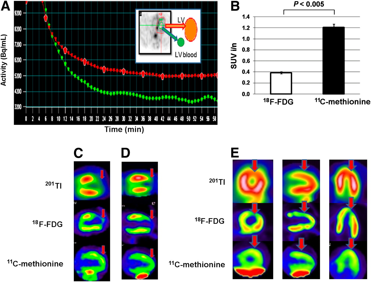
Before the patients had been examined, 3 healthy volunteers (2 men and 1 woman) were examined to determine the optimal timing of myocardial imaging after 11C-methionine injection; dynamic data was acquired for up to 60 min after 11C-methionine injection, and PET images of 11C-methionine uptake in normal myocardium were thus obtained (Fig. 1A). In the normal myocardium, 11C-methionine uptake was almost homogeneous, with a tendency toward a slight elevation in the basal anteroseptal area. In 2 volunteers, the average standardized uptake values (SUVs) of the basal anterior wall were 2.0 and 1.96 and those of the lateral wall were 1.71 and 1.78; the ratios of uptake in the basal anterior wall to the lateral wall were 1.17 and 1.10. Physiologic uptake in the liver was high, and this uptake masked some segments of the inferior wall. Patients with inferior-wall myocardial infarctions were also examined but were not included in the present study; instead, only cases of left anterior descendent infarction were chosen.

(A) Time–activity curves of regions of interest after tracer administration: whole left ventricle (red) and left ventricular blood pool (green). Blood-pool activity decreased from initially steep curve to stable lower level at around 20 min after injection, resulting in higher and stable myocardium–to–left-ventricle contrast at 20 min and thereafter. Then, we determined imaging time of myocardium with 11C-methionine at 20 min after injection. (B) Comparison of infarction-to-normal SUV ratios (SUV i/n) on 18F-FDG and 11C-methionine images (P < 0.005 vs. 11C-methionine imaging: mean ± SD, unpaired t test). (C and D) 201Tl SPECT, 18F-FDG PET, and 11C-methionine PET in 53-y-old man (patient 7) (C) and 71-y-old man (patient 9) (D). In 11C-methionine images, physiologic uptake in liver was high. Infarction zone in anterior wall exhibited decreased uptake on 201Tl and 18F-FDG images and increased uptake on 11C-methionine images. 11C-methionine uptake increased more in middle anterior wall than in apex: highest zone of uptake was almost next to spare zone in 201Tl and 18F-FDG images. 201Tl images were similar to 18F-FDG images visually. However, direct comparisons are difficult because of the different modalities. (E) Another instance of 3 orthogonal views of patient with segment 9 AMI (patient 8). Relative increase in11C-methionine uptake is also apparent in this patient.
Analysis of SPECT and PET Images
First, we visually assessed uptake of the 3 tracers semiquantitatively by categorizing the regional uptake as greater than, less than, or similar to uptake in normal cardiac tissue. Next, to analyze 18F-FDG and 11C-methionine uptake in the PET images quantitatively, we determined the average SUVs of infarcted areas (infarction SUVs) and normal areas (normal SUVs) by placing regions of interest at corresponding places on short axial and long vertical views. The lateral wall was defined as a normal area, since stenosis of the coronary artery was not apparent on coronary angiography in any patient. The mean infarction and normal SUVs were calculated from 3 regions of interest in each area. The infarction-to-normal SUV ratio was also calculated. For 201Tl, the average counts per pixel were determined in the infarction and normal zones, and the infarction-to-normal count ratio was calculated. All data were presented as the mean ± SD, and statistical differences were examined using an unpaired t test.
RESULTS
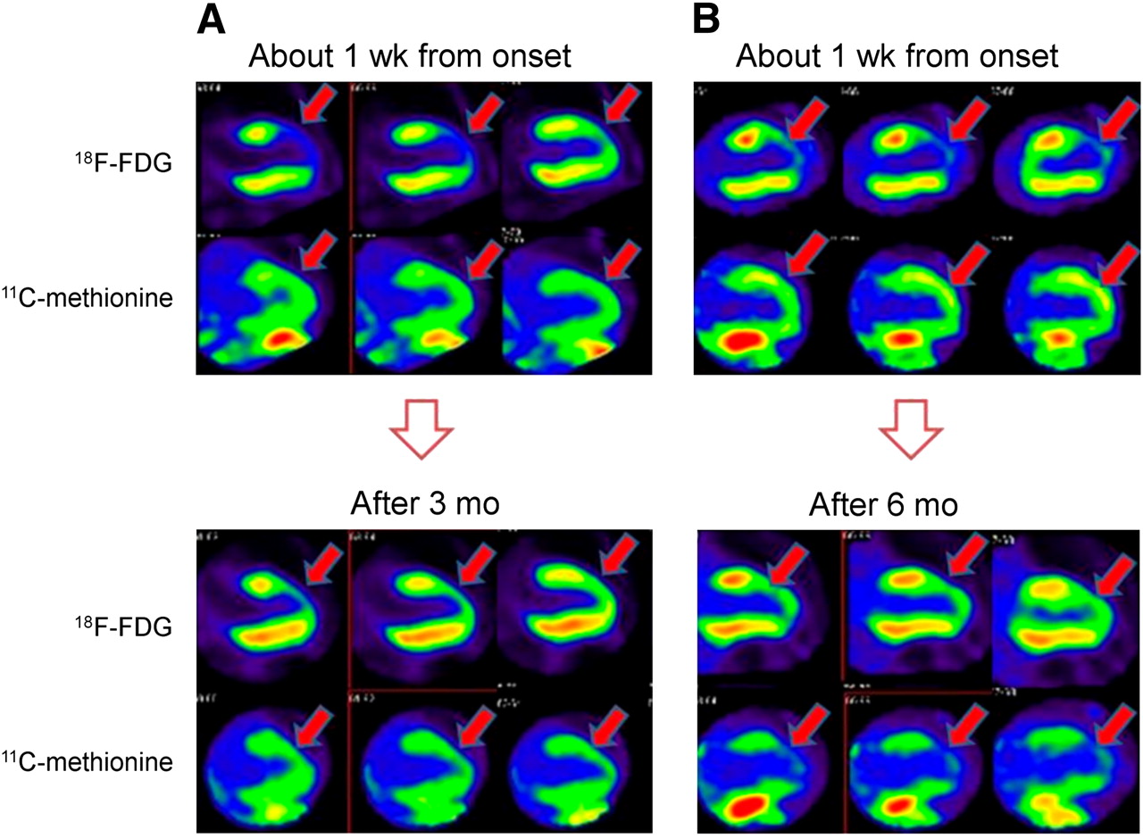
201Tl and 18F-FDG uptake was decreased, whereas 11C-methionine uptake was similar or increased, relative to the normal region on semiquantitative visual analysis (Table 1). Table 1 summarizes the clinical information and analyzed data of the 9 patients (mean age, 57.1 ± 15.1 y; female-to-male ratio, 0:9). All infarction-to-normal count ratios were lower than 1.0 in the 201Tl images. All infarction SUVs were lower than normal SUVs in the 18F-FDG images, whereas all infarction SUVs were similar to or higher than normal SUVs in the 11C-methionine images. Using the SUVs for all 9 patients, we determined the infarction-to-normal SUV ratio (mean ± SD) to be 0.39 ± 0.128 for the 18F-FDG images and 1.207 ± 0.095 for the 11C-methionine images (Fig. 1B). Representative patients are shown in Figures 1C–1E. Two patients with segment 6 AMI (Figs. 1C and 1D) exhibited increased uptake of 11C-methionine in the infarct zone, although uptake of the other tracers was decreased in the same zone. The greatest 11C-methionine uptake was not in the core of the infarct region but in the border region between the infarct and normal tissue. Visually, relative 18F-FDG uptake was reduced in the infarction zone, and the 201Tl uptake was the same. Three orthogonal views of another patient with no. 9 AMI are shown in Figure 1E. A relative increase in 11C-methionine uptake is visible. Figure 2 shows the changes in 11C-methionine uptake in studies performed 1 wk and 3 mo (patient 3) and 6 mo (patient 7) after AMI onset. At 3 mo after onset, uptake of 11C-methionine was similar to that observed during the acute phase, whereas uptake was decreased or showed a partial defect at 6 mo after onset. The 11C-methionine infarction-to-normal SUV ratio also decreased in 2 patients (Table 1). Thus, 11C-methionine uptake changed during imaging studies performed over the course of 6 mo after AMI onset.

Vertical long-axis images obtained 1 wk and 3 mo after AMI onset in 60-y-old man (patient 3) (A) and 1 wk and 6 mo after AMI onset in 53-y-old man (patient 7) (B). In both patients, 11C-methionine uptake in region was higher than that in lateral wall at 1 wk after AMI onset (infarction-to-normal ratios, 1.12 and 1.10). However, uptake became similar to 18F-FDG uptake at 3 mo after AMI onset and almost undetectable after 6 mo (infarction-to-normal ratios, 0.80 and 0.77). Uptake was slightly increased around infarction zone in 18F-FDG images obtained at 3 mo after AMI onset, whereas decreased 18F-FDG uptake was observed at 6 mo.
Clinical Information and Quantitative Data of Patients
DISCUSSION
11C-methionine PET has been used for diagnosing brain tumors and evaluating their treatment (1–4). Because of the low physiologic uptake of 11C-methionine in the brain, 11C-methionine PET is currently used mainly to assess brain tumors. Numerous studies have examined 11C-methionine uptake, and 11C-methionine uptake is widely known to reflect amino acid transport, transmethylation, and protein synthesis. In particular, Ishiwata et al. reported that the amino acid transport system was dominant in the brain and in tumors (2). Kracht et al. demonstrated a correlation between 11C-methionine uptake and microvessel density and showed a strong correlation between increased amino acid uptake and angiogenesis in evolving gliomas (4). In studies on brain infarction, Jacobs observed an increase in 11C-methionine uptake in peripheral areas of acute cerebral infarction (5), and Nagano-Saito et al. concluded that 11C-methionine uptake by damaged but viable brain tissue represented gliosis and angiogenesis (6).
However, few studies have examined the application of 11C-methionine PET to myocardial imaging, especially using clinical PET images. Here, we presented 201Tl SPECT and 18F-FDG/11C-methionine PET images of AMI after successful reperfusion. All the 201Tl and 18F-FDG images were typical of myocardial ischemia and exhibited similar uptake profiles. However, the 11C-methionine images showed a different pattern of uptake. An increase in 11C-methionine uptake was observed in regions exhibiting reduced or the absence of 201Tl and 18F-FDG uptake.
As we had suspected on the basis of the pattern of 11C-methionine uptake in the brain, 11C-methionine uptake was observed in regions of myocardial infarction. Blankesteijn et al. separated the process of healing after myocardial infarction into 4 phases: myocardial cell death, acute inflammation, the formation of granulation tissue, and scar formation (10). 11C-methionine uptake was observed in the second and third of these phases.
During the acute inflammatory reactions that occur during the early phase of healing, many inflammatory cells in the damaged region produce cytokines, which degrade the extracellular collagen matrix (11). In tumor tissue, 11C-methionine uptake increases in granulation tissue and macrophages, but not as much as does 18F-FDG (1). Thus, 11C-methionine uptake may reflect the activity of these inflammatory cells.
On the other hand, numerous small blood vessels are present during the formation of granulation tissue. Angiogenesis, a repair process after ischemic injuries (12), recently has become a field of molecular imaging research (13–17). 111In-RP748 SPECT (13) and 18F-galacto-RGD PET (14) targeting αvβ3 integrin, an angiogenic factor, have been used to study animal models of myocardial infarction; these studies showed overexpression of αvβ3 integrin in activated endothelial cells during angiogenesis after hypoxia, and increased uptake was observed in infarcted areas. The time course of the 11C-methionine images obtained after AMI was similar to the acute phase of angiogenesis described in these reports. Thus, the increased uptake of 11C-methionine might reflect some angiogenic factors, as mentioned in the above studies on cerebral infarction and gliomas, suggesting a relation between 11C-methionine uptake and angiogenesis. Possibly, the amino acid transport system might become active during remodeling of the heart, just as it does in the brain and in tumors.
This study had some limitations. 11C-methionine uptake was elevated during early AMI. This elevated uptake may gradually decrease until it disappears in scar tissue. 11C-methionine uptake after infarction may be useful for assessing the process of remodeling after AMI and for monitoring the effects of therapy. Further studies on 11C-methionine uptake after myocardial infarction are needed.
The present findings were limited to only the early acute phase of myocardial infarction, and more data on long-term changes in 11C-methionine uptake should be obtained.
In addition, although we compared 201Tl uptake for perfusion images, 18F-FDG for glucose metabolism, and 11C-methionine for amino acid metabolism, differences between the SPECT and PET modalities prevented us from determining whether the region of 11C-methionine uptake better matched the region of low 18F-FDG uptake or the region of 201Tl hypoperfusion.
Furthermore, because our 18F-FDG PET images were obtained after the oral administration of glucose, a comparison of 18F-FDG after fasting with heparin injection and 11C-methionine uptake would be useful to confirm whether 11C-methionine uptake actually reflects acute inflammation or ischemia.
Finally, from a technical viewpoint, the considerable physiologic uptake of 11C-methionine in the liver makes a detailed assessment of the right coronary artery area difficult.
CONCLUSION
We have presented the first (to our knowledge) images of 11C-methionine uptake in damaged myocardium after reperfusion for AMI. These images differ greatly from those of 201Tl and 18F-FDG uptake. 11C-methionine uptake might reflect an aspect of dynamic myocardial changes, such as remodeling during healing, after reperfusion for AMI.
Acknowledgments
We thank Drs. Yuriko Tanaka, Hiroyuki Soejima, Yuiichi Tamori, Munehiro Kamimura, Tomohiro Yamazaki, Junko Shibata, Kazuhiko Nakajima, and Ai Hori. Part of this study was supported by Grant-in-Aid 17-12 for cancer research from the Ministry of Health, Labor, and Welfare.
Footnotes
-
COPYRIGHT © 2009 by the Society of Nuclear Medicine, Inc.
References
- Received for publication December 17, 2008.
- Accepted for publication May 7, 2009.


Jump to section
Related Articles
Cited By...
- Imaging Inflammation Past, Present, and Future: Focus on Cardioimmunology
- Molecular Imaging of Myocardial Inflammation With Positron Emission Tomography Post-Ischemia: A Determinant of Subsequent Remodeling or Recovery
- 11C-Methionine PET of Myocardial Inflammation in a Rat Model of Experimental Autoimmune Myocarditis
- 62Cu-Diacetyl-Bis (N4-Methylthiosemicarbazone) PET in Human Gliomas: Comparative Study with [18F]Fluorodeoxyglucose and L-Methyl-[11C]Methionine PET
- 14C-Methionine Uptake as a Potential Marker of Inflammatory Processes After Myocardial Ischemia and Reperfusion
- Targeted Metabolic Imaging to Improve the Management of Heart Disease
- Methylphenidate-Elicited Dopamine Increases in Ventral Striatum Are Associated with Long-Term Symptom Improvement in Adults with Attention Deficit Hyperactivity Disorder