


Abstract
Our aim was to evaluate the role of SPECT/CT for the diagnosis of diabetic foot infection by labeled leukocytes. Methods: Seventeen patients with 19 clinically suspected sites of infection were included. After leukocyte labeling and administration, planar scans were acquired at 30 min, 4 h, and 24 h for 18 consecutive patients. SPECT/CT was obtained at 6 h. The final diagnosis was established by clinical follow-up (24 mo) in all cases and by bone biopsy for 14 sites. Results: Leukocyte scanning was positive in 16 of 19 lesions and negative in 3. SPECT/CT changed the interpretation of the planar and SPECT images for 10 of 19 suspected sites (52.6%): it excluded osteomyelitis in 6 cases, revealed bone infection in 1 case, and revealed both bone and soft-tissue infection in 3 cases. The hybrid device did not significantly contribute to the evaluation of patients with negative scan results. Conclusion: SPECT/CT can be useful for a more accurate diagnosis of diabetic foot infection by labeled leukocyte imaging.
Foot infection is a serious and relatively frequent complication in diabetic patients. Defining the location (i.e., bone or soft tissue) and extent of the infectious process is of paramount value in deciding on the duration of antibiotic therapy and the need for surgery (1).
Several imaging techniques have been proposed to image foot infection in diabetic patients. CT can be used but is not sufficiently accurate (2), MRI has high sensitivity and specificity (3) but has some limitations in differentiating osteomyelitis from neuropathic osteoarthropathy, from stress changes related to altered weight bearing, and from alterations of signal due to surgery (4). 99mTc-hexamethylpropylene amine oxime (HMPAO)–labeled leukocyte scanning has been also used to image diabetic foot infection (5) but is often unable to discriminate bone from soft-tissue involvement. 99mTc-methylene diphosphonate (MDP) is often used in association with labeled leukocyte scintigraphy to confirm or exclude the diagnosis of osteomyelitis (6).
SPECT/CT technology increases the accuracy of conventional scintigraphic imaging in many oncologic and nononcologic scenarios (7,8). In particular, published papers have suggested that SPECT/CT can be useful for imaging infectious processes, particularly when osteomyelitis is suspected (8).
The aim of this study was to evaluate the additional contribution of SPECT/CT to 99mTc-HMPAO–labeled leukocyte imaging of foot infection in diabetic patients.
MATERIALS AND METHODS
Patient Population
Eighteen consecutive patients (10 men and 7 women; mean age ± SD, 55 ± 4 y) with 20 foot lesions suggestive of infection were recruited as part of a research protocol. All had diabetes (10 type 1 and 8 type 2), and all presented with clinical signs of infections (i.e., ulceration, pain, or edema) and abnormal results on laboratory tests (white blood cell count, percentage of neutrophils, erythrocyte sedimentation rate). This prospective study was approved by the institutional ethics committee, and all subjects gave written informed consent to be included in the study. Antibiotic therapy was withdrawn at least 20 d before scintigraphy.
Imaging Protocol
Leukocyte isolation and labeling were performed as previously described (9). The labeling yield averaged 70%−85%. The labeled cells were reinjected into each patient, and the administered activity ranged from 400 to 555 MBq.
A SPECT/CT system (Millenium VG or Hawkeye; GE Healthcare) was used (10). Planar images of the feet (anterior, plantar, and lateral views) were acquired at 30 min (early images), 4 h, and 24 h (late images) after injection. The images were acquired in a 128 × 128 matrix using an imaging time of 10 min for the 30-min and the 4-h views; for the late scans, an imaging time of 15 min per view was used. SPECT/CT was performed 6 h after tracer injection. The CT data were acquired according to previously described protocols (9). The SPECT data were acquired in a 128 × 128 matrix, obtaining multiple views over 360° at a 30-s acquisition time per projection with an angular step of 3°. Images were reconstructed using Butterworth filtered backprojection (cutoff, 0.5; order, 10); transverse, sagittal, and coronal slices were generated. Scintigraphic results were considered to be positive when one or more areas of increased uptake, compared with background activity, were identified.
Transmission data were reconstructed on a nuclear medicine workstation (eNTEGRA; GE Healthcare) to obtain cross-sectional attenuation images (256 × 256 matrix) in which each pixel represented the attenuation value of the corresponding tissue. The reconstructed x-ray images and the nuclear medicine data were transmitted to a nuclear medicine database. Matching emission and transmission data were fused to obtain images of SPECT slices superimposed on the corresponding anatomic planes.
Data Analysis
First, scintigraphic data, including SPECT and planar images, were assessed for the presence, location, and extent of infection by an experienced nuclear medicine physician supported by a team of colleagues, who were unaware of the results of any prior radiologic investigations (CT or MRI). Afterward, the reader evaluated in the same manner the fusion images, and the SPECT/CT findings were compared with the findings obtained from SPECT and planar imaging alone.
SPECT/CT images were evaluated for possible misalignment, which was found in one patient. This patient was excluded from the study: the overall number of patients included, therefore, was 17.
Compared with planar imaging and SPECT, SPECT/CT was considered contributory when, by revealing a precise anatomic location, it radically changed the diagnosis (i.e., distinguished between soft-tissue and bone infection) or more accurately defined the extent of the infection (i.e., revealed both soft-tissue and bone infection).
The final diagnosis was established by correlation with clinical follow-up (24 mo) in all patients and by bacteriologic and histologic findings after bone biopsy for 14 sites.
RESULTS
99mTc-HMPAO–labeled leukocyte scanning was true-positive for infection in 14 of 17 patients at a total of 16 sites of cell accumulation; true-negative results were obtained for 3 patients, each with a single lesion suspected of infection (i.e., total number of clinically suspected lesions, 19).
Clinical features, final diagnosis, and a comparison of SPECT and planar imaging versus SPECT/CT are reported in Table 1.
Results of Labeled Leukocyte Scintigraphy (Planar plus SPECT) and SPECT/CT: Data on 15 Patients with Suspected Diabetic Foot Infection and Positive Scan Findings
The final diagnosis was osteomyelitis for 8 of 16 sites of leukocyte uptake. Compared with planar imaging and SPECT, SPECT/CT better assessed the extent of disease at 3 sites, demonstrating infection in bone coupled with neighboring soft tissue: all cases were confirmed by histologic examination and cultures. In one patient with a focus of uptake in the lateral aspect of the left hind foot, SPECT/CT localized infection to the lateral malleolus, and osteomyelitis was confirmed by histologic examination and cultures (isolation of polymicrobial agents). For the other 4 sites of osteomyelitis, SPECT/CT did not provide any significant contribution, because planar imaging and SPECT were concordant in localizing leukocyte uptake to bone: 2 cases were diagnosed on the basis of histologic examination and cultures, and the remaining 2 were diagnosed on the basis of clinical observation and serial radiologic studies that showed bone alterations typical of osteomyelitis. The final diagnosis was soft-tissue infection at 8 of 16 positive sites. Among these, SPECT/CT was able to exclude osteomyelitis at 6 sites. In such cases, both planar imaging and SPECT strongly suggested the presence of infection in bone. In contrast, fusion images localized cell uptake to soft tissue, thereby excluding bone involvement. All these cases were treated for soft-tissue infection and showed an adequate clinical response during follow-up.
In 3 patients, each with a single site of suspected infection (i.e., ulcer or nonhealing wound), planar imaging and SPECT were negative and SPECT/CT did not substantially contribute. One patient showed MRI bone alterations consistent with Charcot neuroarthropathy. All patients with negative scan results were free of infection at clinical follow-up.
The interpretation of planar and SPECT images was substantially changed on the basis of SPECT/CT in 10 of 19 lesions suspected of infection (52.6%), excluding osteomyelitis in 6 cases, localizing infection to bone at 1 site, and better defining the extent of disease (osteomyelitis and soft-tissue infection) in 3 cases.
DISCUSSION
99mTc-HMPAO–labeled leukocyte scintigraphy proved useful for the diagnosis of diabetic foot infection (5). Published papers indicated that SPECT/CT can improve the accuracy of scintigraphic images, particularly when highly specific tracers are used and background activity is low and anatomic information poor (11).
In our series, SPECT/CT was of paramount value in excluding osteomyelitis in 6 of 19 lesions suspected of infection (31.5%) and in better defining the extent of disease in 3 patients (15.7%). This high specificity and sensitivity may be due to the small cohort of patients in this preliminary study.
Devillers et al. (5) evaluated 99mTc-HMPAO–labeled leukocyte scanning in 42 patients with diabetes and a total of 56 foot ulcers: sensitivity was 88.4% and specificity 96.6%. To discriminate bone infection from soft-tissue infection, they used a combination of labeled leukocyte scintigraphy and 99mTc-MDP bone scanning. Although this procedure represents the currently best method of imaging bone infection, it is quite time-consuming for patients. The overall radiation burden to the patients must receive particular consideration because the dose is due to both 99mTc-HMPAO–labeled scintigraphy and 99mTc-MDP scanning. The hybrid technology can be useful in discriminating bone infection from soft-tissue infection with high accuracy, and the radiation burden due to the CT component is relatively low (0.5 mSv), considering the diagnostic contribution of fusion imaging. However, the radiation dose delivered by the CT component of SPECT/CT varies with the specific device used.
Bar-Shalom et al. (11) recently evaluated the additional value of SPECT/CT in 82 patients with clinical symptoms, laboratory tests, or imaging results suggesting infection but of uncertain location. Planar imaging plus SPECT and SPECT/CT had concordant results for diagnosis and location in 50% of patients. In the remaining 50%, SPECT/CT was discordant with conventional imaging for diagnosis (5 patients) or location (36 patients). When compared with the results of Bar-Shalom's series, our series confirmed that SPECT/CT had added value mainly in gaining the precise anatomic location and therefore the correct diagnosis (i.e., osteomyelitis, cellulitis, or both).
Keidar et al. (12) investigated the role of using 18F-FDG PET/CT to diagnose infections in the diabetic foot. They found that the precise anatomic correlation provided by PET/CT enabled accurate differentiation between osteomyelitis and soft-tissue infection. Although representing a more complex and time-consuming procedure than 18F-FDG PET, labeled leukocyte scanning can more specifically assess cell-mediated infectious processes.
SPECT/CT avoided bone amputation in 6 patients with planar and SPECT images showing infection of uncertain location (Fig. 1). The management of diabetic foot infection still remains a serious challenge for physicians: SPECT/CT may help to diagnose and better characterize infectious processes. In fact, its value is paramount for assessing the depth of infection and the specific tissues involved in order to plan treatment: for mild or severe infections, 12–24 wk of antibiotic therapy may be sufficient, whereas if osteomyelitis is suspected at least 46 wk of therapy is required (1). Surgery is another therapeutic option: amputation may be performed when the foot is gangrenous or septic or when the infection does not respond to drainage or antibiotics. Amputation may involve 1 or 2 toes, part of the foot, or part of the leg. The decision to amputate is based on assessment of the extent of disease (ischemia/infection), the entity of tissue perfusion, and the general condition of the patient (13). In such cases, SPECT/CT may help support treatment planning by accurately characterizing the location and extent of infectious processes.
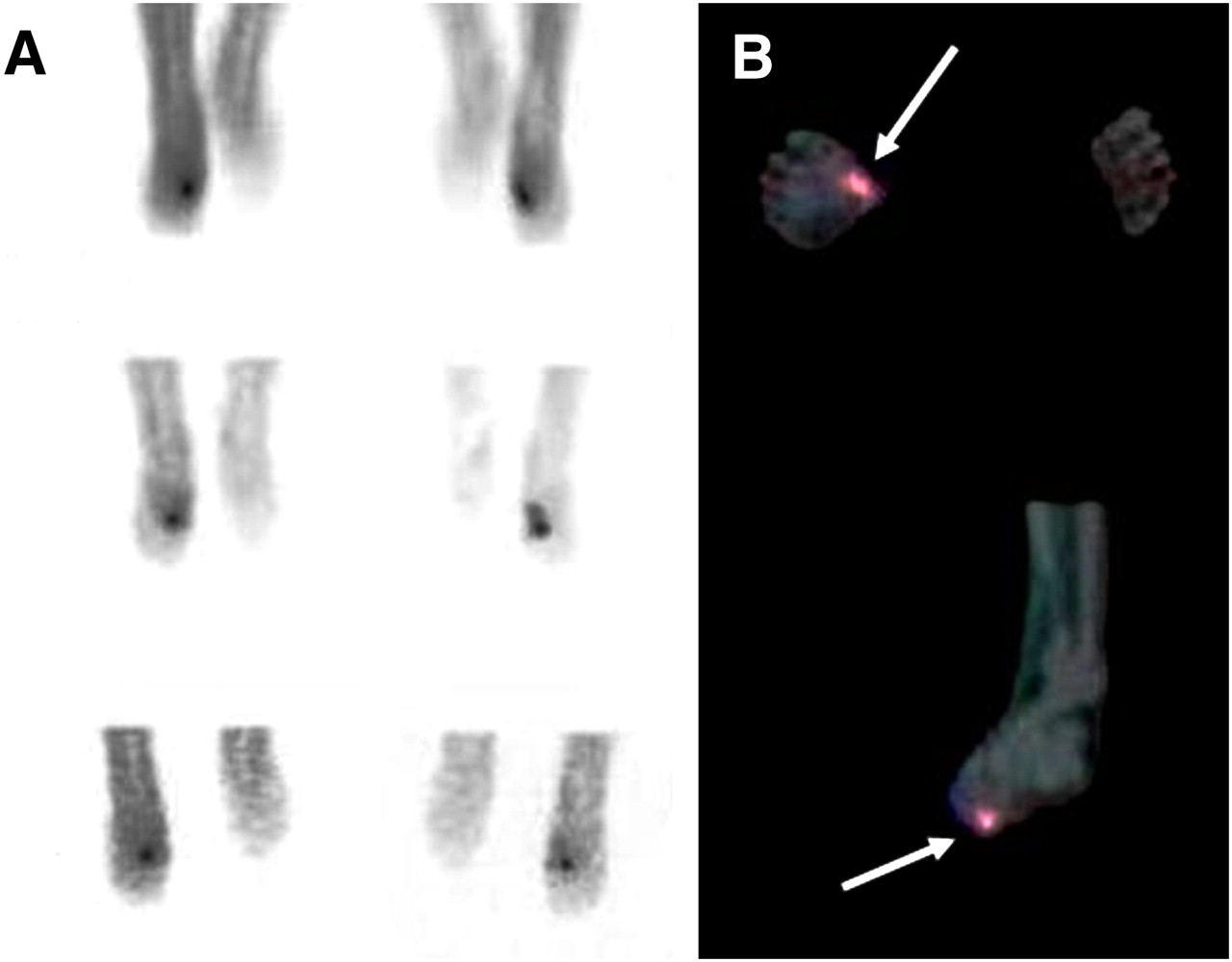
SPECT/CT-based exclusion of osteomyelitis in 55-y-old patient with nonhealing ulcer and cellulitis in right mid foot. (A) Planar images showed area of increased uptake strongly suggestive of osteomyelitis. (B) SPECT/CT allowed localization of infection (violet area in superimposition images) to soft tissues (arrow). No evidence of osteomyelitis was found at histopathologic diagnosis.
Hybrid imaging was not contributory to the evaluation of patients with negative scan results. As suggested by a previously published paper (14), SPECT/CT was particularly useful in increasing the accuracy of nuclear imaging and interobserver agreement.
SPECT/CT may be affected by misalignment due to a patient's involuntary movements. We did not obtain precise CT and SPECT coregistration in one patient, who was excluded from the study. Dedicated physical restraints might be used to immobilize lower limbs and reduce movement artifacts.
A limitation of the present study was its design. The review of the 2 sets of images (planar imaging plus SPECT and SPECT/CT) should have been masked and performed separately, preferably at a distance of several days or weeks. This approach would have removed any possible reader bias. Another limitation was the few patients included, reducing statistical power. Further studies with larger series are needed to better define the role of SPECT/CT in diabetic foot infection, to confirm our preliminary results, and to make comparisons with 99mTc-MDP bone scanning (15).
CONCLUSION
SPECT/CT can represent a potential tool for diagnosing diabetic foot infection by 99mTc-HMPAO–labeled leukocytes by differentiating between bone and soft-tissue involvement and by more precisely defining the extent of the disease, thus supporting treatment planning and avoiding more invasive procedures.
Footnotes
-
COPYRIGHT © 2009 by the Society of Nuclear Medicine, Inc.
References
- Received for publication October 22, 2008.
- Accepted for publication March 18, 2009.


Jump to section
Related Articles
Cited By...
- Detection of Osteomyelitis in the Diabetic Foot by Imaging Techniques: A Systematic Review and Meta-analysis Comparing MRI, White Blood Cell Scintigraphy, and FDG-PET
- Radionuclide Imaging of Musculoskeletal Infection: A Review
- Value of a Lower-Limb Immobilization Device for Optimization of SPECT/CT Image Fusion
- SPECT-CT: applications in musculoskeletal radiology
- Diagnosing Diabetic Foot Osteomyelitis in Patients Without Signs of Soft Tissue Infection by Coupling Hybrid 67Ga SPECT/CT With Bedside Percutaneous Bone Puncture
- Indexing Severity of Diabetic Foot Infection With 99mTc-WBC SPECT/CT Hybrid Imaging