


Abstract
Peptide-targeted α-therapy with 7.4 MBq of 212Pb-[1,4,7,10-tetraazacyclododecane-1,4,7,10-tetraacetic acid]-ReO-[Cys3,4,10,d-Phe7,Arg11]α-MSH3–13 (212Pb-DOTA-Re(Arg11)CCMSH) cured 45% of B16/F1 murine melanoma–bearing C57 mice in a 120-d study, highlighting its melanoma treatment potential. However, there is a need to develop an imaging surrogate for patient-specific dosimetry and to monitor the tumor response to 212Pb-DOTA-Re(Arg11)CCMSH therapy. The purpose of this study was to evaluate the potential of 203Pb-DOTA-Re(Arg11)CCMSH as a matched-pair SPECT agent for 212Pb-DOTA-Re(Arg11)CCMSH. Methods: DOTA-Re(Arg11)CCMSH was labeled with 203Pb in 0.5 M NH4OAc buffer at pH 5.4. The internalization and efflux of 203Pb-DOTA-Re(Arg11)CCMSH were determined in B16/F1 melanoma cells. The pharmacokinetics of 203Pb-DOTA-Re(Arg11)CCMSH was examined in B16/F1 melanoma–bearing C57 mice. A micro-SPECT/CT study was performed with 203Pb-DOTA-Re(Arg11)CCMSH in a B16/F1 melanoma–bearing C57 mouse at 2 h after injection. Results: 203Pb-DOTA-Re(Arg11)CCMSH was easily prepared in NH4OAc buffer and completely separated from the excess nonradiolabeled peptide by reversed-phase high-performance liquid chromatography (RP-HPLC). 203Pb-DOTA-Re(Arg11)CCMSH displayed fast internalization and extended retention in B16/F1 cells. Approximately 73% of 203Pb-DOTA-Re(Arg11)CCMSH activity internalized after a 20-min incubation at 25°C. After incubation of the cells in culture medium for 20 min, 78% of internalized activity remained in the cells. 203Pb-DOTA-Re(Arg11)CCMSH exhibited a biodistribution pattern similar to that of 212Pb-DOTA-Re(Arg11)CCMSH in B16/F1 melanoma–bearing mice. 203Pb-DOTA-Re(Arg11)CCMSH exhibited a peak tumor uptake of 12.00 ± 3.20 percentage injected dose per gram (%ID/g) at 1 h after injection. The tumor uptake gradually decreased to 3.43 ± 1.12 %ID/g at 48 h after injection. 203Pb-DOTA-Re(Arg11)CCMSH exhibited a peak tumor-to-kidney uptake ratio of 1.53 at 2 h after injection. The absorbed doses to the tumor and kidneys were 4.32 and 4.35 Gy, respectively, per 37 MBq. Whole-body clearance of 203Pb-DOTA-Re(Arg11)CCMSH was fast, with approximately 89% of the injected activity cleared through the urinary system by 2 h after injection. 203Pb showed 1.6-mm SPECT resolution, which was comparable to 99mTc. Melanoma lesions were visualized through SPECT/CT images of 203Pb-DOTA-Re(Arg11)CCMSH at 2 h after injection. Conclusion: 203Pb-DOTA-Re(Arg11)CCMSH exhibited favorable pharmacokinetic and tumor imaging properties, highlighting its potential as a matched-pair SPECT agent for 212Pb-DOTA-Re(Arg11)CCMSH melanoma treatment.
Investigators can predict the usefulness of new imaging and therapy agents based on the results of matched pairs of identical or near-identical radiopharmaceuticals. The advantage of taking a matched-pair approach is that the imaging agent can be used to demonstrate selective tumor targeting and to obtain patient-specific dosimetry, allowing optimal and safe deployment of the therapeutic counterpart. Some examples of matched-pair approaches have been the use of 11lIn- and 90Y-radiolabeled compounds such as octreotide (1–4) as well as 99mTc- and 188Re-radiolabeled α-melanocyte–stimulating hormone (α-MSH) peptides (5–7). However, in the cases mentioned earlier, similar but not chemically identical radiometals were used. Even though the radiometals may have similar coordination chemistries, small differences among radionuclides can result in different pharmacokinetics (8,9). Therefore, it is desirable to radiolabel the targeting compound with 2 chemically identical radioisotopes to extract the maximum benefit from the matched-pair approach to radiopharmaceutical design.
Peptide-targeted α-particle therapy for melanoma using 212Pb-[1,4,7,10-tetraazacyclododecane-1,4,7,10-tetraacetic acid]-ReO-[Cys3,4,10,d-Phe7,Arg11]α-MSH3–13 (212Pb-DOTA-Re(Arg11)CCMSH) was reported in our previous publication (10). The results demonstrated that the peptide-targeted α-particle therapy was effective in increasing the mean survival times of mice initially bearing melanoma tumors. Treatment with single doses of 3.7 or 7.4 MBq of 212Pb-DOTA-Re(Arg11)CCMSH resulted in, respectively, 20% and 45% of animals with complete cures. The development of a matched-pair imaging agent counterpart for 212Pb-DOTA-Re(Arg11)CCMSH would be useful to demonstrate tumor uptake and to calculate the dose to healthy tissues and vital organs. Patient-specific dosimetry determined from the imaging studies would be useful for treatment planning so that the maximum tolerable activity of the α-particle–emitting radiolabeled peptide could be administered for safe and effective melanoma treatment. Moreover, a matched-pair imaging agent could be used to further monitor the patients' response to the targeted α-radiation therapy.
A potential matched-pair imaging radioisotope for the therapeutic radionuclide 212Pb is 203Pb (11–14). On decay, 203Pb (half-life, 51.9 h) emits a 279-keV γ-ray (81% abundance) suitable for SPECT. 203Pb can be produced via the 203Tl(d,2n)203Pb reaction by irradiating natural Tl2O3 or an enriched Tl2O3 (203Tl) target with 13.7-MeV deuterons (11). A simple and rapid procedure was reported for purifying cyclotron-produced 203Pb via the 203Tl(d,2n)203Pb reaction (11). High specific activity and radiochemical purity of 203Pb are essential for radiolabeling peptides that target lower-copy-number cellular receptors. Low radiochemical purity of 203Pb can dramatically reduce if not eliminate the radiolabeling yields caused by the competition of the existing contaminating metals. Likewise, low specific activity of 203Pb preparations may result in the saturation of the targeted receptors with nonradioactively labeled peptides. Purified 203Pb was used to label the monoclonal antibody trastuzumab (Herceptin; Genentech), which was shown to be immunoreactive and displayed favorable biodistribution properties in vivo, demonstrating the suitability and feasibility of 203Pb-labeled biomolecules to target cellular antigens (11).
In this study, DOTA-Re(Arg11)CCMSH (5,8) was radiolabeled with 203Pb to evaluate its potential as a matched-pair imaging agent for 212Pb-DOTA-Re(Arg11)CCMSH. The spatial resolution of 203Pb was examined through hot-rod phantom imaging by small-animal SPECT and compared with the spatial resolution of 99mTc. The melanocortin-1 (MC1) receptor–mediated uptake and efflux of 203Pb-DOTA-Re(Arg11)CCMSH were examined in vitro. Biodistribution and SPECT studies were performed to demonstrate the potential of 203Pb-DOTA-Re(Arg11)CCMSH to image the melanoma lesions.
MATERIALS AND METHODS
Chemicals and Reagents
DOTA-Re(Arg11)CCMSH was purchased from Bachem Inc., and 203Pb was obtained from AlphaMed, Inc. All other chemicals used in this study were purchased from Fischer Scientific and used without further purification. The B16/F1 murine melanoma cell line was obtained from American Type Culture Collection.
Calibration of SPECT Detector with 203Pb
A high-count flood image was acquired with 0.37 MBq of 203Pb and placed in the central axis above the detector face, using uncollimated detectors. Energy discriminating windows were used for photopeak isolation of the 203Pb spectrum. Detector nonuniformities arise because of the detector crystal-to-crystal detection efficiency variability and pinhole sensitivity changes associated with the angle of acceptance of the pinhole aperture. To correct the detector nonuniformity, all projection images were normalized to 203Pb with a correction matrix derived from the collected 203Pb uniform flood image.
Jaszczak SPECT Phantom Imaging
SPECT volumetric performance for 203Pb was assessed using a micro-Deluxe ECT hot-insert phantom (Data Spectrum Inc.). The phantom had an inner diameter of 4.4 cm, with 6 equally sized rod quadrants. The rod diameters were 1.2, 1.6, 2.4, 3.2, 4.0, and 4.8 mm. The phantom was filled with 74 MBq of 203Pb and was imaged with a 1.0-mm pinhole and a magnification factor of 2 at a distance of 4.5 cm. The SPECT scan images were acquired for 60 frames over 360°, and the projection data were reconstructed using a 3-dimensional ordered-subset expectation maximization (OSEM) algorithm. The phantom SPECT data were reconstructed using 12 iterations and 4 subsets. The reconstructed images were smoothed after reconstruction with a 3-dimensional gaussian kernel. A parallel study was performed with 99mTc for comparison.
Synthesis of 203Pb-DOTA-Re(Arg11)CCMSH
DOTA-Re(Arg11)CCMSH was radiolabeled with 203Pb in 0.5 M NH4OAc at pH 5.4. Briefly, 50 μL of 203PbCl2 in 0.5 M HCl (∼37 MBq), 500 μL of 0.5 M NH4OAc (pH 5.4), and 20 μL of 1 mg/mL DOTA-Re(Arg11)CCMSH were added into a reaction vial and incubated at 75°C for 40 min. 203Pb-DOTA-Re(Arg11)CCMSH was purified to single species by HPLC (Waters) on a C-18 reverse-phase analytic column (Vydac), using a 20-min linear gradient of 16%–26% acetonitrile in 20 mM HCl aqueous solution with a flow rate of 1.5 mL/min. The stability of 203Pb-DOTA-Re(Arg11)CCMSH was monitored up to 24 h for degradation by HPLC in 0.1% bovine serum albumin (BSA) in 10 mM phosphate-buffered saline (PBS) at 37°C. HPLC-purified peptide samples were purged with N2 gas for 20 min to remove the acetonitrile. The pH of the final solution was adjusted to 5 with 0.1N NaOH and diluted with normal saline for animal studies.
Cellular Internalization and Efflux of 203Pb-DOTA-Re(Arg11)CCMSH
B16/F1 murine melanoma cells were obtained from American Type Culture Collection and cultured in RPMI 1640 medium containing NaHCO3 (2 g/L), supplemented with 10% heat-inactivated fetal calf albumin, 2 mM L-glutamine, and 48 mg of gentamicin. The cells were incubated at 37°C in 75-cm3 tissue culture flasks under a humidified 5% CO2 atmosphere. The culture medium was changed every 2 d. Cellular internalization and efflux of 203Pb-DOTA-Re(Arg11)CCMSH were evaluated in B16/F1 murine melanoma cells. B16/F1 cells (5 × 10⁵/well) were seeded into a 24-well cell culture plate and incubated at 37°C overnight. After being washed once with binding medium (minimal essential medium with 25 mM N-(2-hydroxyethyl)piperazine-N′-(2-ethanesulfonic acid), pH 7.4 0.2% BSA/0.3 mM 1,10-phenathroline), the cells were incubated at 25°C for 20, 40, 60, 90, and 120 min (n = 4) in approximately 100,000 counts per minute (cpm) of HPLC-purified 203Pb-DOTA-Re(Arg11)CCMSH. After incubation, the reaction medium was aspirated and cells were rinsed with 2 × 0.5 mL of ice-cold, pH 7.4 0.2% BSA/0.01 M PBS. Cellular internalization of 203Pb-DOTA-Re(Arg11)CCMSH was assessed by washing the cells with acidic buffer (40 mM sodium acetate [pH 4.5] containing 0.9% NaCl and 0.2% BSA) to remove the membrane-bound radioactivity. The remaining internalized radioactivity was obtained by lysing the cells with 0.5 mL of 1N NaOH for 5 min. Membrane-bound and internalized 203Pb activity was counted in a γ-counter. Cellular efflux of 203Pb-DOTA-Re(Arg11)CCMSH was determined by incubating B16/F1 cells with 203Pb-DOTA-Re(Arg11)CCMSH for 2 h at 25°C, removing nonspecific-bound activity with 2 × 0.5 mL of ice-cold, pH 7.4 0.2% BSA/0.01 M PBS rinse, and monitoring radioactivity released into the cell culture medium. The radioactivity in the medium, on the cell surface, and in cells was separately collected and counted in a γ-counter 20, 40, 60, 90, and 120 min after incubation in the culture medium.
Biodistribution Studies
Animal studies were conducted in compliance with Institutional Animal Care and Use Committee approval. Pharmacokinetic studies were performed on C57 mice that were inoculated subcutaneously with 1 × 10⁶ B16/F1 murine melanoma cells in the right flank. When the weight of tumors reached approximately 0.2 g, 7.4 × 10⁻³ MBq of 203Pb-DOTA-Re(Arg11)CCMSH was injected into each mouse through the tail vein. Groups of 5 mice per each time point were used for the biodistribution studies. The mice were sacrificed at 5 and 30 min and at 1, 2, 4, 24, and 48 h after injection, and tumors and organs of interest (whole organs except muscle, bone, and skin) were harvested, weighed, and counted in a Wallac 1480 automated γ-counter (PerkinElmer). The results were expressed as percentage injected dose per gram (%ID/g) and as percentage injected dose (%ID). Blood values were taken as 6.5% of the whole-body weight. Portions of muscle, bone, and skin were collected and weighed for calculating %ID/g in those organs. The tumor uptake specificity of 203Pb-DOTA-Re(Arg11)CCMSH was determined by blocking tumor uptake at 2 h after injection with the coinjection of 10 μg of unlabeled [Nle4,D-Phe7]α-MSH (NDP-MSH), a linear α-MSH peptide analog with picomolar affinity for the α-MSH receptor present on murine melanoma cells.
Dosimetry Calculation
The biodistribution of 203Pb-DOTA-Re(Arg11)CCMSH over time was determined to evaluate uptake and retention and to calculate absorbed radiation doses from 203Pb-DOTA-Re(Arg11)CCMSH in tumors, healthy organs, and tissues using methods described previously (15–17). Time–activity curves were generated for 16 organs and tissues (blood, bone, brain, heart, lung, liver, skin, spleen, stomach, kidney, large intestine, small intestine, muscle, pancreas, carcass, and tumor). Cumulative activity of 203Pb was determined for each organ by integrating the area under the time–activity curves. The cumulative activity was then used with a dosimetric model (15,16) developed specifically for the laboratory mouse.
Melanoma Imaging with 203Pb-DOTA-Re(Arg11)CCMSH
One B16/F1 melanoma–bearing C57 mouse was injected with 6.29 MBq of HPLC-purified 203Pb-DOTA-Re(Arg11)CCMSH via the tail vein 14 d after cell implantation. The mouse was euthanized by CO2 inhalation for micro-SPECT/CT at 2 h after injection. The SPECT data were collected right after CT data collection. Approximately 0.3 MBq of 203Pb-DOTA-Re(Arg11)CCMSH activity was left in the mouse at the moment of acquisition. Micro-SPECT scans of 60 frames per animal were acquired for a total count acquisition of 0.5 million counts for 203Pb-DOTA-Re(Arg11)CCMSH. A pinhole magnification factor of 2.2 was used in the experiments. The micro-SPECT/CT images were obtained using high-resolution SPECT pinhole collimators (MicroCAT II SPECT/CT; Siemens Pre-Clinical Solutions). The SPECT projection data (78 × 78 × 102 matrix) were reconstructed using a 3-dimensional OSEM algorithm with geometric misalignment corrections, and the CT raw data were reconstructed via a cone beam (Feldkamp) filtered backprojection algorithm. Reconstructed data from SPECT and CT were visualized and coregistered using Amira 3.1 (TGS).
RESULTS
Tomographic spatial resolution of 1.6 mm was achieved with the micro-Deluxe ECT hot-rod 203Pb SPECT phantom at a distance of 4.5 cm (Fig. 1). The results of the transaxial slice of the 203Pb-reconstructed volumetric SPECT data with the same phantom filled with 74 MBq of 99mTc were comparable when considering the 4.8- to 1.6-mm quadrant regions and the differentiation of rods within the phantom quadrants.
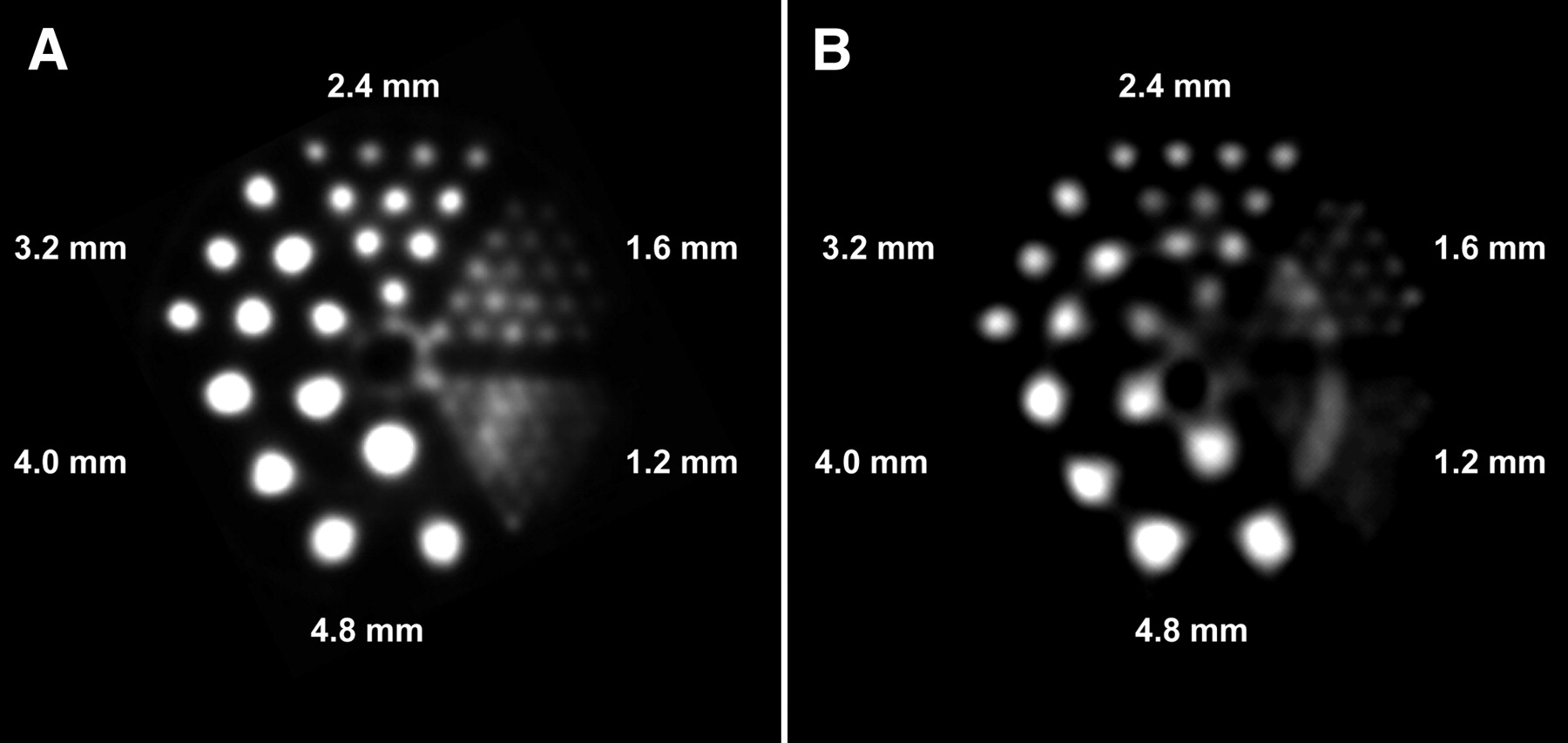
Phantom imaging of 99mTcO4− (A) and 203PbCl2 (B).
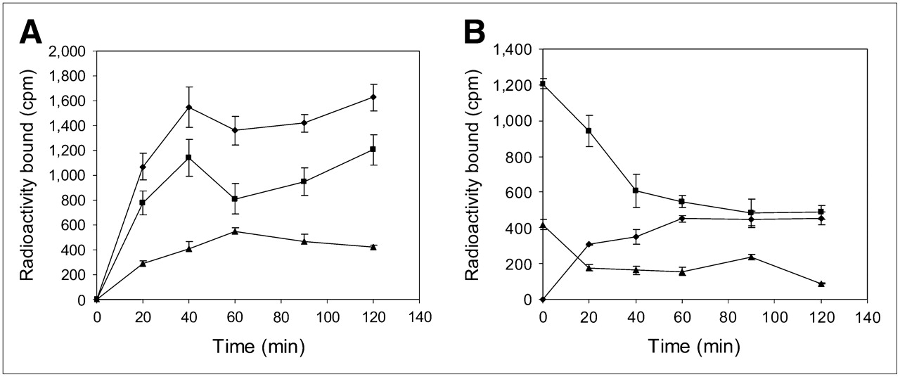
DOTA-Re(Arg11)CCMSH was labeled with 203Pb using a 0.5 M NH4OAc-buffered solution at pH 5.4. 203Pb-DOTA-Re(Arg11)CCMSH was completely separated from its nonradiolabeled counterpart by reversed-phase high-performance liquid chromatography (RP-HPLC). The stability of 203Pb-DOTA-Re(Arg11)CCMSH was determined by incubation in 0.1% BSA in 10 mM PBS (pH 7.4) at 37°C. Only the 203Pb-labeled peptide was detected by RP-HPLC after 24 h incubation. A schematic structure of 203Pb-DOTA-Re(Arg11)CCMSH is presented in Figure 2. Cellular internalization and efflux of 203Pb-DOTA-Re(Arg11)CCMSH were evaluated in B16/F1 cells at 25°C (Fig. 3). 203Pb-DOTA-Re(Arg11)CCMSH exhibited rapid cellular internalization. Approximately 73% and 74% of 203Pb-DOTA-Re(Arg11)CCMSH activity were internalized in the B16/F1 cells after 20 min and 2 h of incubation, respectively. Cellular efflux of 203Pb-DOTA-Re(Arg11)CCMSH demonstrated that 78% and 40% of the 203Pb activity remained inside the cells 20 min and 2 h, respectively, after incubating cells in culture medium at 25°C.

A schematic structure of the rhenium-cyclized peptide, 203Pb-DOTA-Re(Arg11)CCMSH.

Cellular internalization (A) and efflux (B) of 203Pb-DOTA-Re(Arg11)CCMSH in B16/F1 murine melanoma cells at 25°C. Total bound radioactivity (♦), internalized activity (▪), cell membrane activity (▴), and cell culture medium activity (•) were presented as cpm.
The pharmacokinetics and tumor-targeting properties of 203Pb-DOTA-Re(Arg11)CCMSH were determined in B16/F1 murine melanoma–bearing C57 mice. The biodistribution of 203Pb-DOTA-Re(Arg11)CCMSH is shown in Table 1. 203Pb-DOTA-Re(Arg11)CCMSH exhibited high uptake and long retention in the tumor. At 1 h after injection, 203Pb-DOTA-Re(Arg11)CCMSH reached its peak tumor uptake value of 12.00 ± 3.20 %ID/g. There remained 9.86 ± 1.86 %ID/g of 203Pb-DOTA-Re(Arg11)CCMSH activity in the tumor at 4 h after injection. The tumor uptake value gradually decreased to 4.35 ± 0.24 %ID/g at 24 h and 3.43 ± 1.12 %ID/g at 48 h after injection. Tumor uptake specificity of 203Pb-DOTA-Re(Arg11)CCMSH was examined by coinjecting 10 μg of the high-affinity α-MSH peptide analog NDP-MSH. The tumor uptake of 203Pb-DOTA-Re(Arg11)CCMSH with NDP coinjection was only 7.4% of the tumor uptake without NDP coinjection at 2 h after dose administration (P < 0.01), demonstrating that tumor uptake was specific and receptor-mediated. Whole-body clearance of 203Pb-DOTA-Re(Arg11)CCMSH was rapid, with approximately 90% of the injected dose washed out of the body by 2 h after injection. Ninety-four percent of the injected dose was washed out of the body by 24 h after injection. Normal organ uptakes of 203Pb-DOTA-Re(Arg11)CCMSH were generally very low (<1 %ID/g) at 2 h after injection except for the kidneys. High tumor-to-blood and tumor–to–normal-organ uptake ratios were demonstrated as early as 30 min after injection (Table 1). The kidneys appeared to be the major excretion organ of 203Pb-DOTA-Re(Arg11)CCMSH. The kidney uptake values of 203Pb-DOTA-Re(Arg11)CCMSH were 7.78 ± 1.42 %ID/g and 3.69 ± 0.58 %ID/g at 2 and 24 h after injection, respectively. Bone uptake values of 203Pb-DOTA-Re(Arg11)CCMSH activity were less than 1.4 %ID/g at all time points after 1 h after injection in this study.
Pharmacokinetics of 203Pb-DOTA-Re(Arg11)CCMSH in B16/F1 Murine Melanoma–Bearing C57 Mice
The absorbed radiation doses to tumors and healthy organs from 203Pb-DOTA-Re(Arg11)CCMSH were determined in this study from the biodistribution data in B16/F1 murine melanoma–bearing mice (Table 2). The absorbed dose from 203Pb-DOTA-Re(Arg11)CCMSH in the B16/F1 mouse tumors was 4.32 Gy per 37 MBq. The relatively high tumor dose was directly related to the rapid uptake kinetics and retention of the 203Pb-labeled peptide. Normal-tissue doses were low except for the kidneys, which were estimated at 4.35 Gy per 37 MBq. These results suggest that the kidneys may be the dose-limiting normal organ for radionuclide therapy. One B16/F1 murine melanoma–bearing C57 mouse was injected with 203Pb-DOTA-Re(Arg11)CCMSH to visualize the tumor at 2 h after dose administration (Fig. 4). Although there was substantial activity in the kidneys, melanoma tumors in the right flank were visualized clearly at 2 h after injection. 203Pb-DOTA-Re(Arg11)CCMSH exhibited high tumor–to–normal-organ uptake ratios except for the kidney in the SPECT image, which was coincident with the trend observed in the biodistribution of 203Pb-DOTA-Re(Arg11)CCMSH.
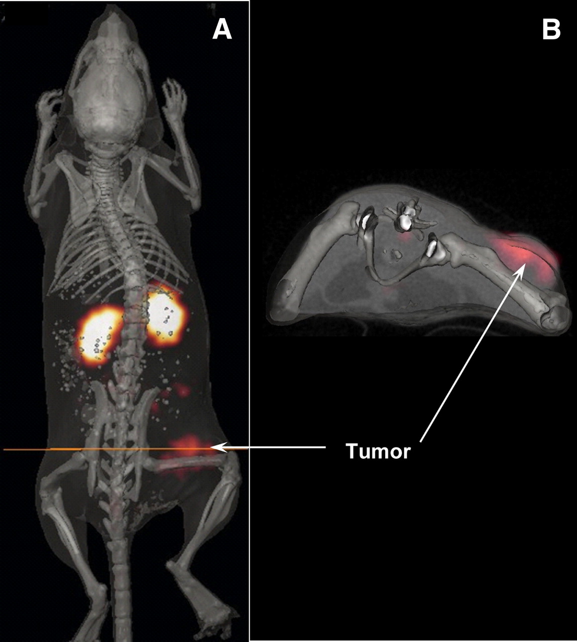
Whole-body (A) and transaxial (B) images with 203Pb-DOTA-Re(Arg11)CCMSH at 2 h after injection in B16/F1 murine melanoma–bearing C57 mouse.
Absorbed Radiation Doses per Unit Administered Activity from 203Pb-DOTA-Re(Arg11)CCMSH in B16/F1 Murine Melanoma–Bearing C57 Mice
DISCUSSION
Radiolabeled α-MSH peptide analogs as imaging agents may have their greatest utility when used in a matched-pair approach for melanoma radionuclide therapy. In a matched-pair approach to radionuclide imaging and therapy, the same melanoma-targeting peptide can be labeled with radioisotopes possessing diagnostic imaging or therapeutic decay properties. The advantage of this approach is that patient-specific dosimetry can be determined using the imaging agent so that the optimal dose of the peptide labeled with the therapeutic radioisotope can be administered. Moreover, a matched-pair imaging agent could be used to further monitor the patients' response to the targeted radionuclide therapy. 111In-labeled conjugates are often used as imaging surrogates for dosimetric calculation of 90Y-labeled conjugates based on the assumption that 90Y- and 111In-labeled conjugates are chemically and biologically equivalent. However, 90Y- and 111In-labeled monoclonal antibody and peptide showed differences in their biologic properties (8,9,18), which raised some concerns about the validity of using 111In-labeled conjugates as imaging surrogates for their 90Y-labeled conjugates. The atomic radius of 90Y fits nearly perfectly into the cavity of DOTA, whereas 111In has a smaller atomic radius than that of 90Y. The biodistribution differences between 111In- and 90Y-labeled conjugates are likely to be related to the different coordination chemistries in solution (9,19,20). Hence, radiolabeling the targeting compound with 2 radioisotopes of the same metal will maximally extract the benefit from the matched-pair approach to radiopharmaceutical development for cancer imaging and therapy.
212Pb-DOTA-Re(Arg11)CCMSH was used for targeted α-radiation therapy for melanoma in our previous studies (10). DOTA-Re(Arg11)CCMSH exhibited nanomolar MC1 receptor–binding affinity and specifically targeted 212Pb to melanoma cells. 212Pb decays via β-emission to 212Bi, which subsequently decays via a branched pathway to stable 208Pb, yielding high-energy α-particles and β-particles (10). High cyctotoxic ionizing radiation from α-particles results in irreparable DNA double-strand breaks, causing cell death. A major advantage of administering 212Pb-DOTA-Re(Arg11)CCMSH is that the radiolabeled peptide will circulate, target melanoma tumor cells, and be cleared from the body as the 212Pb-labeled peptide rather than the α-emitting 212Bi compound, minimizing normal-tissue exposures. Peptide-targeted 212Pb internalized and retained by tumor cells will decay to the α-particle–emitting 212Bi and serve as an in vivo generator of α-particles, localizing the highly toxic short-ranged α-radiation within the tumor. Meanwhile, the 10.6-h half-life of 212Pb makes dose preparation and administration easier and more efficient than the short half-life (half-life, 60.6 min) of 212Bi. 212Pb-DOTA-Re(Arg11)CCMSH exhibited remarkable therapeutic efficacy in B16/F1 melanoma–bearing mice in our previous report (10). The treatment of 7.4 MBq of 212Pb-DOTA-Re(Arg11)CCMSH cured 45% of B16/F1 murine melanoma–bearing C57 mice in a 120-d study and highlighted its potential as a novel agent for targeted radionuclide therapy of melanoma.
203Pb and 212Pb are 2 isotopes with diagnostic and therapeutic properties (11,12). The favorable decay and imaging properties of 203Pb make it an ideal matched-pair radioisotope for 212Pb for targeted radionuclide therapy (11,12). 203Pb is a manageable radioisotope with respect to dose preparation and waste disposal because of its half-life of 51.9 h. 203Pb displayed a spatial resolution (1.6 mm) comparable to that of 99mTc (Fig. 1), demonstrating the suitability and feasibility of 203Pb as a SPECT isotope. Moreover, 203Pb can be produced via the 203Tl(d,2n)203Pb reaction by a cyclotron and can be easily purified to achieve high specific activity for radiolabeling of antibodies or peptides for antigen or receptor targeting (11). In this study, 203Pb-DOTA-Re(Arg11)CCMSH was prepared and evaluated in vitro and in melanoma-bearing mice to examine its potential as a matched-pair imaging agent for 212Pb-DOTA-Re(Arg11)CCMSH.
In vitro, 203Pb-DOTA-Re(Arg11)CCMSH exhibited rapid cellular internalization and moderate retention. Tumor cell retention of 203Pb-DOTA-Re(Arg11)CCMSH at 40 min was approximately 50%, compared with 87% and 90% for 90Y- and 177Lu-labeled DOTA-Re(Arg11)CCMSH, respectively (9). The greater efflux rate for 203Pb-DOTA-Re(Arg11)CCMSH was potentially caused by the 2+ oxidation state of 203Pb, coupled with the presence of the metal chelator phenathroline in the cell-binding and efflux medium, which led to its accelerated release in vitro. However, in vivo higher tumor efflux kinetics for 203Pb-DOTA-Re(Arg11)CCMSH were not observed from 1 to 4 h after injection. The tumor uptake reached its peak value of 12.00 ± 3.20 %ID/g at 1 h after injection. In vivo tumor retention of 203Pb-DOTA-Re(Arg11)CCMSH remained constant at 2 h after injection (Table 1), leading to the high imaging contrast between the tumor and the healthy tissues. The tumor uptake value at 4 h after injection was 82.2% of the tumor uptake value at 1 h after injection (Table 1). Clearance of activity from the healthy organs and tissues was rapid, which resulted in high tumor-to-blood and tumor–to–normal-organ uptake ratios as early as 30 min after injection (Table 1). The majority of the administered activity cleared through the kidneys, with 89% of the injected dose being excreted in the urine by 2 h after injection. Coinjection of excess nonradioactive NDP-MSH peptide dramatically reduced tumor uptake but did not affect radioactivity in the kidneys, demonstrating that radioactivity in the tumor was receptor-mediated whereas the renal radioactivity was nonspecific. The dosimetry results (Table 2) demonstrated that the absorbed dose to tumor and kidneys was approximately 8 times or greater than the absorbed dose to other healthy organs, suggesting that the kidneys would be the dose-limiting normal organ for targeted radionuclide therapy. The statistical analyses of the tumor and kidney uptake values were performed between 203Pb-DOTA-Re(Arg11)CCMSH and 212Pb-DOTA-Re(Arg11)CCMSH to confirm the matched-pair properties between 203Pb and 212Pb (data not shown). There were no significant differences (P > 0.05) in the tumor uptake values between 203Pb-DOTA-Re(Arg11)CCMSH and 212Pb-DOTA-Re(Arg11)CCMSH 1, 2, 4, 24, and 48 h after injection and no significant differences (P > 0.05) in the kidney uptake values between 203Pb-DOTA-Re(Arg11)CCMSH and 212Pb-DOTA-Re(Arg11)CCMSH 1, 2, and 24 h after injection. Overall, the in vivo biodistribution and clearance kinetics of 203Pb-DOTA-Re(Arg11)CCMSH were nearly identical to 212Pb-DOTA-Re(Arg11)CCMSH (10) in B16/F1 melanoma–bearing mice, confirming its matched-pair properties.
LS-174T colon carcinoma xenografts were successfully imaged by others with a 203Pb-DOTA-B72.3 antibody conjugate by a γ-camera at 120 h after injection (12), highlighting the potential of radiolabeling the antibody with 203Pb to target the antigen for tumor imaging. In our study, dual-modality micro-SPECT/CT was used to evaluate the potential of 203Pb-DOTA-Re(Arg11)CCMSH as a melanoma-imaging probe in a melanoma mouse model. Coregistration of high-spatial-resolution micro-CT anatomic data combined with molecular imaging data obtained using micro-SPECT allowed accurate identification and localization of the melanoma tumors. Flank melanoma tumors were clearly visualized with 203Pb-DOTA-Re(Arg11)CCMSH at 2 h after injection by SPECT/CT images (Fig. 4). The SPECT images of tumors accurately matched the anatomic information from CT images. 203Pb-DOTA-Re(Arg11)CCMSH displayed high tumor–to–normal-organ uptake ratios except for the kidneys in the SPECT/CT images, which was coincident with the trend observed in the biodistribution results (Table 1). High melanoma uptake and tumor–to–normal-organ uptake ratios in SPECT/CT images validated the feasibility of 203Pb-DOTA-Re(Arg11)CCMSH as a matched-pair imaging agent for 212Pb-DOTA-Re(Arg11)CCMSH, which appears to be a promising peptide radiopharmaceutical for targeted radionuclide therapy of melanoma.
CONCLUSION
203Pb-DOTA-Re(Arg11)CCMSH exhibited high melanoma uptake and a biodistribution pattern similar to that of 212Pb-DOTA-Re(Arg11)CCMSH, highlighting its potential as a matched-pair imaging probe for 212Pb-DOTA-Re(Arg11)CCMSH. In combination with SPECT/CT equipment, 203Pb-DOTA-Re(Arg11)CCMSH could provide an effective approach to optimize therapeutic doses using patient-specific dosimetry calculations and monitoring patient response to targeted radionuclide therapy with 212Pb-DOTA-Re(Arg11)CCMSH.
Acknowledgments
We thank Drs. Wynn A. Volkert and Susan L. Deutscher for their helpful discussions and Katherine Benwell and Tiffani Shelton for their technical assistance. This work was supported by National Cancer Institute grant 1R43 CA11492 and P50 Imaging Center grant P50-CA-103130, the VA Biomolecular Imaging Center at the Harry S Truman VA Hospital and the University of Missouri-Columbia, University of New Mexico–Los Alamos National Laboratory MOU on Research and Education grant 2R76T, American Foundation Pharmaceutical Education grant 3R48E, and American Cancer Society Institutional Research grant IRG-92-024.
Footnotes
-
COPYRIGHT © 2008 by the Society of Nuclear Medicine, Inc.
References
- Received for publication October 23, 2007.
- Accepted for publication January 24, 2008.


Jump to section
Related Articles
Cited By...
- Preclinical Applications of Multi-Platform Imaging in Animal Models of Cancer
- Preclinical Evaluation of 203/212Pb-Labeled Low-Molecular-Weight Compounds for Targeted Radiopharmaceutical Therapy of Prostate Cancer
- High-Contrast PET of Melanoma Using 18F-MEL050, a Selective Probe for Melanin with Predominantly Renal Clearance
- PET of Malignant Melanoma Using 18F-Labeled Metallopeptides