

FIGURE 4.

FIGURE 4.
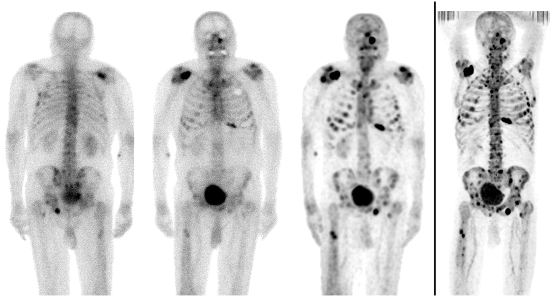
From left to right: posterior and anterior 99mTc-MDP planar scintigraphy, 99mTc-MDP multiple-field-of-view SPECT, and 18F-fluoride PET of 82-y-old patient with numerous bone metastases. As in this patient, more lesions are typically detected by SPECT than by planar imaging, and 18F-fluoride PET detects more lesions than does SPECT.
In this issue


Related Articles
Cited By...
- 18F-NaF PET/CT of Obese Patients on a Lutetium-Yttrium Oxyorthosilicate PET/CT System: Patient Dosimetry, Optimization of Injected Activity, and Acquisition Time
- 18F-Sodium Fluoride PET: History, Technical Feasibility, Mechanism of Action, Normal Biodistribution, and Diagnostic Performance in Bone Metastasis Detection Compared with Other Imaging Modalities
- Characterization of Bone Lesions in Myeloma Before and During Anticancer Therapy Using 18F-FDG-PET/CT and 18F-NaF-PET/CT
- Three-Hour Delayed Imaging Improves Assessment of Coronary 18F-Sodium Fluoride PET
- Comparison of the Variability of SUV Normalized by Skeletal Volume with the Variability of SUV Normalized by Body Weight in 18F-Fluoride PET/CT
- Assessment of Physiologic Intracranial Calcification in Healthy Adults Using 18F-NaF PET/CT
- Hospice Admission and Survival After 18F-Fluoride PET Performed for Evaluation of Osseous Metastatic Disease in the National Oncologic PET Registry
- The Role of 18F-Sodium Fluoride PET/CT Bone Scans in the Diagnosis of Metastatic Bone Disease from Breast and Prostate Cancer
- Evaluation of Prostate Cancer Bone Metastases with 18F-NaF and 18F-Fluorocholine PET/CT
- Bone-Targeted Imaging and Radionuclide Therapy in Prostate Cancer
- Prospective Study Evaluating Na18F PET/CT in Predicting Clinical Outcomes and Survival in Advanced Prostate Cancer
- 18F-Fluoride PET in the Assessment of Malignant Bone Disease
- Prognostic Factors in Patients Treated with 223Ra: The Role of Skeletal Tumor Burden on Baseline 18F-Fluoride PET/CT in Predicting Overall Survival
- Semiquantitative Analysis of the Biodistribution of the Combined 18F-NaF and 18F-FDG Administration for PET/CT Imaging
- Evaluation of 18F-Fluoride PET/MR and PET/CT in Patients with Foot Pain of Unclear Cause
- 18F-Fluoride PET Used for Treatment Monitoring of Systemic Cancer Therapy: Results from the National Oncologic PET Registry
- AEG-1 Promoter-Mediated Imaging of Prostate Cancer
- Impact of 18F-Fluoride PET on Intended Management of Patients with Cancers Other Than Prostate Cancer: Results from the National Oncologic PET Registry
- Impact of 18F-Fluoride PET in Patients with Known Prostate Cancer: Initial Results from the National Oncologic PET Registry
- Reply: Regarding Dynamic Bone Imaging with 99mTc-Labeled Diphosphonates and 18F-NaF: Mechanisms and Applications
- Bisphosphonate-Induced Osteonecrosis of the Jaw: Comparison of Disease Extent on Contrast-Enhanced MR Imaging, [18F] Fluoride PET/CT, and Conebeam CT imaging
- Dynamic Bone Imaging with 99mTc-Labeled Diphosphonates and 18F-NaF: Mechanisms and Applications
- Clinical utility of fluoride-18 positron emission tomography/CT in temporomandibular disorder with osteoarthritis: comparisons with 99mTc-MDP bone scan
- Combined 18F-Fluoride and 18F-FDG PET/CT Scanning for Evaluation of Malignancy: Results of an International Multicenter Trial
- Biodistribution, Tumor Detection, and Radiation Dosimetry of 18F-DCFBC, a Low-Molecular-Weight Inhibitor of Prostate-Specific Membrane Antigen, in Patients with Metastatic Prostate Cancer
- Validation of a Paper Chromatographic Methodology as an Alternative for Determination of the Radiochemical Purity of Na18F
- Utility of 18F-Fluoride PET/CT and 18F-FDG PET/CT in the Detection of Bony Metastases in Heightened-Risk Head and Neck Cancer Patients
- Radiation Exposure Should Not Limit Bone Scintigraphy with 18F-NaF
- The Kinetics and Reproducibility of 18F-Sodium Fluoride for Oncology Using Current PET Camera Technology
- Coronary Arterial 18F-Sodium Fluoride Uptake: A Novel Marker of Plaque Biology
- Assessment of Valvular Calcification and Inflammation by Positron Emission Tomography in Patients With Aortic Stenosis
- Nuclear Medicine in the First Year of Life
- Molecular Mechanisms of Bone 18F-NaF Deposition
- SNM Practice Guideline for Sodium 18F-Fluoride PET/CT Bone Scans 1.0
- In Vivo Molecular Imaging Analysis of a Nasal Vaccine That Induces Protective Immunity against Botulism in Nonhuman Primates
- Imaging of Back Pain in Children
- Potential Iatrogenic Alteration to 18F-Fluoride Biodistribution
- Dosimetry of Pediatric PET/CT
- Bone Metastases in Patients with Neuroendocrine Tumor: 68Ga-DOTA-Tyr3-Octreotide PET in Comparison to CT and Bone Scintigraphy
- The Role of Radiotracer Imaging in the Diagnosis and Management of Patients with Breast Cancer: Part 1--Overview, Detection, and Staging
- Radiodefluorination of 3-Fluoro-5-(2-(2-[18F](fluoromethyl)-thiazol-4-yl)ethynyl)benzonitrile ([18F]SP203), a Radioligand for Imaging Brain Metabotropic Glutamate Subtype-5 Receptors with Positron Emission Tomography, Occurs by Glutathionylation in Rat Brain