

FIGURE 3.
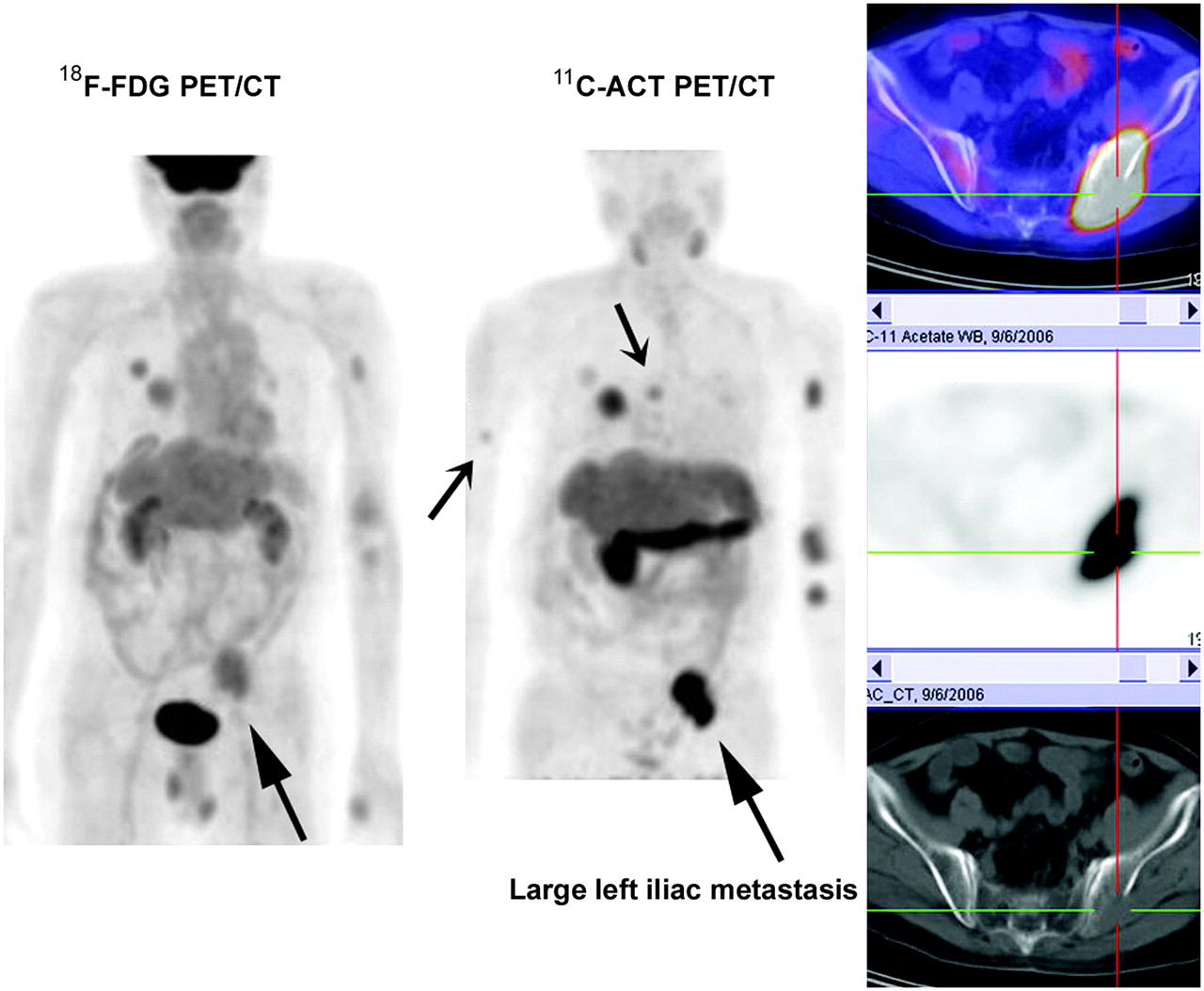
FIGURE 3.
Multifocal bone metastases: a 62-y-old patient with previous liver resection for HCC. 18F-FDG and 11C-ACT PET/CT showed multifocal lung and bone metastases. Note that 11C-ACT revealed more bone metastasis than 18F-FDG (e.g., right humeral and thoracic lesions [small arrows]), and 11C-ACT lesions are significantly more intense. Largest left iliac lesion (large arrows) showed typical osteolytic pattern on CT bone window.
In this issue


Related Articles
Cited By...
- Radiation Dosimetry of Whole-Body Dual-Tracer 18F-FDG and 11C-Acetate PET/CT for Hepatocellular Carcinoma
- Reply: Underestimated Role of 18F-FDG PET for HCC Evaluation and Promise of 18F-FDG PET/MR Imaging in This Setting
- 11C-Acetate and 18F-FDG PET/CT for Clinical Staging and Selection of Patients with Hepatocellular Carcinoma for Liver Transplantation on the Basis of Milan Criteria: Surgeon's Perspective
- Detection of Hepatocellular Carcinoma with PET/CT: A Prospective Comparison of 18F-Fluorocholine and 18F-FDG in Patients with Cirrhosis or Chronic Liver Disease
- Hepatocellular Carcinoma: Consensus Recommendations of the National Cancer Institute Clinical Trials Planning Meeting
- A Prospective Evaluation of 18F-FDG and 11C-Acetate PET/CT for Detection of Primary and Metastatic Hepatocellular Carcinoma
- Detection of Hepatocellular Carcinoma Using 11C-Choline PET: Comparison with 18F-FDG PET
- A Multicenter Clinical Trial on the Diagnostic Value of Dual-Tracer PET/CT in Pulmonary Lesions Using 3'-Deoxy-3'-18F-Fluorothymidine and 18F-FDG