


Abstract
Targeting extracellular structures that are involved in angiogenic processes, such as the extra domain B of fibronectin, is a promising approach for the diagnosis of solid tumors. The aim of this study was to determine uptake of the 18F-labeled PET tracers 18F-fluorocholine (N,N-dimethyl-N-18F-fluoromethyl-2-hydroxyethylammonium), 18F-fluoro-ethyl-l-tyrosine (FET), and 18F-FDG in C6 gliomas of the rat and to correlate it with uptake of the anti–extra domain B antibody 131I-SIP(L19) as a marker of neoangiogenesis. Methods: C6 gliomas were orthotopically induced in 17 rats. Uptake of all tracers was measured using quantitative autoradiography, and uptake of 18F-fluorocholine, 18F-FET, and 18F-FDG was correlated with uptake of 131I-SIP(L19) on a pixelwise basis. Results: The mean 131I-SIP(L19), 18F-fluorocholine, 18F-FET, and 18F-FDG standardized uptake values in the tumor and the contralateral normal cortex (in parentheses) were 0.31 ± 0.22 (not detectable), 2.00 ± 0.53 (0.49 ± 0.07), 3.67 ± 0.36 (1.42 ± 0.22), and 7.23 ± 1.22 (3.64 ± 0.51), respectively. The 131I-SIP(L19) uptake pattern correlated best with 18F-fluorocholine uptake (ż = 0.80, averaged ż-transformed Pearson correlation coefficient) and 18F-FET uptake (ż = 0.79) and least with 18F-FDG (ż = 0.37). Conclusion: One day after intravenous injection, 131I-SIP(L19) displayed a very high tumor-to-cortex ratio, which may be used in the diagnostic work-up of brain tumor patients. Of the 3 investigated 18F tracers, 18F-fluorocholine and 18F-FET correlated better with the pattern of 131I-SIP(L19) uptake than did 18F-FDG. Whether this means that 18F-fluorocholine and 18F-FET are better suited than 18F-FDG to monitor antiangiogenic therapy should be investigated in future studies.
Vascular targeting and antiangiogenic strategies have been propagated as promising tools for diagnostic and therapeutic purposes (1). Angiogenesis, the development of new blood vessels from preexisting ones, is a typical feature of many malignant tumors and of benign disorders such as arthritis, diabetic retinopathy, and several others. The fact that tumor growth depends mainly on blood supply from newly built vessels (2) leads to the investigation of angiogenic markers. Today, the extra domain B of fibronectin is one of the most promising (3). It is present in the perivascular space of many aggressive solid tumors but not in normal tissue (except the ovaries and the endometrium). Extra domain B is identical in sequence in different species including rat, mouse, and man and has been successfully targeted with anti–extra domain B antibodies including the human recombinant antibody fragment scFv(L19) (4). In recent years, various L19 formats have been labeled with radioisotopes such as 123I and 125I for quantitative biodistribution analysis in murine tumor models and for immunoscintigraphic SPECT imaging of cancer patients (5–7). In accordance with the results of an immunohistochemical analysis of high- and low-grade astrocytomas (8), a study of Santimaria et al. (6) indicated a promising potential for radiolabeled L19 antibodies for diagnosis, grading, and therapy of human glioma patients.
In neurooncology, 18F-FDG and 18F-fluoro-ethyl-l-tyrosine (FET) are established tracers. 18F-FET has the advantage of displaying a higher tumor-to-background ratio and of not accumulating in inflammatory lesions (9,10). On the other hand, 18F-FDG is more widely available. The value of 18F-fluorocholine in the evaluation of brain tumors is yet less clear, although a high tumor-to-background ratio has been demonstrated (11).
In a previous study, we showed that microvascular density influences the uptake of 18F-fluorocholine but not of 18F-FET or 18F-FDG (10). The purpose of the present study was to correlate the uptake of 18F-fluorocholine, 18F-FET, and 18F-FDG with that of 131I-SIP(L19) and therefore with the degree of neoangiogenesis. We hypothesized that a correlation of 18F uptake with 131I-SIP(L19) could point to a possible use of the 18F tracer for the monitoring of antiangiogenic therapy warranting further studies in this field.
MATERIALS AND METHODS
Animals
Seventeen male Wistar rats weighing 250–350 g were used in this study. The experiments were approved by the local veterinary authorities. All animal work was performed by licensed investigators.
Radiopharmaceuticals
18F-Labeled Tracers.
18F-FDG was obtained from the commercial 18F-FDG production of the University Hospital Zurich. The production of 18F-FET and 18F-fluorocholine was described in detail elsewhere (12).
131I-SIP(L19).
The SIP(L19) antibody was prepared according to a published procedure (5). The carrier-free radionuclide 131I (98.0% radiochemical purity, 99% radionuclidic purity, with a specific activity of 675 GBq/mg) was obtained from Perkins Elmer as a solution of Na131I in 0.1 M NaOH. The protein was radioiodinated using a modified chloramine-T labeling procedure in a ventilated hood. Typically, the antibody solution (200 μL, 100 μg of protein, 1.25 × 10⁻⁹ mol) was introduced into the vial with the starting Na131I solution (10 μL) with 370 MBq (10 mCi, 3.56 × 10⁻⁹ mol of Na131I) for the autoradiography experiments. Then, 5 μL of 0.5% aqueous solution of chloramine-T (25 μg, 1.17 × 10⁻⁷ mol) were added, and after 5 min at room temperature, the mixture was introduced into a Sephadex G-25 column (PD-10, GE Healthcare) previously treated with 1 mL of a 2% aqueous solution of bovine serum albumin for occupation of the nonspecific binding sites followed by equilibration with 25 mL of phosphate-buffered saline (pH 7.4). The column was eluted with phosphate-buffered saline. Fractions of 1 mL were collected. The labeled antibody was eluted from the column usually within the second or third fraction, whereas the unreacted 131I remained at the column.
The labeling efficiency was usually about 75%, giving radioiodinated antibodies with specific activities of 222 GBq/μmol.
Cell Culture
The rat glioma cell line C6 was kindly provided by W. Risau (Bad Nauheim, Germany). C6 rat glioma cells were grown in Dulbecco's modified Eagle medium containing 4,500 mg of d-glucose (GIBCO, Life Technologies) per liter, supplemented with 10% fetal calf serum, 2 mM N-acetyl-l-alanyl-l-glutamine (Biochrom AG), and 20 μg of gentamycin (Sigma-Aldrich) per milliliter. For injection, the cells were harvested by trypsinization, washed 3 times with phosphate-buffered saline, and resuspended at a final concentration of 108 cells/mL.
Inoculation Procedure
The rats were anesthetized with an intraperitoneal injection of a mixture of ketamine (50 mg/kg; Ketasol 100, Dr. E. Graeub AG) and medetomidine (1 mg/kg; Domitor, Dr. E. Graeub AG) and placed in a stereotactic frame (David Kopf Instruments). We used the inoculation method of Ambar et al. with few modifications (13). After preparation of the inoculation area, a small hole was drilled into the skull. Then, 3 × 10⁵ C6 cells in 3 μL were implanted into the left parietal cortex at a depth of 2 mm below the dura mater using a 10-μL, 26-gauge syringe (Hamilton). After a short incubation time, the syringe was slowly removed, the hole was closed with bone wax, and the skin was sutured. The well-being of the animals was monitored daily using a score sheet that included weight, behavior, posture, gait, and breathing rate.
Dual-Tracer Autoradiographic Studies with 18F-Tracers and 131I-SIP(L19)
Tracer uptake studies were performed 13–22 d after inoculation (based on preliminary MRI studies for tumor growth control; data not shown). One day before the autoradiographic experiments, 6–50 MBq of 131I-SIP(L19) were administered via the tail vein to the conscious rats. The following day, a catheter was placed in the tail vein under isoflurane inhalation anesthesia, allowing intravenous application of 50–150 MBq of the 18F-tracers. 18F-Fluorocholine (14) and 18F-FET (15) were injected 15 min, and 18F-FDG 45 min, before removal of the brain (16). The animals were sacrificed using an overdose of intravenous pentobarbital. The brains were removed and instantly frozen in isopentane cooled to −50°C. For quantification of the 18F tracers, 10-μm brain slices (100-μm slice distance) were placed on a phosphor imaging screen together with 14C-standards and left for 240 min. Tritium-sensitive screens (Fuji TR2025) were used, because their uncoated, thin, sensitive layer yields higher-resolution 18F-autoradiograms than do ordinary screens. After decay of the 18F activity, the identical slices were again placed on the screens to get the 131I-SIP(L19) autoradiograms. Both datasets were then scanned (Fuji BAS 1800 II; pixel size, 50 μm) and converted to kBq/cm3. For this conversion, the 14C-standards had previously been calibrated using a brain homogenate containing a defined amount of 18F and 131I.
For the quantitative analysis, the activities were decay-corrected to the time of screen exposure. Dividing these values by the amount of injected activity per gram of body weight yielded standardized uptake values (SUVs). Regions of interest were subsequently placed over the tumors and over the contralateral healthy cortex using the software PMOD (17). In addition, tumor uptake was divided by uptake in the contralateral cortex to obtain ratios of tumor to normal brain.
Morphologic Characterization
For morphologic analysis, the brain slices were fixed in 4% formalin/phosphate-buffered saline, followed by hematoxylin/eosin conventional staining and glial fibrillary acidic protein (GFAP) immunohistochemistry. In short, slides were incubated with anti-GFAP antibody (1:1000, DAKO Z0334) without pretreatment.
On adjacent slides, immunohistochemical labeling of the von-Willebrand factor was performed for visualization of blood vessels. Brain slices were pretreated with protease I for 4 min and stained with polyclonal antibody against von-Willebrand factor (1:1000, DAKO A00802). Finally, slides were developed using a Ventana machine and the iView DAB (DAKO) development kit.
Statistics
The corresponding autoradiographs were coregistered using the software PMOD. Regions of interest encompassing the whole tumor were then defined and applied to the 18F tracers and 131I-SIP(L19) autoradiographs. To investigate the correlation of the uptake pattern of each 18F tracer with that of 131I-SIP(L19), each 100th pixel was chosen for a regression analysis. Individual Pearson regression coefficients were then ż-transformed (Fisher's ż-transformation) and averaged for the correlations 18F-FDG vs. 131I-SIP(L19), 18F-fluorocholine vs. 131I-SIP(L19), and 18F-FET vs. 131I-SIP(L19). These transformed average regression coefficients were compared pairwise according to Sachs (18) to assess differences in the correlations between each 18F tracer and 131I-SIP(L19).
RESULTS
Animals
No animal experienced tumor-induced side effects during the observation period.
Morphologic Characterization
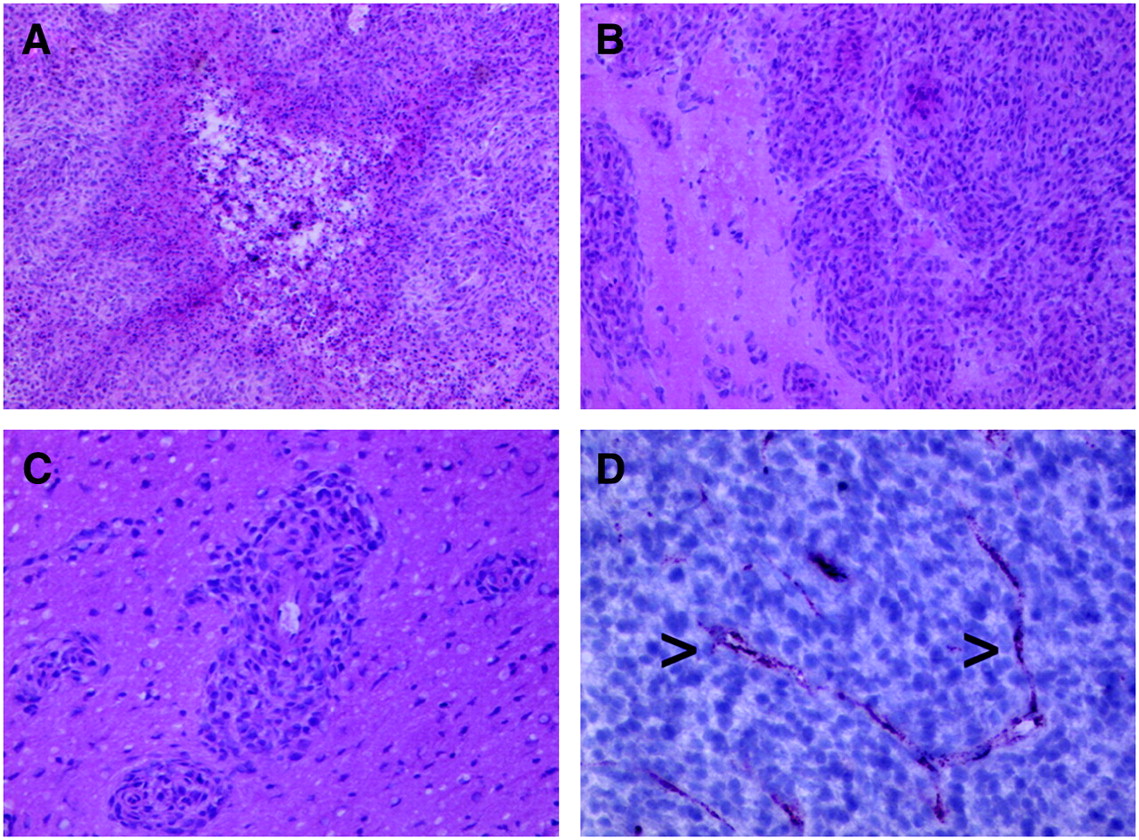
The orthotopically grown tumors presented mostly in a compact glioblastomalike formation with or without necrosis (Fig. 1A). However, a subset of cells was found to invade the adjacent brain parenchyma or perivascular Virchow-Robin spaces (Figs. 1B and 1C). Immunohistochemical stains for GFAP revealed the astrocytic character of the tumor cells, although most did not express GFAP. The surrounding parenchyma presented with reactive, highly GFAP-positive astrocytes. Inflammatory cells such as lymphocytes or macrophages were not prominent. Transplanted tumor cells exhibited a homogeneous tumor mass with an evenly distributed microvascular network containing small arterioles and capillaries. A representative intratumoral microvessel stained against von-Willebrand factor is demonstrated in Figure 1D.

Morphologic characterization of transplanted glioma cells and developing tumor: central necrotic area (hematoxylin/eosin, ×60) (A), parenchymal infiltration zone of glioma cells (hematoxylin/eosin, ×100) (B), infiltration in Virchow-Robin perivascular space (hematoxylin/eosin, ×130) (C), and representative intratumoral microvessels (arrowheads; anti–von-Willebrand factor, ×200) (D).
Tumoral Tracer Uptake
Examples of dual-tracer autoradiographs and corresponding correlations are demonstrated in Figures 2–4⇓. In each tumor, the uptake of all tracers was substantially higher than in cortical gray matter. The uptake values are summarized in Table 1. 18F-FDG displayed the highest SUV, followed by 18F-FET, 18F-fluorocholine, and 131I-SIP(L19). Ratios of tumor to normal brain were highest for 131I-SIP(L19), because of the low uptake in normal brain.
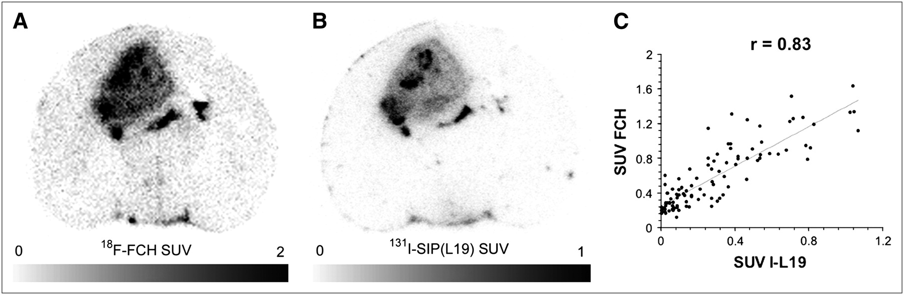
(A and B) Dual-tracer autoradiographs of 18F-fluorocholine (FCH) (A) and corresponding 131I-SIP(L19) (B). (C) Pixelwise correlation of each 100th pixel in tumor region of interest and Pearson correlation coefficient.

(A and B) Dual-tracer autoradiographs of 18F-FET (A) and corresponding 131I-SIP(L19) (B). (C) Pixelwise correlation of each 100th pixel in tumor region of interest and Pearson correlation coefficient.

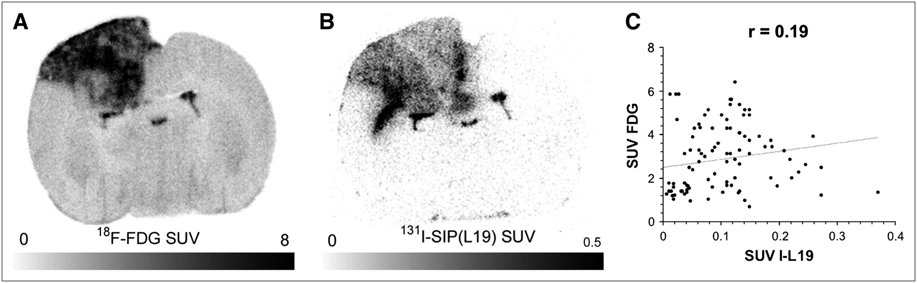
(A and B) Dual-tracer autoradiographs of 18F-FDG (A) and corresponding 131I-SIP(L19) (B). (C) Pixelwise correlation of each 100th pixel in tumor region of interest and the Pearson correlation coefficient.
SUVs in Animals with Induced C6 Gliomas
Correlation Between Uptake Pattern of 18F-Tracers and 131I-SIP(L19)
Regression analysis of tumor uptake of the 18F-tracers and 131I-SIP(L19) revealed the highest correlation for 18F-fluorocholine (ż = 0.80, averaged ż-transformed Pearson correlation coefficient, n = 6), and 18F-FET (ż = 0.79, n = 6). For 18F-FDG, the correlation was poorer (ż = 0.37, n = 5). Pairwise comparison of the ż-transformed and averaged Pearson correlation coefficients revealed that 18F-fluorocholine and 18F-FET correlated significantly better with 131I-SIP(L19) than did 18F-FDG, whereas no significant difference was found in the correlations of 18F-fluorocholine and 18F-FET with 131I-SIP(L19) (18F-fluorocholine/131I-SIP(L19) vs. 18F-FDG/131I-SIP(L19): P < 0.01, ż = 2.98; 18F-FET/131I-SIP(L19) vs. 18F-FDG/131I-SIP(L19): P < 0.01, ż = 2.86; 18F-FET/131I-SIP(L19) vs. 18F-fluorocholine/131I-SIP(L19): P > 0.05, ż = 0.12).
The order of the correlations is already evident in the autoradiographs depicted in Figures 2–4⇑. The pattern of 18F-fluorocholine and 18F-FET uptake seems to correspond better with 131I-SIP(L19) uptake (Figs. 2 and 3) than did the pattern of 18F-FDG uptake (Fig. 4).
DISCUSSION
The oncofetal domain, the so-called extra domain B, of fibronectin is one of the few markers of angiogenic processes known thus far and has frequently been studied in the past 2 decades (1). Because malignant tumor growth is often characterized by an increased development of new blood vessels, angiogenic markers seem promising for tumor diagnosis therapy and therapy evaluation (1,2). During the last few years, immunoconjugates such as SIP(L19), with promising characteristics regarding molecular stability, clearance rate, and tumor accumulation (5,7), have been introduced in clinical research. Santimaria et al. and Castellani et al. demonstrated the potential of L19 antibodies for brain tumor diagnosis (6,8). Encouraged by these studies, we investigated uptake of 131I-SIP(L19) in an orthotopically inoculated C6 rat glioma model and compared the results with three 18F-labeled established oncologic PET tracers.
C6 Rat Glioma Model
The C6 rat glioma model adequately reflects the growth and invasion of the human glioblastoma (19). The implantation of the tumor cells into animal brain tissue as performed in the present study closely mimics the natural behavior of brain tumors and, unlike simpler models (e.g., subcutaneous tumor models), has the advantage of activating vascular mechanisms. Accordingly, we found morphologic characteristics similar to those present in human glioblastomas. For instance, a high tendency to infiltrate the adjacent brain tissue and areas of necrosis could be detected (Fig. 1). These findings support the relevance of our results to PET of human glioma patients.
Tumor Tracer Uptake
Of all the 18F tracers, 18F-FDG displayed the highest tumor SUV, followed by 18F-FET and 18F-fluorocholine. This order is similar to the one we found in another cell line (F98 gliomas), although the SUV for all tracers was somewhat higher in the F98 gliomas (10). Because of the low accumulation in normal brain tissue, 18F-fluorocholine displayed the highest tumor-to-cortex ratio of the 18F tracers.
The highest tumor-to-cortex ratio was achieved with 131I-SIP(L19), although an exact value cannot be indicated because the uptake in normal cortex was so low it could not adequately be measured. Nevertheless, the high tumor-to-cortex ratio is evident in Figures 2B, 3B, and 4B. This high tumor-to-cortex ratio opens the possibility of using iodine-labeled SIP(L19) for the work-up of brain tumor patients. A promising application might be the differentiation of tumor recurrence from postoperative changes or radiation injury. For this purpose, the value of 18F-FET and other amino acid analogs and also 18F-FDG has already been established (12,20–23). Furthermore, SIP(L19) labeled with 131I or 123I opens the opportunity of using SPECT, which is more widely available than PET and potentially less expensive.
Another application of 131I-SIP(L19) is radioimmunotherapy of brain tumors. In animal models, such an approach has proven promising. Using the same C6 cell line, radioimmunotherapy with 131I-SIP(L19) seemed to prolong survival (24). A similar result was published in a study on mice bearing head and neck cancer xenografts (25).
Correlation of Pixelwise Uptake of 18F Tracers and 131I-SIP(L19)
Pixelwise correlation of each 18F tracer and 131I-SIP(L19) uptake revealed higher correlation coefficients for 18F-fluorocholine and 18F-FET than for 18F-FDG. These findings may be explained by the different metabolic pathways.
18F-Fluorocholine is a choline analog and has uptake characteristics similar to those of radiolabeled natural choline (11,26). Choline, an essential compound of the cell membrane, is transported into mammalian cells and then phosphorylated by choline kinase (27). In a further step, it is metabolized to phosphatidylcholine, which is incorporated into the cell membrane. In previous studies, the increased choline uptake in tumor cells was explained mainly by the upregulation of choline kinase due to an increased demand of membrane constituents (28,29). Consequently, further in vitro and in vivo studies postulated the use of choline analogs as a marker for tumor proliferation (9,29,30). Because 18F-fluorocholine mimics natural choline, it might be a potential tracer of proliferation, too. This possibility with regard to brain tumors has not yet been adequately explored. There have been only anecdotal investigations of 18F-fluorocholine uptake in human brain tumors (14,31,32). The positive correlation between 18F-fluorocholine and 131I-SIP(L19) may be due to several factors. Neither tracer penetrates an intact blood–brain barrier. Therefore, disruption of the latter in the neoangiogenic vessels may increase leakage of both tracers to a similar degree. However, this is not likely to be the main reason, because our previous results did not demonstrate an effect of the degree of brain barrier disruption on 18F-fluorocholine uptake in F98 glioma cells (10). Another, more likely, possibility is that a combination of increased blood flow and blood–brain barrier disruption leads to the positive correlation.
The amino acid analog 18F-FET is taken up by a specific amino acid carrier (L-system) located in the tumor cell membrane but is not metabolized or incorporated into proteins (33,34). In brain tumors, 18F-FET has to cross the blood–brain barrier via endothelial L-transporters or diffusion through the impaired blood–brain barrier of tumor vessels (10,12,35). The latter mechanism can also be assumed to be the main route of 131I-SIP(L19) into tumors after intravenous application and may thus explain the positive correlation between these tracers in our study. This explanation is in line with our previous study (10), which demonstrated a trend toward a positive correlation between 18F-FET uptake and the degree of blood–brain barrier disruption. In contrast to 18F-fluorocholine, 18F-FET as evaluated by SUVs does not seem to have the same potential for the grading of brain tumors (22,36,37). It remains to be confirmed that the kinetics of 18F-FET may allow some degree of grading (38). Nevertheless, the still decent correlation of 18F-FET uptake with that of 131I-SIP(L19) suggests that 18F-FET may also be useful for the assessment of treatment response to antiangiogenic therapy.
Finally, 18F-FDG, the most widely used tracer in oncologic PET today, is taken up into cells by glucose transporters and then phosphorylated by the enzyme hexokinase, the first enzyme of glycolysis. Both glucose transporter expression and hexokinase activity are upregulated in tumor cells (39), as explains the differential uptake of 18F-FDG in different tumors. In addition, the 18F-FDG uptake is not substantially affected by perfusion or disruption of the blood–brain barrier (10,40). This suggestion is in accordance with our findings and may explain the poor correlation between 18F-FDG and 131I-SIP(L19) uptake, although we cannot exclude that the relatively low 131I-SIP(L19) uptake in the 18F-FDG group contributes to the worse correlation. The lower mean 131I-SIP(L19) uptake in the 18F-FDG group itself is probably coincidental, because we found no specific reason.
An interesting question concerns the suitability of the investigated 18F-tracers for monitoring antiangiogenic therapy. Does the higher correlation of 18F-fluorocholine and 18F-FET with 131I-SIP(L19) indicate that these tracers are more suitable than 18F-FDG for that purpose? This possibility would be an interesting subject of future studies. In such studies, the change of tracer uptake after antiangiogenic treatment could be correlated with long-term outcome.
Comparison of 18F Tracer Uptake in C6 and F98 Glioma
In a previous study, we used the same methodology to measure the uptake of the same 18F tracers in rats bearing F98 gliomas (10). It is interesting to note that the mean 18F-FDG and 18F-fluorocholine uptakes were substantially higher (52% and 49%, respectively) in the biologically more aggressive F98 cell line whereas 18F-FET uptake was only 14% higher. This result suggests that 18F-fluorocholine, like 18F-FDG, may also be useful for grading brain tumors, whereas 18F-FET seems less suitable for that purpose. This suggestion is in line with the finding of previous studies demonstrating that 18F-FET SUV was not suitable for tumor grading (22,36,37).
CONCLUSION
One day after intravenous injection, 131I-SIP(L19) displayed a high tumor-to-cortex ratio, which may be used in the diagnostic work-up of brain tumor patients. Of the 3 investigated 18F tracers, 18F-fluorocholine and 18F-FET correlated better with the pattern of 131I-SIP(L19) uptake than did 18F-FDG. Whether this means that 18F-fluorocholine and 18F-FET are better suited than 18F-FDG for monitoring antiangiogenic treatment should be investigated in future studies.
Acknowledgments
This study was supported by the Wilhelm-Sander-Stiftung in Zurich. The authors thank Tibor Cservényak and Konstantin Drandarov for tracer production, Kerstin Goepfert and Amélie Lutz for technical assistance with MRI, and Florent Haiss for important laboratory work. Furthermore we appreciate the valuable discussions with Gustav K. von Schulthess and Gerrit Westera. Three of the authors were supported by the Swiss National Science Foundation.
Footnotes
-
↵* Contributed equally to this work.
-
COPYRIGHT © 2007 by the Society of Nuclear Medicine, Inc.
References
- Received for publication September 4, 2006.
- Accepted for publication December 15, 2006.


Jump to section
Related Articles
Cited By...
- Evidence of 18F-FCH Uptake in Human T98G Glioblastoma Cells
- Multimodal Elucidation of Choline Metabolism in a Murine Glioma Model Using Magnetic Resonance Spectroscopy and 11C-Choline Positron Emission Tomography
- A Dose-Escalation and Signal-Generating Study of the Immunocytokine L19-IL2 in Combination with Dacarbazine for the Therapy of Patients with Metastatic Melanoma
- Switching on the Lights for Real-Time Multimodality Tumor Neuroimaging: The Integrated Positron-Emission Tomography/MR Imaging System