


Abstract
Apoptosis, or programmed cell death (PCD), contributes to the decline in ventricular function in heart failure. Because apoptosis comprises a programmed cascade of events, it is potentially reversible, and timely intervention should delay the development of cardiomyopathy. 99mTc-Labeled annexin A5 has successfully been used for the noninvasive detection of PCD in myocardial infarction and heart transplant rejection. The present study evaluated the role of annexin A5 imaging for detection of PCD in heart failure patients. Methods: Annexin A5 imaging was performed on 9 consecutive heart failure patients with advanced nonischemic cardiomyopathy (dilated, n = 8; hypertrophic, n = 1) and in 2 relatives having the same genetic background as the hypertrophic cardiomyopathy patient but no heart failure. Results: Four of the patients with dilated cardiomyopathy and the 1 with hypertrophic cardiomyopathy and heart failure showed focal, multifocal, or global left ventricular uptake of annexin A5. No uptake was visualized in the remaining 4 patients or in the 2 controls. All cases showing annexin A5 uptake within the left ventricle experienced significant reduction in left ventricular function or functional class. In cases with no annexin A5 uptake, left ventricular function and clinical status remained stable. Conclusion: These data indicate the feasibility of noninvasive PCD detection with annexin imaging in heart failure patients. Annexin A5 uptake is associated with deterioration in left ventricular function, and this association may lend itself to the development of novel management strategies.
Heart failure is becoming the most important cardiovascular health problem (1), and strategies that allow recognition of potentially reversible myocardial damage may have a significant clinical impact. Heart failure is characterized by inexorable deterioration in ventricular function (2,3). Apoptosis, or programmed cell death (PCD), of cardiomyocytes has been proposed as an important process that mediates the slow, ongoing loss of heart muscle cells and ventricular dysfunction (4–7). Antiapoptotic intervention is known to delay and prevent the occurrence or minimize the severity of heart failure in animal models (8,9). Because apoptosis is genetically programmed and can be modified, it is important to develop techniques for noninvasive detection of PCD in heart failure (10).
Activation of caspase 3, the hallmark of PCD, leads to alterations in the assortment of phospholipids in the sarcolemmal lipid bilayer, resulting in externalization of phosphatidyl serine (PS) to the outer surface of the cell membrane (11,12). PS externalization has successfully been detected noninvasively by radionuclide imaging with 99mTc-labeled annexin A5 (13,14). The clinical feasibility of imaging with annexin A5 has been demonstrated in patients presenting with acute myocardial infarction (13), cardiac allograft rejection (15), or malignant intramyocardial masses (14). We studied the feasibility of annexin A5 imaging for the detection of PCD in a small group of patients with advanced heart failure.
MATERIALS AND METHODS
Patients
Annexin A5 imaging was performed on 9 consecutive patients admitted with nonischemic cardiomyopathy and advanced heart failure. Their ages ranged from 35 to 64 y, and 6 of the 9 patients were male. New York Heart Association class II, III, and IV symptoms were reported for 1, 5, and 3 patients, respectively. Eight patients had idiopathic dilated cardiomyopathy (patients 1–8), and failure had recently worsened in 4 (patients 3, 6, 7, and 8), with a reduction by at least 1 New York Heart Association functional class during the past 3 mo. In the remaining patient (patient 9) heart failure had developed secondary to familial hypertrophic cardiomyopathy caused by a mutation in the myosin gene at locus 14q11–q12. Two relatives of patient 9 (aged 33 and 36 y, both women) with the same myosin gene mutation, a hypertrophic echocardiographic phenotype, and normal left ventricular ejection fraction (LVEF) also underwent annexin A5 imaging as hypertrophic but nonfailing controls. Patient characteristics are summarized in Table 1.
Patient Characteristics and Outcome of Annexin A5 SPECT
Echocardiography was performed on all patients at the time of imaging. LVEF was assessed by 2-dimensional echocardiography. The inner myocardial wall of the left ventricle was traced in both the end-diastolic phase and the end-systolic phase. Using modified Simpson's analysis (16), we assessed LVEF. Patients were followed up routinely in the cardiology department of our hospital as outpatients. LVEF was reassessed by echocardiography after 1 y in all patients except the hypertrophic controls.
Annexin A5 Labeling and SPECT Study Protocol
Human recombinant annexin A5 (Theseus Imaging Corp.) was labeled with 1 GBq of 99mTc for imaging. Six hours before imaging, 0.25 mg of human recombinant 99mTc-annexin A5 was administered intravenously. In addition, 32–48 MBq of 201Tl were administered 30 min before imaging. All scintigraphic studies were performed using a MultiSPECT2 dual-head γ-camera (Siemens). A dual-isotope imaging protocol was used to acquire 99mTc and 201Tl data simultaneously. For 99mTc data, an energy peak of 140 keV with a window of 10% was used. 201Tl data were acquired using peaks of 166 keV and 70 keV and windows of 15% and 20%, respectively. We used a 64 × 64 matrix and 64 angled views, counting each angle for 60 s. Studies were reconstructed with a backprojection method. Standard views of the left ventricle were constructed using the 201Tl dataset. Limits and orientation of the left ventricle were transferred onto the 99mTc-annexin dataset. Because of simultaneous acquisition of these data, we were able to precisely localize myocardial uptake of 99mTc-annexin A5. Radiation exposure was calculated to between 3.4 and 4.5 mSv. Two readers, unaware of the clinical information, assessed the SPECT data independently. The study complied with the Declaration of Helsinki and was approved by the institutional review committee of the University Hospital of Maastricht. All subjects gave written informed consent.
RESULTS
Patients 1–9 had advanced heart failure due to nonischemic cardiomyopathy. Before a patient was entered into the study, the absence of coronary artery disease was confirmed by coronary angiography. Standard 2-dimensional echocardiography did not show regional wall motion abnormalities. LVEF ranged from 15% to 31% at the time of annexin A5 imaging. LVEF in the 2 control subjects was 52% and 73%, respectively.
Of the 9 congestive heart failure patients, 5 showed annexin A5 uptake in the left ventricular myocardium; no uptake was observed in the right ventricle. The uptake was focal in 1 patient (patient 6), multifocal in 2 patients (patients 3 and 7) (Fig. 1A), and diffuse in 1 patient (patient 9) (Fig. 1B). Myocardial perfusion was essentially normal in these patients, and the areas of annexin A5 uptake did not correspond to a single coronary territory as often observed in myocardial infarction. All 4 dilated cardiomyopathy patients with annexin A5 uptake had experienced a significant worsening or the onset of heart failure in the past 3 mo. Similarly, the patient with the myosin gene mutation demonstrated positive, diffuse uptake and had experienced a substantial decrease in LVEF in the past 6 mo.

Dual-isotope imaging using 201Tl for left ventricular contour detection and, simultaneously, radiolabeled annexin A5 in patients with dilated cardiomyopathy. (A) Dilated cardiomyopathy patient with rapid deterioration of left ventricular function. Note focal uptake in apex and lateral wall, and slight septal uptake. (B) Dilated cardiomyopathy patient in acute heart failure. Note global uptake of radiolabeled annexin A5. (C) Dilated cardiomyopathy patient in stable clinical condition. Uptake is absent even when image is enhanced to the extent that background radioactivity can be observed. (D) Family member of patient in panel B. No clinical evidence is seen of dilated cardiomyopathy. Note absence of uptake of radiolabeled annexin A5. ANT = anterior; INF = inferior; LAT = lateral; SEPT = septal.
The remaining 4 heart failure patients did not show uptake of the radiotracer (Fig. 1C). These patients had poor left ventricular function (LVEF, 25%–31%) but had no recent evidence of worsening of heart failure. The 2 family members of the hypertrophic cardiomyopathy patient, with the myosin gene mutation and echocardiographic evidence of left ventricular hypertrophy and preserved LVEF, did not show radiolabeled annexin uptake (Fig. 1D).
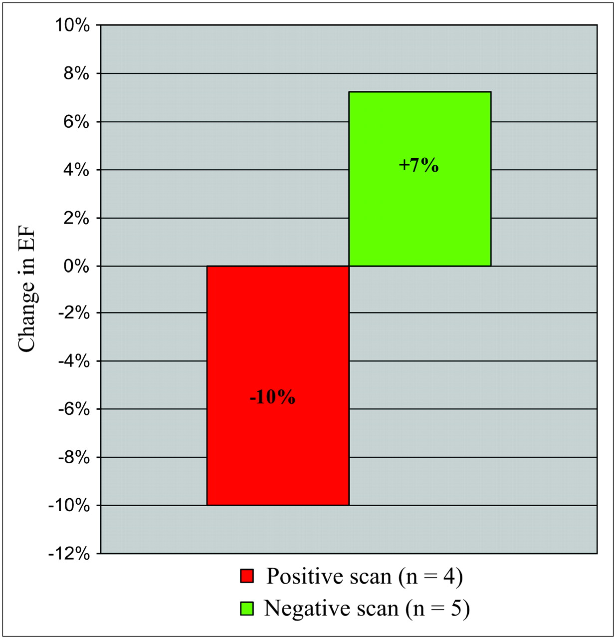
During a follow-up of 1 y, the 4 patients with annexin A5 uptake showed, on average, a decline in LVEF. On the other hand, in the annexin A5–negative patients, LVEF remained stable or increased somewhat after 1 y of follow-up. Figure 2 depicts the change in ejection fraction, subtracting LVEF at the time of imaging from LVEF 1 y after imaging. Student t testing for paired samples showed a significant difference between patients positive and negative for annexin A5 uptake (P = 0.038).

Change in LVEF 1 y after annexin imaging. Green bar shows patients with negative scan findings (mean LVEF increase, 7%); red bar shows patients with positive scan findings (mean LVEF decrease, 10%). P = 0.038.
DISCUSSION
Apoptosis in Heart Failure
PCD contributes to slow, ongoing myocardial dysfunction in heart failure (17). Cytokinemia and ischemic/oxidative stress have been shown to lead to a release of cytochrome c from the mitochondria into the cytoplasmic compartment and to the activation of caspase 3 (18). Active caspase 3 cleaves contractile proteins and activates DNA fragmentation enzymes. The loss of cytochrome c (hence the loss of the energy production mechanism in mitochondria) and the fragmentation of contractile proteins contribute to a decline in left ventricular function. Activation of caspase 3 also results in scrambling of cell membrane phospholipids, thereby expressing PS (target for annexin imaging). However, the simultaneous activation of various antiapoptotic factors in the failing myocardium inhibits caspase-mediated activation of DNases. Such protective steps represent the survival instinct of the apoptotic myocardium and interrupt the apoptotic process (19–21). The greater the number of antiapoptotic factors, the better should be the survival of cardiomyocytes. On the other hand, more activation of caspase 3 results in more PS exposure and, presumably, a worse prognosis.
Noninvasive Imaging of PCD
Radionuclide imaging targets cell-surface alterations to noninvasively recognize various forms of cell death associated with cardiovascular diseases (13,15,22). Myocardial necrosis is characterized by a loss of sarcolemmal integrity, with the cell membrane allowing free access to radiotracers such as glucarate (binding to positively charged histones in the disintegrating nuclei) and antimyosin antibody (binding to fragmenting, insoluble heavy-chain myosin molecules) (23). On the other hand, the sarcolemma remains intact in PCD, but the asymmetry of the phospholipid distribution in the lipid bilayer of the membrane is lost (24). As such, abundant PS is exteriorized to the outer surface of the membrane that serves as a marker for macrophage phagocytosis (24). As an endogenous protein, annexin A5 has high affinity for PS and has therefore successfully been used for the detection of PCD in vivo (13,22). PS exposure is closely linked to the activation of the executioner caspase 3 and can be prevented by administration of caspase inhibitors (25). These cell death inhibitors have been shown to reduce the extent of myocardial damage in various myocardial diseases (8,25). The present study confirmed the feasibility of annexin A5 imaging for the detection of PCD in heart failure patients. Annexin A5 uptake was seen predominantly in those patients who had demonstrated a recent worsening in their left ventricular function or functional class. In addition, these patients on average continued to show a decrease in LVEF for up to 1 y during follow-up.
Other Noninvasive Imaging Studies of Heart Failure
Previous imaging studies for detection of cell death in heart failure were performed using antimyosin antibodies and showed evidence of myocardial necrosis in such patients (23,26). Patients with antimyosin uptake were shown to have a high likelihood of myocarditis or evidence of noninflammatory myocyte degeneration in their endomyocardial biopsy samples. Patients with scans positive for antimyosin showed functional improvement over time, in contrast to those with scans negative for antimyosin. The functional improvement in antimyosin-positive patients appears to be counterintuitive. It was proposed that the antimyosin positivity in dilated cardiomyopathy represented merely the extent of acute myocardial insult and that the accompanying irreversibly damaged, antimyosin-negative myocytes were responsible for functional resolution (23). In contrast, in annexin A5 imaging, annexin-positive patients continue to show a decrease in left ventricular function, and annexin-negative patients show improved left ventricular function. We can only surmise that the antimyosin-positive cells were an indirect marker of reversible cells, whereas the annexin-positive cells represented the true state of balance of proapoptotic and antiapoptotic factors in the cardiomyocytes and should be more predictive of prognosis. It is apparent that the cells with lower amounts of antiapoptotic factors (hence more PS expression and annexin A5 positivity) would be better candidates for exogenous antiapoptotic therapy. These findings have been translated into Figure 3.
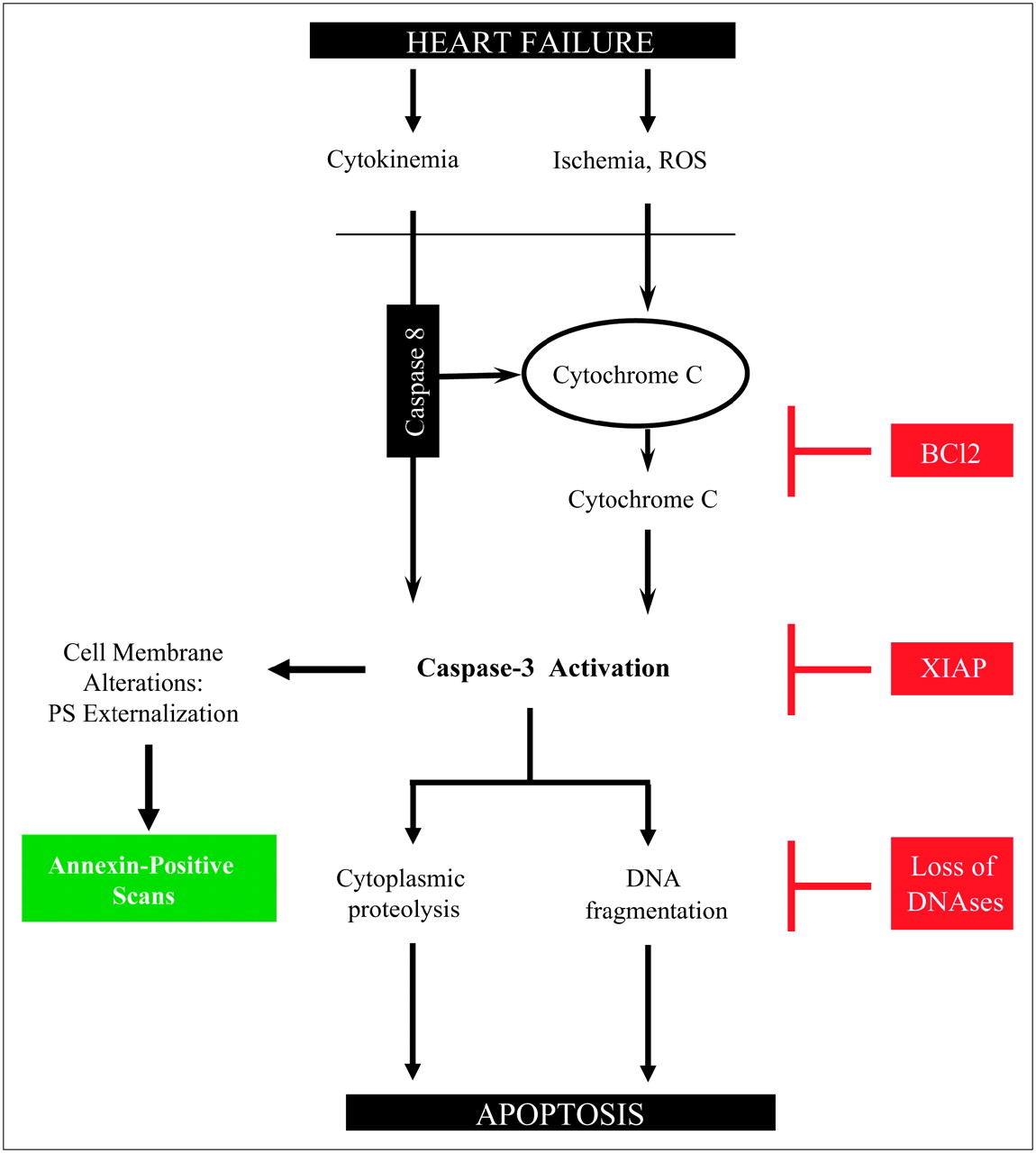
Cytokine and oxidative stress (reactive oxygen species) in heart failure lead to caspase 3 activation by release of cytochrome c from mitochondria into cytoplasmic compartment. Activation of caspase 3 results in cytoplasmic proteolysis and DNA fragmentation and, hence, apoptosis. Endogenous upregulation of BCl2- and XIAP-like proteins and loss of DNA fragmentation factors prevent completion of apoptotic process (apoptosis interruptus). Amount of activated caspase 3 is determined by balance of antiapoptotic and proapoptotic factors. The fewer the endogenous antiapoptotic factors, the greater is the residual caspase, the PS externalization, the likelihood of an annexin-positive scan, and the necessity for apoptosis inhibition therapy and the poorer is the prognosis.
Limitations of Study
These data should be interpreted carefully because of the small number of patients in the study. Further prospective trials including larger numbers of dilated cardiomyopathy patients with sequential scans and functional follow-ups may clarify the current observations. Because 201Tl γ-photons also have an energy peak at 166 keV, there may be some downscatter into the 140-keV window of 99mTc. However, this downscatter was not observed in the family members of the hypertrophic cardiomyopathy patient (Fig. 1D). In addition, we did not observe downscatter in patients included in other studies (unpublished data, November 2006). Furthermore, annexin A5 uptake is focal in most patients, whereas downscatter from 201Tl should appear throughout the left ventricle. Endomyocardial biopsies were not performed in the present study. Because biopsies are not likely to influence management strategy, the ethical committee did not allow biopsies for research purposes and merely for comparison with the results of annexin A5 scans. In addition, serial annexin A5 scans were not allowed. The lack of endomyocardial biopsies precludes the diagnosis of myocarditis in scan-positive patients. Because none of the scan-positive patients showed an improvement in LVEF, the likelihood of myocarditis in those cases is low.
CONCLUSION
This proof-of-principle study suggests that annexin A5 imaging may identify accelerated myocardial cell loss in nonischemic dilated cardiomyopathy patients with a recent worsening of heart failure. Such a strategy may offer a new possibility for studying interventions to minimize the progression of myocardial dysfunction.
Footnotes
-
COPYRIGHT © 2007 by the Society of Nuclear Medicine, Inc.
References
- Received for publication July 18, 2006.
- Accepted for publication January 10, 2007.


Jump to section
Related Articles
Cited By...
- Molecular Imaging of Apoptosis in Ischemia Reperfusion Injury With Radiolabeled Duramycin Targeting Phosphatidylethanolamine: Effective Target Uptake and Reduced Nontarget Organ Radiation Burden
- Noninvasive Imaging in the Assessment of the Cardiopulmonary Vascular Unit
- Noninvasive Imaging of Myocyte Apoptosis Following Application of a Stem Cell-Engineered Delivery Platform to Acutely Infarcted Myocardium
- Molecular Imaging of Coronary Atherosclerosis and Myocardial Infarction: Considerations for the Bench and Perspectives for the Clinic
- Molecular MRI Detects Low Levels of Cardiomyocyte Apoptosis in a Transgenic Model of Chronic Heart Failure
- Serial Noninvasive Assessment of Apoptosis During Right Ventricular Disease Progression in Rats
- Molecular Imaging of Macrophage Cell Death for the Assessment of Plaque Vulnerability
- Multimodality Cardiovascular Molecular Imaging, Part II
- From Better Understood Pathogenesis to Superior Molecular Imaging, and Back...
- Broad and Specific Caspase Inhibitor-Induced Acute Repression of Apoptosis in Atherosclerotic Lesions Evaluated by Radiolabeled Annexin A5 Imaging