

FIGURE 2.

FIGURE 2.
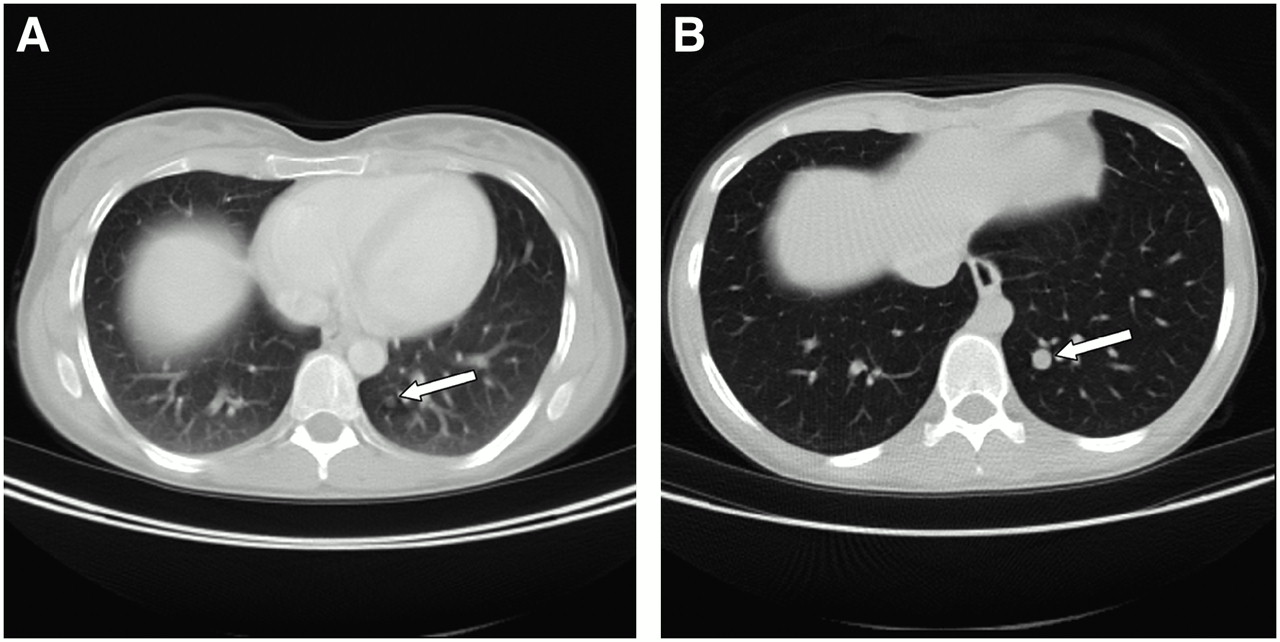
Comparison of chest scanning during shallow breathing (A) and chest scanning with additional low-dose CT during maximal inspiration (B). (A) Lung metastasis from colorectal cancer is only barely visible during shallow breathing (arrow). Also note blurred lung vessels and congested lung parenchyma in this image. (B) Metastasis can be clearly detected by low-dose CT during maximal inspiration (arrow). Additionally, lung parenchyma is well inflated, and lung vessels are displayed sharply.
In this issue


Related Articles
Cited By...
- Weight-Based Protocols Offer Significant Reduction in Radiation Dose Without Affecting PET-CT Image Quality
- Variations in Clinical PET/CT Operations: Results of an International Survey of Active PET/CT Users
- Integrated PET/CT in the staging of nonsmall cell lung cancer: technical aspects and clinical integration
- SPECT/CT
- Introduction