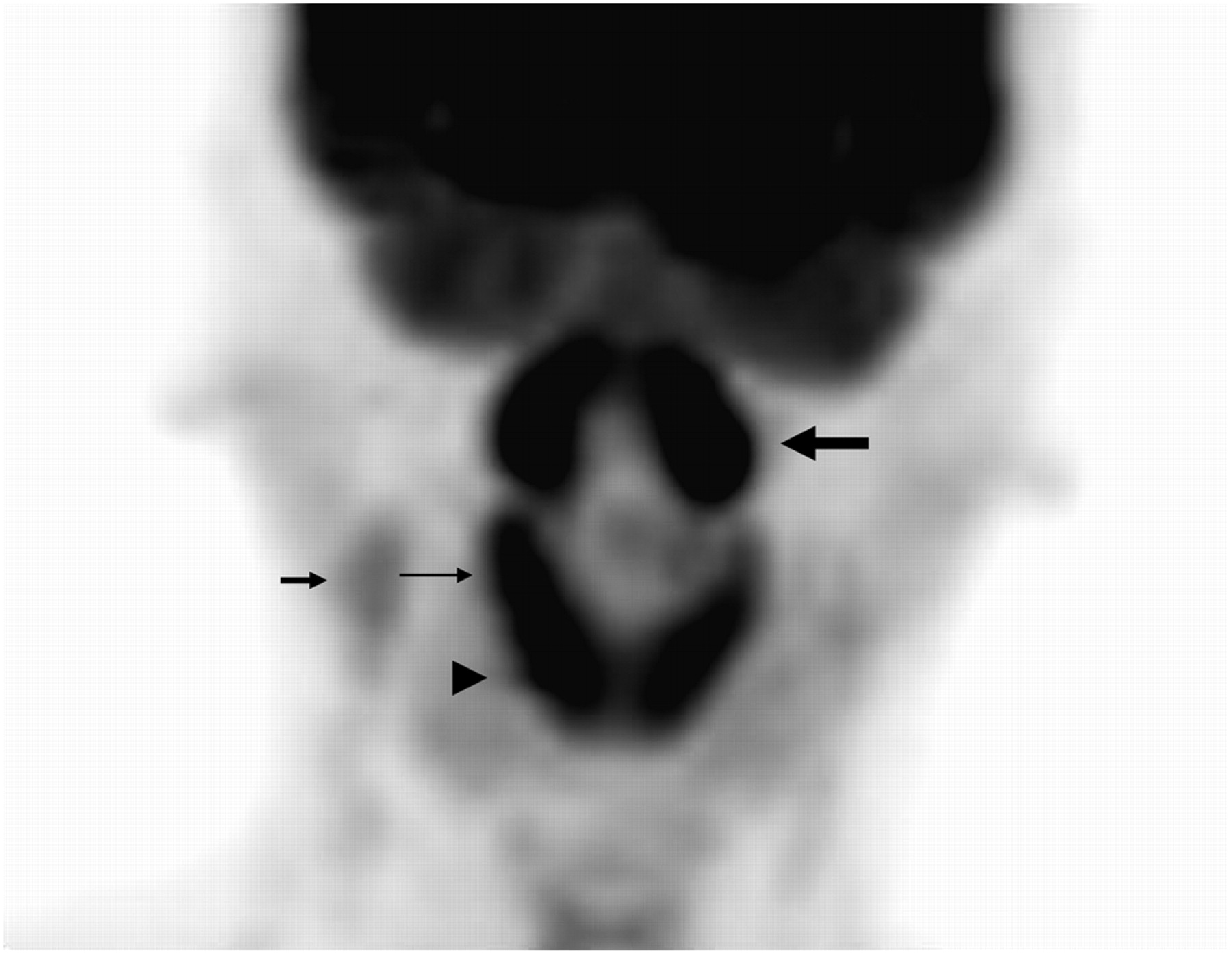
JNM
HOME HELP FEEDBACK SUBSCRIPTIONS ARCHIVE SEARCH TABLE OF CONTENTS
 QUICK SEARCH:   [advanced]


Right arrow Help viewing high resolution images
Right arrow Return to article
Click on image to view larger version.


Figure 2


FIGURE 2.  Maximum-intensity-projection PET image reveals symmetrically intense 18F-FDG uptake in LPR (thick long arrow), palatine tonsil (thin long arrow), and lingual tonsil (arrowhead) in 39-y-old man with upper respiratory airway infection. Mild 18F-FDG uptake in right high jugular lymph nodes (short arrow) is also seen.


Right arrow Return to article


HOME HELP FEEDBACK SUBSCRIPTIONS ARCHIVE SEARCH TABLE OF CONTENTS
JOURNAL OF NUCLEAR MEDICINE TECHNOLOGY THE JOURNAL OF NUCLEAR MEDICINE