

FIGURE 4.

FIGURE 4.
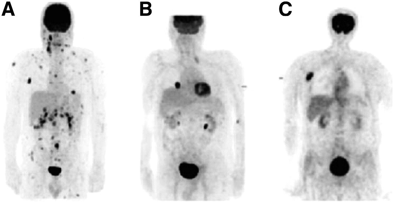
(A) A 45-y-old-man with history of melanoma of right neck presented with palpable lymph nodes in right neck. 18F-FDG PET showed extensive metastatic disease. (B) A 73-y-old-man with history of melanoma of left scalp presented with undetermined lung nodule in right lower lobe. 18F-FDG PET scan showed single metastasis in right lung; lesion was suitable for resection. (C) A 61-y-old-man with history of melanoma on back presented with posterior neck lesion. 18F-FDG PET scan showed only right axillary mass and no other sites of metastatic disease.
In this issue


Related Articles
Cited By...
- Ultrasensitive detection of malignant melanoma using PET molecular imaging probes
- Assessment of Patient Exposure to X-Radiation from SPECT/CT Scanners
- PET of Malignant Melanoma Using 18F-Labeled Metallopeptides
- Amelanotic Malignant Melanoma Mimicking Hemangioma of the Hand: One Case Report and Literature Review
- Melanin-Targeted Preclinical PET Imaging of Melanoma Metastasis
- Monitoring Caspase-3 Activation with a Multimodality Imaging Sensor in Living Subjects