

FIGURE 1.

FIGURE 1.
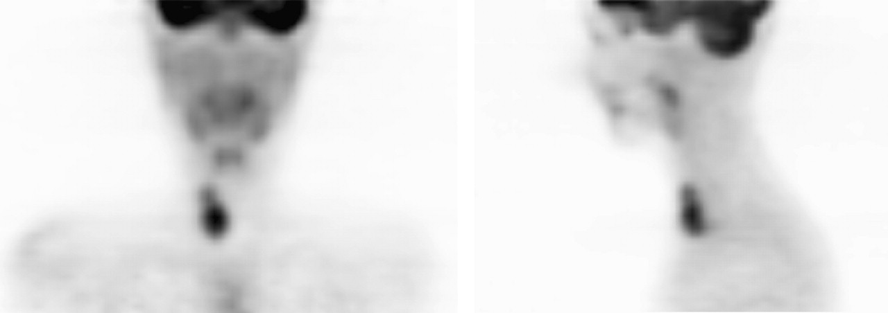
In this 38-y-old woman, FNAB showed follicular proliferation. 18F-FDG PET demonstrated 2 lesions in right thyroid lobe. Final histopathologic diagnosis revealed right-sided pT3 follicular thyroid carcinoma and pT1 papillary thyroid carcinoma cranial from this lesion.
In this issue


Related Articles
Cited By...
- Thyroid nodules with indeterminate cytology: prospective comparison between 18F-FDG-PET/CT, multiparametric neck ultrasonography, 99mTc-MIBI scintigraphy and histology
- IMAGING IN ENDOCRINOLOGY: 2-[18F]-fluoro-2-deoxy-D-glucose positron emission tomography/computed tomography in differentiated thyroid carcinoma: clinical indications and controversies in diagnosis and follow-up
- The role of 18F-fluorodeoxyglucose positron emission tomography in differentiated thyroid cancer before surgery
- The Role of 18F-Fluorodeoxyglucose Positron Emission Tomography in Thyroid Neoplasms
- Hybrid SPECT-CT and PET-CT imaging of differentiated thyroid carcinoma
- Incidental thyroid nodule
- The role of F-18-fluorodeoxyglucose positron emission tomography in the postoperative evaluation of differentiated thyroid cancer
- Reply: 18F-FDG PET of Thyroid Nodules with Inconclusive Cytologic Results.
- 18F-FDG PET of Thyroid Nodules with Inconclusive Cytologic Results