

FIGURE 1.

FIGURE 1.
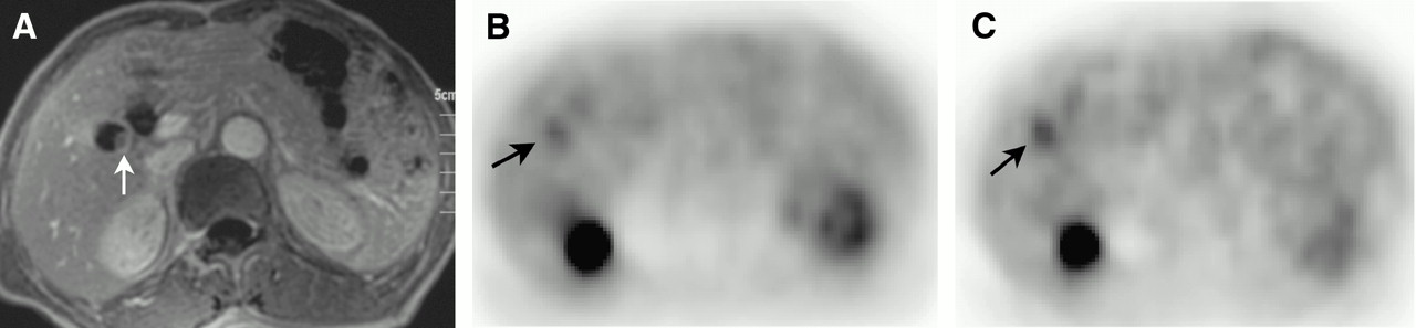
Radiologic findings in 66-y-old man with gallbladder carcinoma. (A) MR image shows small polypoid lesion projecting into lumen of gallbladder (arrow). (B) Early 18F-FDG PET image (SUVearly, 2.72; T/L(E) ratio, 1.10) shows slightly increased uptake at tumor site (arrow). (C) Delayed 18F-FDG PET image (SUVdelayed, 3.21; T/L(D) ratio, 1.52) shows more definite uptake (arrow) than does early image. CRP was 0.25 mg/dL.
In this issue


Related Articles
Cited By...
- Risk Stratification of Gallbladder Polyps (1-2 cm) for Surgical Intervention with 18F-FDG PET/CT
- The value of dual-time-point 18F-FDG PET/CT for identifying axillary lymph node metastasis in breast cancer patients
- Time and Again, Children Resemble Their Parents
- Evaluation of Dual-Time-Point 18F-FDG PET for Staging in Patients with Lung Cancer