

FIGURE 1.

FIGURE 1.
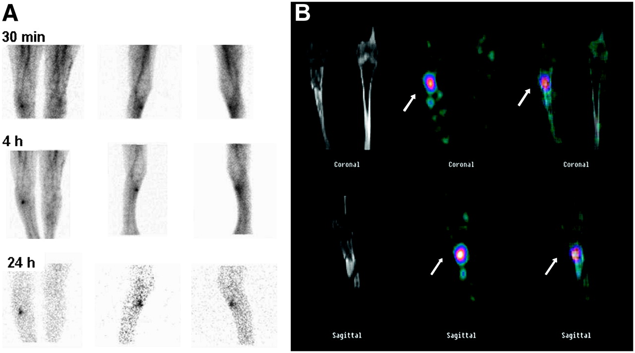
99mTc-labeled leukocyte scintigraphy of 50-y-old woman in whom posttraumatic osteomyelitis of right tibia was suspected. (A) Planar and SPECT images show focal area of tracer uptake in lower right limb. (B) SPECT/CT precisely localizes this focus to corresponding structural alteration in right tibia. Final diagnosis (made by surgery) was posttraumatic tibial osteomyelitis.
In this issue


Related Articles
Cited By...
- 68Ga-Pentixafor PET/CT Imaging of Chemokine Receptor CXCR4 in Chronic Infection of the Bone: First Insights
- Radionuclide Imaging of Musculoskeletal Infection: A Review
- SPECT-CT: applications in musculoskeletal radiology
- Indexing Severity of Diabetic Foot Infection With 99mTc-WBC SPECT/CT Hybrid Imaging
- Leukocyte SPECT/CT for Detecting Infection of Left-Ventricular-Assist Devices: Preliminary Results
- Diabetic Foot Infection: Usefulness of SPECT/CT for 99mTc-HMPAO-Labeled Leukocyte Imaging
- Incremental Value of 131I SPECT/CT in the Management of Patients with Differentiated Thyroid Carcinoma
- SPECT/CT
- Diagnosis of Vascular Prosthesis Infection: PET or SPECT?