

FIGURE 1.
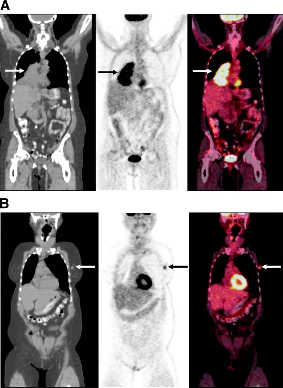
FIGURE 1.
Coronal PET/CT images of 73-y-old woman with recently diagnosed cancer in right lung. (A) Images show large 18F-FDG-avid mass in right lung (arrow), consistent with patient’s known lung cancer. Biopsy revealed small cell lung cancer. (B) Anterior slices from same PET/CT study showed 18F-FDG-avid nodule in left breast (arrow), highly suggestive of malignancy. Pathology revealed infiltrating ductal carcinoma. Thus, this case was true-positive for an additional primary malignancy.
In this issue


Related Articles
Cited By...
- Detection of Additional Primary Neoplasms on 18F-Fluciclovine PET/CT in Patients with Primary Prostate Cancer
- Multiple primary tumours: challenges and approaches, a review
- Assessment of incidental and clinically unsuspected fluorodeoxyglucose-avid foci detected on oncological positron emission tomography/CT
- The role of the breast radiologist in evaluation of breast incidentalomas detected on 18-fludeoxyglucose positron emission tomography/CT
- Incidental findings on positron emission tomography/CT scans performed in the investigation of lung cancer
- Significance of incidental focal uptake in prostate on 18-fluoro-2-deoxyglucose positron emission tomography CT images
- Incidental findings in imaging diagnostic tests: a systematic review
- Comparison of Whole-Body PET/CT, Dedicated High-Resolution Head and Neck PET/CT, and Contrast-Enhanced CT in Preoperative Staging of Clinically M0 Squamous Cell Carcinoma of the Head and Neck
- Prospective Evaluation of Whole-Body Cancer Screening With Multiple Modalities Including [18F]Fluorodeoxyglucose Positron Emission Tomography in a Healthy Population: A Preliminary Report
- Incidental Detection of Concurrent Extramedullary Plasmacytoma and Amyloidoma of the Nasopharynx on [18F]Fluorodeoxyglucose Positron Emission Tomography/Computed Tomography
- Detection of extrapulmonary lesions with integrated PET/CT in the staging of lung cancer
- Role of Nuclear Medicine in the Management of Cutaneous Malignant Melanoma
- Focal Thyroid Lesions Incidentally Identified by Integrated 18F-FDG PET/CT: Clinical Significance and Improved Characterization