


Abstract
Hepatic ischemia/reperfusion injury occurs in numerous clinical situations including liver transplantation and hepatic resection. Therefore, accurate functional assessment of hepatocytes and prevention of ischemia/reperfusion injury to hepatocytes would be important. 99mTc-Galactosyl-human serum albumin is a liver scintigraphic agent that binds to asialoglycoprotein receptors (ASGP-R) on hepatocytes. We determined the number of ASGP-R during hypoxic conditions in primary cultured rat hepatocytes. Methods: We used 3 durations of hypoxia (1, 2, and 3 h) for the cultured rat hepatocytes. The control incubation was performed under normoxic conditions (humidified 5% CO2 in air) for the entire experiment. The maximal binding of 99mTc-galactosyl-human serum albumin (Bmax) to the hepatocytes (plasma membrane and endocytosis) and ketone body ratio (KBR) in the medium were estimated. Results: The Bmax to hepatocytes and the KBR significantly decreased with time under the 3 different hypoxic conditions, whereas the cell counts of the hepatocytes did not decrease. Three hours after reoxygenation, the Bmax and KBR values that were decreased in the first 2 h of hypoxia reversed to control levels, but those Bmax and KBR values that were decreased after 3 h of hypoxia were irreversible. Conclusion: We conclude that the decrease in the number of ASGP-R per hepatocyte appears to be more significant than the decrease in the number of hepatocytes. Therefore, measurement of ASGP-R may provide an accurate assessment of hepatic function in the clinical setting of hepatic injury and recovery.
Hepatic ischemia/reperfusion (I/Rp) injury occurs in numerous clinical situations including liver transplantation and hepatic resection (1,2). Therefore, accurate functional assessment of hepatocytes and prevention of I/Rp injury to hepatocytes would be important. Asialoglycoprotein receptors (ASGP-R) are present only on the hepatocyte cell surface and are specific for a galactose-terminated glycoprotein in the sinusoidal membranes of hepatocytes. Sawamura et al. (3,4) reported that a decrease in the number of ASGP-R led to an accumulation of asialoglycoprotein in the sera of galactosamine-treated rats, and the number of these receptors decreases in patients with chronic liver diseases.
99mTc-Diethylenetriamine-pentaacetic acid-galactosyl-human serum albumin (GSA) is a new liver scintigraphic agent that binds to ASGP-R on hepatocytes (5). The maximal removal rate of 99mTc-GSA reflects the total number of ASGP-R in the whole liver and represents, therefore, liver function. Thus, the maximal removal rate of 99mTc-GSA can be measured to estimate the functioning hepatocyte mass during a routine clinical evaluation (6,7). Using a rat I/Rp injury model, Toyama et al. (8) reported that ASGP-R binding provided valuable information on I/Rp injury and recovery. However, it has still been unclear whether a decrease in the maximal removal rate of GSA reflects a decrease in the number of hepatocytes or a decrease in the density of ASGP-R per hepatocyte. In this study, we determined the number of ASGP-R during hypoxic conditions in primary cultured rat hepatocytes.
MATERIALS AND METHODS
Radiopharmaceuticals
We used 99mTc-GSA (Nihon Medi-Physics). The GSA was obtained by combining 1 molecule of human serum albumin with 30–44 molecules of galactose, and diethylenetriaminepentaacetic acid was used as the chelating agent for labeling with 99mTc. The radiochemical purity was confirmed by thin-layer chromatography and was always 98% or higher.
Primary Culture of Rat Hepatocytes
Hepatocytes were obtained from adult male Wistar rats (6–7 wk old, 180–240 g) fed ad libitum. The cells were isolated by perfusing the liver with collagenase under sterile conditions (9). The isolated hepatocytes were suspended in culture medium at a density of 0.5–0.6 × 10⁶ cells/mL and were inoculated into plastic culture dishes (2 mL per well, 35 mm × 10 mm; Falcon Plastic) and cultured as monolayers in a CO2 incubator (humidified 5% CO2 in air) at 37°C. Unless otherwise specified, the hepatocytes were maintained at a concentration of 0.8–1.0 × 10⁶ cells per well in Williams’ medium E supplemented with 10% fetal bovine serum, 2-[4-(2-hydroxyethyl)-1-piperazinyl-ethanesulfonic acid] (5 mmol/L), penicillin (100 U/mL), streptomycin (0.1 mg/mL), dexamethasone (108 mol/L), insulin (108 mol/L), and glucagon (109 mol/L). After 2 h, the initial medium was replaced by a serum-free medium (1.5 mL per dish), and the culture medium was then changed daily. After 2 d in culture, the cell culture medium was changed to a new medium and then used in the experiments. This study was approved by the animal welfare committee of Kansai Medical University.
Preparation of Hypoxic Conditions
We maintained hypoxic conditions using the Anaeropack for Cell (Mitsubishi Gas Chemical Co.) according to methods previously reported (10). The Anaeropack for Cell contains sodium ascorbate as the principal ingredient, which absorbs oxygen and generates carbon dioxide by oxidative degradation. Another reagent (magnesium hydroxide) is used as a carbon dioxide absorber to scavenge the carbon dioxide. These reagents were packed into a paper sachet. The culture dishes were placed into a 1.6-L airtight jar (Mitsubishi Gas Chemical) with the paper sachet, and the jar was then incubated in a CO2 incubator at 37°C. We used 3 durations of hypoxia (1, 2, and 3 h) for the cultured rat hepatocytes. The control incubation was performed under normoxic conditions (humidified 5% CO2 in air) for the entire experiment. A set of dishes was used for counting the viable cells. Hepatocyte viability was determined by the trypan blue exclusion method and leakage of lactate dehydrogenase.
Measurement of Surface-Bound Ligand and Endocytosis on Cultured Hepatocytes
The concentration of 99mTc-GSA (3 mg/185 MBq) for the binding assays was adjusted with saline just before each experiment. To perform the binding assay, various doses of 99mTc-GSA in 20 μL of saline were added to each dish (n = 3) for all experiments. For experiments designed to measure cell surface–bound ligand, the cells were preincubated at 4°C for 15 min before the binding with 99mTc-GSA. The binding to a monolayer of cultured hepatocytes was performed on ice for 3 h with gentle swirling every 15 min. For the measurement of endocytosis, the binding to a monolayer of hepatocytes was performed for 30 min in a 37°C incubator. The internalization was stopped by chilling the hepatocytes on ice and by replacing with ice-cold medium. The unbound ligand was then removed by washing with saline at 4°C.
In both experiments, the nonspecific binding of 99mTc-GSA was assessed by including an excess of unlabeled GSA (at least 500-fold). Finally, the unbound ligand was removed by careful aspiration of the medium and 3 washes with fresh medium. The cells were scraped off the dish with a rubber policeman and were collected into tubes containing 2 mL of saline. The radioactivity was then determined using an automatic γ-counter (model B5003, COBRA; Packard Instrument Co.). The maximal binding of 99mTc-GSA (Bmax) was analyzed by Scatchard plots using Rosenthal’s graphic method (11) and was expressed as nanograms of ligand per 106 cells and as the number of binding sites (ASGP-R) per cell.
Total Ketone Body (TKB) Concentration and Ketone Body Ratio (KBR) in Hepatocyte Culture Medium
The concentration of TKB (acetoacetate + β-hydroxybutyrate) in the medium was measured to assess hepatic mitochondrial function. Immediately on removal from the hypoxic airtight jar, the culture dish was placed on crushed ice and the culture medium was collected into a cold glass test tube. The TKB concentration of the medium was measured using a Ketolex kit (Sanwa Kagaku Co., Ltd.), and the KBR was calculated as the ratio of acetoacetate to β-hydroxybutyrate (12).
Statistical Analysis
All data are expressed as mean ± SD. Differences between groups were evaluated by F ratio using a 1-way ANOVA, and a P value less than 0.05 was considered to be statistically significant.
RESULTS
Confirmation of Hypoxic Conditions
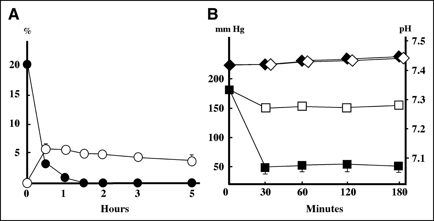
The concentration of oxygen in the jar dropped to less than 1% within 1 h after the induction of hypoxia and reached its lowest level (<0.1%) after 3 h. The concentration of carbon dioxide in the jar rose to about 5% at 30 min after the induction of the hypoxia. Both oxygen and carbon dioxide concentrations were maintained at these levels for up to 5 h after hypoxia began, when the sachet was initially placed into the airtight jar. There was no detectable difference in the pH value of the medium between the hypoxic and control incubations. Moreover, the partial oxygen pressure in the cell culture medium decreased to 50 mm Hg within 30 min of being in the jar (Fig. 1).

Confirmation of hypoxic conditions. Concentration of oxygen (•) in jar dropped to less than 1% within 1 h after induction of hypoxia, whereas concentration of carbon dioxide (○) in jar rose to about 5% at 30 min after induction of hypoxia (A). No difference in pH value of medium was detectable between hypoxia (♦) and control incubation (⋄). Partial oxygen pressure in culture medium decreased to 50 mm Hg within 30 min of being in the jar (▪), whereas culture medium partial oxygen pressure in incubator (□) was maintained at 150 mm Hg (B). All data are expressed as mean ± SD (n = 3).
Binding of Hepatocyte Plasma Membrane and Endocytosis Assay of 99mTc-GSA to Cultured Hepatocytes During Hypoxic Conditions
The Bmax to the hepatocyte plasma membrane significantly decreased with time under the 3 different hypoxic conditions (P < 0.0001), whereas the cell counts of the hepatocytes, which indicate the number of viable cultured hepatocytes, were not influenced by the hypoxic conditions. Therefore, the decrease in the number of the ASGP-R binding sites on the plasma membrane per hepatocyte was dependent on the duration of the hypoxia (Table 1). Similarly, the endocytotic Bmax significantly decreased according to the duration of the hypoxia (P < 0.0001). Because the cell counts of the viable hepatocytes were not influenced by the duration of the hypoxic conditions, we concluded that the hypoxic conditions decreased the number of ASGP-R binding sites involved in endocytosis per hepatocyte (Table 2). Three hours after reoxygenation, the Bmax values that were decreased in the first 2 h of hypoxia reversed to control levels, but those Bmax values that were decreased after 3 h of hypoxia were irreversible (Tables 1 and 2).
Number of ASGP-R on Hepatocyte Plasma Membrane Under Hypoxic Conditions and Reoxygenation
Number of ASGP-R Involved in Hepatocyte Endocytosis Under Hypoxic Conditions and Reoxygenation
TKB Concentration and KBR in Hepatocyte Culture Medium
The TKB concentrations in the medium were not influenced by the duration of the hypoxic conditions. However, the KBR in the hepatocyte culture medium significantly decreased according to the duration of the hypoxia (P < 0.0001). The cell counts of the viable hepatocytes were not influenced by the duration of the hypoxic conditions, in contrast to the decreased Bmax values seen under those conditions. Three hours after reoxygenation, the KBR seen after the first 2 h of hypoxia appeared to reverse to prehypoxic values, but the KBR found after 3 h of hypoxia remained at a lower level (Table 3).
TKB and KBR in Culture Medium Under Hypoxic Conditions and Reoxygenation
DISCUSSION
Several pathways for I/Rp injury have been postulated, such as oxygen-derived free radicals (13), calcium influx (14), adenosine triphosphate depletion and mitochondrial dysfunction (15), activation of lysosomal enzymes (16), and disturbance of microcirculation (2). However, the precise mechanisms of hepatic injury remain obscure. Extensive investigation of pathogenesis in I/Rp injury and its estimation would be potentially important in the clinical setting. Hepatic I/Rp injury consists of 2 phases. An early phase of injury that occurs during the first 1–3 h after reperfusion is mediated through an oxygen-free radical mechanism, which is correlated with Kupffer’s cell activation. A late phase of injury that occurs between 6 and 24 h after reperfusion is associated with hepatic neutrophil infiltration. The GPT levels transiently increased in the case of reversible liver damage but remained high and increased continuously when the liver injury was irreversible (17). The tissue blood flow gradually recovered until 120 min after reperfusion with a 30-min I/Rp model in rat liver (18). In a 90-min I/Rp model, liver uptake of 125I-GSA was significantly decreased but blood uptake was significantly increased at 1 and 3 h after declamping (8). Our findings of the decrease in the ASGP-R per hepatocyte under hypoxic conditions suggests that the impaired liver function reflects the impaired hepatocyte that has fewer ASGP-R rather than a decrease in the number of intact hepatocytes. Burgess et al. (19) demonstrated that an abnormal cell surface distribution of ASGP-R commonly occurs in cirrhosis and that this abnormality might result in the impaired clearance of desialylated glycoproteins from the plasma. This abnormality of the ASGP-R might also contribute to generation of impaired hepatocytes, resulting in impaired liver function.
Receptor-mediated endocytosis is common to many cell types, including hepatocytes, and molecules in the extracellular fluid bind to cell surface receptors and are internalized as receptor–ligand complexes via a clathrin-coated pit/coated vesicle pathway. Within this pathway, the vesicles lose the clathrin coats, form endosomes, and are sorted in the acidic compartment of uncoupling receptor and ligand. Once separated, ligands and receptors can be routed to the lysosomes for degradation or can be recycled back to the cell surface (20,21). The ASGP-R is almost exclusively distributed between the microsomal fraction (72%) and the plasma membrane fraction (23%) (22). Both the binding and the uptake of asialoglycoprotein occur at 37°C, and only the binding to the cell surface occurs at 4°C. Freshly isolated rat hepatocytes have about 7 × 10⁴ surface receptors per cell. However, when cells were incubated at 37°C, the number of surface receptors per cell rapidly increased 2-fold to 3-fold to about 2.2 × 10⁵ (23). In this experimental study, the numbers of ASGP-R (binding sites) for endocytosis and on the plasma membranes were 6.1 × 10⁵/cell and 2.6 × 10⁵/cell, respectively. The uptake and intracellular transport of asialoglycoproteins occurs independently of microfilaments, but the pH gradient in the endocytic compartments is important in the receptor-mediated endocytosis of asialoglycoproteins (24).
The essential hepatic function is the energy produced in the hepatic mitochondria, and the energy-producing capacity of mitochondria may be said to represent the hepatic functional reserve (25). The arterial KBR (acetoacetate/β-hydroxybutyrate) is an index of the liver mitochondrial redox state ([nicotinamide adenine dinucleotide+]/[reduced nicotinamide adenine dinucleotide]), and the depression of ketone body production during the hypoxia indicates the termination of β-oxidation in the hepatic mitochondria (26). The arterial KBR is well correlated with the hepatic energy charge level ([adenosine triphosphate + 0.5 adenosine diphosphate]/([adenosine triphosphate + adenosine diphosphate + adenosine monophosphate]) and is often decreased in jaundiced (27), hepatectomized (28), and shocked animals (29); therefore, the decrease in arterial KBR is accompanied by massive systemic metabolic derangements. Immediately after reoxygenation started, the ketone body production resumed, and the production of ketone bodies correlated with the duration of the hypoxia. The extent of cell injury caused by changes in the duration of the hypoxia was reflected well by the KBR, the concentration of ketone bodies.
Ischemic liver injury in vivo and anoxic hepatocyte injury in vitro have been characterized by morphologic, metabolic, and functional changes in the liver (30,31). Primary cultured hepatocytes are useful for the study of liver function in vitro. However, the conventional method used to produce hypoxic conditions in culture is complex (32), and there is a limit to the number of examination dishes that can be tested in this manner. In the present study, the Anaeropack for Cell method is easy and useful for simulating the conditions of hepatocytes during normothermic ischemia. The carbon dioxide concentration and pH are known to be important factors in primary tissue culture, and the optimum concentration of carbon dioxide is believed to be about 5% for cultured hepatocytes. This was easily obtained with the Anaeropack for Cell method, and the carbon dioxide concentration had no effect on the pH value of the medium. Using this model, we demonstrated that the number of ASGP-R binding sites on the hepatocyte plasma membrane and the number of endocytosis sites per hepatocyte decreased depending on the duration of the hypoxia. Moreover, the KBR in the culture medium also decreased according to the duration of hypoxia. In addition, this elimination of the ASGP-R and the reduction of the KBR were reversible within the first 2 h of hypoxia.
CONCLUSION
When the liver is damaged, the decrease in the number of ASGP-R per hepatocyte appears to be more significant than the decrease in the number of hepatocytes. Therefore, a measurement of ASGP-R using the maximal removal rate of GSA may provide an accurate assessment of hepatic function in the clinical setting of hepatic injury and recovery.
Footnotes
Received Apr. 21, 2004; revision accepted Sep. 13, 2004.
For correspondence or reprints contact: A-Hon Kwon, MD, Department of Surgery, Kansai Medical University, 10-15 Fumizono, Moriguchi, Osaka 570-8507, Japan.
E-mail: kon{at}takii.kmu.ac.jp
REFERENCES
In this issue


Jump to section
Related Articles
Cited By...
- No citing articles found.