


Abstract
Gene therapy trials would benefit greatly from the use of noninvasive imaging to determine the location, magnitude, and time course of gene transfer. Somatostatin receptor subtype 2 (SSTR2) has been used as a reporter probe for γ-camera imaging of gene transfer in animal models. PET has greater sensitivity than γ-camera imaging and therefore would have an advantage for the imaging of SSTR2 gene transfer. Methods: An adenovirus (AdHASSTR2) carrying sstr2, which encodes an N-terminal hemagglutinin epitope, was used for evaluating SSTR2 gene transfer. The somatostatin analog Demotate 1 (Tyr3-octreotate conjugated to the 1,4,8,11-tetraazaundecane chelator) was used for chelation of the positron emitter 94mTc (half-life, 52 min) and targeting to SSTR2. Gene transfer was evaluated in vitro with A-427 non–small cell lung cancer cells after infection with AdHASSTR2 by 94mTc-Demotate 1 binding and internalization assays. In vivo biodistribution and microPET studies were conducted with mice bearing A-427 tumor xenografts directly injected with AdHASSTR2 to determine the tumor localization of 94mTc-Demotate 1. Results: 94mTc-Demotate 1 bound with high affinity and was internalized rapidly into AdHASSTR2-infected A-427 cells. Biodistribution studies showed uptake of 94mTc-Demotate 1 in tumors infected with AdHASSTR2 (4.0 percentage injected dose per gram [%ID/g] at 2 h) and background uptake in tumors infected with a control adenovirus (0.8 %ID/g at 2 h). The uptake of 94mTc-Demotate 1 in AdHASSTR2-infected tumors was greater than the uptake in all other tissues, except for the kidneys and the SSTR2-positive pancreas. MicroPET imaging showed similar results, with clear uptake of 94mTc-Demotate 1 in AdHASSTR2-infected tumors, background uptake in control tumors, and clearance through the kidneys. Conclusion: These studies show that the positron-emitting somatostatin analog 94mTc-Demotate 1 could be used to determine SSTR2 gene transfer by microPET imaging, a result that will improve the sensitivity of the SSTR2 reporter gene system.
In more than 600 clinical gene therapy trials worldwide in the past 15 y, approximately 3,500 patients have been treated (according to the Journal of Gene Medicine Web site, at www.wiley.co.uk/genetherapy/clinical). More than 500 of the trials have been conducted in the United States, and 400 have been carried out in the context of the treatment of cancer. Retroviral vectors were used as gene delivery vehicles in 34% of the protocols, whereas adenoviral vectors were used in 27%. It is clear that gene therapy will be evaluated clinically as an approach for the treatment of numerous human disorders despite obstacles that must be overcome before gene therapy can live up to its potential (1). One obstacle that must be overcome is the ability to determine whether gene transfer has occurred in an individual patient (2). Biopsies are taken to determine whether gene transfer has occurred, but this process is invasive and does not yield spatial or temporal patterns of gene expression. Therefore, noninvasive imaging of gene transfer would determine whether gene transfer has occurred in a patient, what tissues have been transduced, and the magnitude and time course of gene expression (2).
Several groups are developing methods for noninvasively imaging gene transfer. In general, these methods have focused on nuclear imaging, MRI, and optical imaging (3). Each imaging technology has various advantages and limitations. We have focused on nuclear imaging with the human somatostatin receptor subtype 2 (SSTR2) gene as a reporter gene, although many nuclear imaging studies of gene transfer have been carried out with the herpes simplex virus type 1 thymidine kinase (4,5), the dopamine 2 receptor (6,7), and the sodium iodide symporter (8,9) genes as reporter genes. Five somatostatin receptor subtypes (SSTR1–SSTR5) have been cloned, with alternate splicing of SSTR2 to yield SSTR2A and the C-terminally truncated form, SSTR2B (10). All of the receptor subtypes have high binding affinity for the natural somatostatin 14 or somatostatin 28 peptides. Somatostatin receptors are members of the G protein–coupled receptor family and have 7 transmembrane-spanning domains consisting of 3 extracellular loops, termed E1, E2, and E3 (11). The 5 receptor subtypes are highly homologous between mice and humans and are expressed in the brain, gastrointestinal tract, pancreas, kidneys, and spleen (12,13). Human SSTR2A (hereafter referred to as SSTR2) has been used for the imaging of gene transfer by use of γ-camera imaging and SPECT (14–17). These previous studies used the radiolabeled somatostatin analogs 111In-diethylenetriaminepentaacetic acid-octreotide (Octreoscan; Mallinckrodt), 99mTc-P829 (NeoTect; Diatide, Inc.), and 99mTc-P2045 (Diatide, Inc.) to demonstrate localization in a tumor after intratumoral injection with an adenovirus or intraperitoneal injection with a vaccinia virus carrying the sstr2 gene.
Because PET is at least 1 order of magnitude more sensitive than SPECT, in part because of the compromise between spatial resolution and sensitivity attributable to the required collimator for SPECT (18,19), it would be advantageous to image SSTR2 gene transfer by PET. In recent years, there has been interest in somatostatin analogs radiolabeled with PET radionuclides, such as 18F, 86Y, 66,68Ga, and 64Cu (20–24). All of these radionuclides have advantages and disadvantages for use in the labeling of somatostatin analogs. In this study, we used the positron emitter 94mTc (half-life, 52 min; β+, 72%; Eβ+max, 2.5 MeV) produced with a high specific activity on a biomedical cyclotron (25). The somatostatin analog Demotate 1 (Tyr3-octreotate conjugated to the 1,4,8,11-tetraazaundecane chelator) (Fig. 1) was used for chelation of 94mTc and targeting to SSTR2. This analog was previously radiolabeled with 99mTc for γ-camera imaging of SSTR2-positive tumors (26). The binding and internalization of 94mTc-Demotate 1 were evaluated in vitro after infection of cells with an adenovirus (AdHASSTR2) encoding SSTR2 that contains the influenza virus hemagglutinin (HA) sequence at the N terminus of the receptor. The expression of SSTR2 then was evaluated in vivo with tumor-bearing mice in biodistribution and microPET imaging studies. To our knowledge, this is the first study to use PET to evaluate SSTR2 gene transfer.
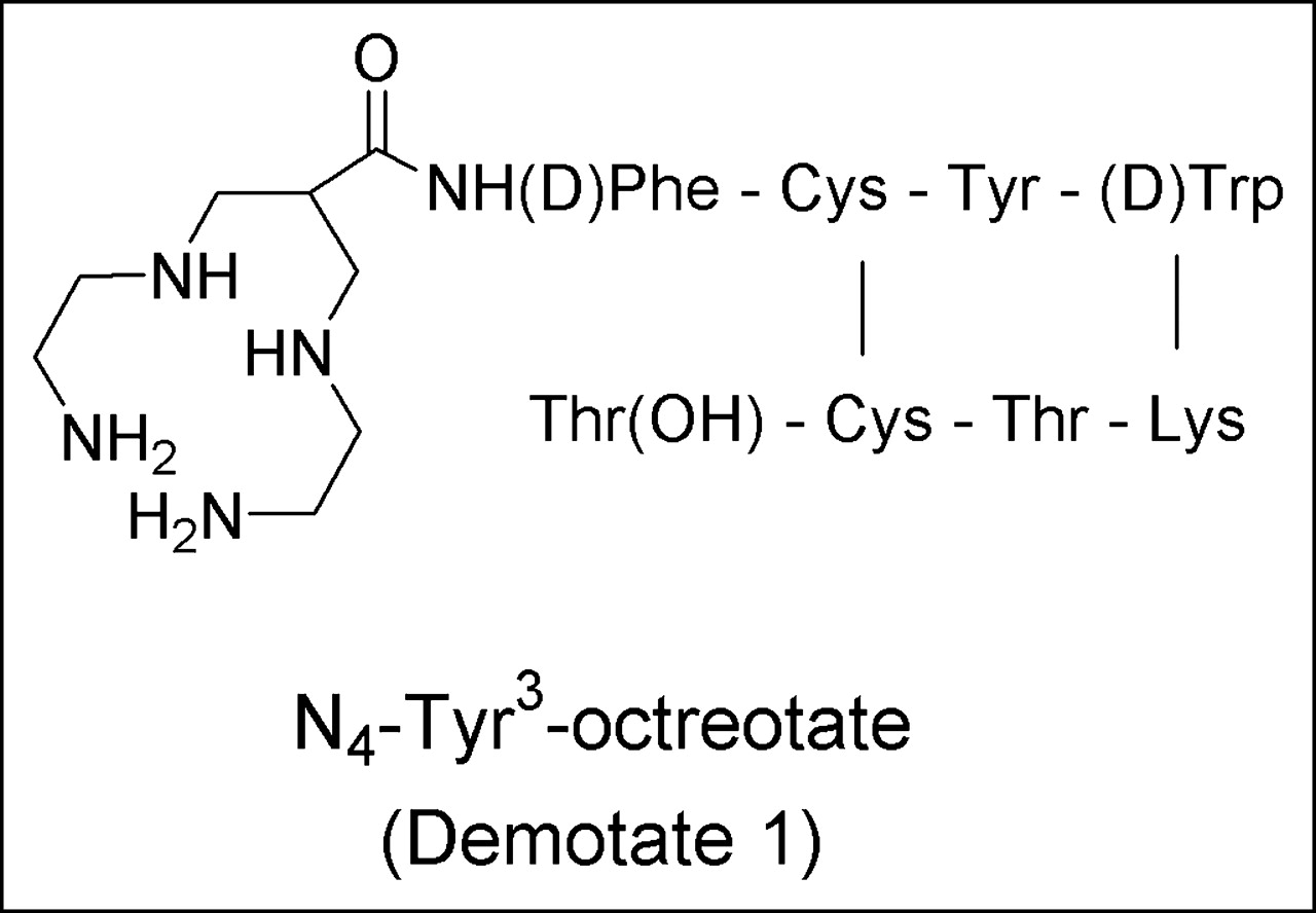
Structure of Demotate 1.
MATERIALS AND METHODS
Cell Line and Adenovirus
The A-427 non–small cell lung cancer cell line was purchased from the American Type Culture Collection and maintained in Eagle minimum essential medium supplemented with 1% nonessential amino acids, 1% sodium pyruvate, and 10% heat-inactivated fetal bovine serum. All cell culture reagents were obtained from Invitrogen, except for fetal bovine serum, which was purchased from Sigma Chemical Co. The cells were maintained in a humidified atmosphere with 5% CO2 at 37°C. AdHASSTR2 was constructed and propagated and virus titers were determined as previously described (27), and the virus was stored at −80°C until use in cell culture and animal studies.
Production of 94mTc
94mTc was produced by a method similar to that previously described (25,28,29). Briefly, MoO3 (Isoflex USA) (∼60 mg), enriched with 91.0% 94Mo, was pressed into a platinum disk and covered with aluminum foil. The disk was irradiated (14.7 MeV, 4 μA, 45 min) on our biomedical cyclotron, and 94mTc was produced by the 94Mo(p,n)94mTc nuclear reaction. After irradiation, the 94mTc was separated by heating of the target in a quartz apparatus at 1,000°C and allowing the 94mTc to condense in the temperature region of 300°C–400°C. The 94mTc was removed in NaOH (0.1 mmol/L) and purified over an Alumina N Sep-Pak Light cartridge (Millipore) to yield the desired 94mTcO4− in saline. The product was >95% 94mTcO4−, as demonstrated by use of instant thin-layer chromatography (ITLC) silica gel plates (Pall Corp.) with acetone as the solvent. The yield of 94mTc was 1,295 MBq (35 mCi)/μA·h at the end of bombardment (EOB), and isotopic impurities were measured on a multichannel analyzing Ge γ-spectrometer (Canberra). The saline containing 94mTcO4− was used immediately after isolation for the radiolabeling of Demotate 1.
Synthesis and Radiolabeling of Demotate 1
Demotate 1 was synthesized as previously described (26). For radiolabeling, Demotate 1 was dissolved in acetic acid (10 mmol/L) at 2 mg/mL, and 15 μg were added to a vial containing 25 μL of phosphate buffer (0.5 mol/L), 3 μL of trisodium citrate buffer (0.1 mol/L), and 196 MBq (5.3 mCi) of 94mTcO4− (200 μL). To this solution, 20 μg of SnCl2 (dissolved in ethanol at 5 mg/mL) were added, and the reaction mixture was incubated for 15 min at room temperature. The reaction was stopped by the addition of 4 μL of HCl (1 mol/L) and 25 μL of ethanol. Radiochemical purity was determined by use of ITLC with acetone and 1:1 methanol:ammonium acetate (1 mol/L) as the solvents. The plates were analyzed by use of an imaging scanner (Bioscan), with 94mTc-Demotate 1 remaining at the origin in the acetone system and moving with the solvent front in the ammonium acetate system.
Saturation Binding Assays
A-427 cells were infected with AdHASSTR2 at 10 plaque-forming units (pfu) per cell and harvested 2 d later to prepare membrane homogenates as previously described (30). Twenty-five micrograms of membrane homogenate were added to each well of a 96-well Multiscreen Durapore filtration plate (Millipore) and rinsed twice with cold buffer [N-(2-hydroxyethyl)piperazine-N′-(2-ethanesulfonic acid) (HEPES) at 10 mmol/L, MgCl2 at 5 mmol/L, ethylenediaminetetraacetic acid at 1 mmol/L, and 0.1% bovine serum albumin (pH 7.4)]. Various concentrations (0.5−100 nmol/L) of 94mTc-Demotate 1 were added to the wells in triplicate in the presence or absence of Tyr1-somatostatin (Sigma Chemical Co.) as an inhibitor such that the final volume in each well was 110 μL. The plate was incubated at room temperature for 30 min with shaking, the wells were rinsed twice with buffer, and the filters were dried and removed. Counts in samples were determined by use of a model 8000 automated well-type γ-counter (Beckman). Data were analyzed by use of the 1-site binding equation in Prism software (GraphPad) to generate equilibrium dissociation constants (Kd) and maximum binding capacities.
Internalization Assays
A-427 cells were infected with AdHASSTR2 at 10 pfu per cell and then used to seed 6-well plates 24 h later. One day after seeding (2 d after adenovirus infection), 94mTc-Demotate 1 (1 nmol/L) in 1 mL of internalization medium (Dulbecco minimum essential medium, HEPES at 30 mmol/L, l-glutamine at 2 mmol/L, sodium pyruvate at 1 mmol/L, and 1% bovine serum albumin) was added to each well and incubated at 37°C for 5, 15, 30, 60, and 120 min. An excess (>1,000 nmol/L) of Tyr1-somatostatin was added to half of the wells to demonstrate specific binding and internalization. The cells were rinsed twice with phosphate-buffered saline and then with Hanks balanced salt solution containing sodium acetate at 20 mmol/L (pH 4.0) to remove surface-bound radioactivity. The cells then were harvested with 1 mL of sodium borate (10 mmol/L) containing 10% sodium dodecyl sulfate and counted in the γ-counter along with the acid washes to determine the internalized radioactivity and the cell surface radioactivity. After 24 h, the amount of protein in each well was determined by use of a bicinchoninic acid (BCA) protein assay kit (Pierce). The data are presented as the specific amount of peptide (Tyr1-somatostatin–blocked wells subtracted from nonblocked wells) bound to the cell surface or internalized, normalized to the amount of protein per well.
Animal Biodistribution Studies
All animal studies were performed in compliance with guidelines for the care and use of research animals established by the Washington University Animal Studies Committee. Homozygous 6- to 8-wk-old female nude (nu/nu) mice (Charles River Laboratories, Inc.) were implanted subcutaneously in each rear flank with 107 A-427 cells mixed 1:1 with Matrigel (Becton Dickinson) such that each mouse had 2 tumors. Three weeks after tumor cell inoculation, tumors on 1 side of the mice were injected directly with 109 pfu (30 μL) of AdHASSTR2, and the other tumors were injected with 109 pfu (30 μL) of a control adenovirus (AdGRPR, encoding the gastrin-releasing peptide receptor), as previously described (31). Two days after the adenovirus injections, 0.6 MBq (17 μCi; 100 ng) of 94mTc-Demotate 1 was injected via the tail vein. The mice (n = 6) were sacrificed by cervical dislocation 1 and 2 h later. In another experiment, the same procedures were followed, except that 1 group of mice was injected in the tail vein with 94mTc-Demotate 1 and another group was coinjected with 50 μg of unlabeled Demotate 1 as an inhibitor. Both groups (n = 5) were sacrificed 1 h after injection. The blood, lungs, liver, spleen, kidneys, muscle, bone, pancreas, and tumors were harvested, weighed, and counted in the γ-counter. Samples were corrected for radioactive decay to calculate the percentage injected dose per gram (% ID/g) of tissue by comparison with a standard representing the injected dose.
MicroPET Imaging
MicroPET imaging was performed with an R4 microPET scanner (Concorde Microsystems Inc.). The R4 microPET scanner has a field of view of 8 cm axially by 11 cm transaxially and is capable of a spatial resolution of 2.3 mm and an absolute sensitivity of 1,020 counts per second per 0.037 MBq in the middle of the field of view. The same animal model as that described above was used for microPET imaging, except that the tumors were implanted in the axillary thorax. In these studies, the mice (n = 3) were injected via the tail vein with 18.5 MBq (500 μCi; 3.4 μg) of 94mTc-Demotate 1, anesthetized 1 and 2 h later with 1%–2% isoflurane, positioned supine, immobilized, and imaged. The tumor and kidney standard uptake values (SUVs) were generated as previously described (32). Briefly, the regions of interest encompassing the entire tumor or tissue on the microPET images were measured to generate the radioactivity concentration, which was decay corrected to the time of injection. This value then was multiplied by the mouse body weight (grams) and divided by the injected dose (Bq) to yield the tissue uptake normalized to injected dose per gram of body weight: (Bq/mL) × (weight [g]/injected dose [Bq]).
Statistical Analysis
All of the data are presented as the mean ± SEM. The Student t test (2 tailed) was performed with Prism software to determine statistical significance at the 95% confidence level, with P < 0.05 being considered significantly different.
RESULTS
Production of 94mTc and Radiolabeling of Demotate 1
The major radionuclide impurity of <7% was from 94gTc (half-life, 293 min; β+, 10.5%; Eβ+max, 811 keV), with all other radionuclide impurities constituting <0.4%. The positron contribution of 94gTc was not greater than 10% up to 4 h after EOB. The processing time from EOB until 94mTcO4− was ready for radiolabeling of Demotate 1 was typically 45 min. This process resulted in a recovery of radioactivity that ranged from 81 to 274 MBq (2.2–7.4 mCi) and an effective specific activity for 94mTc-Demotate 1 of 14,874 ± 4,884 MBq/μmol (402 ± 132 mCi/μmol; n = 8). The radiochemical purity of 94mTc-Demotate 1, as determined with the ITLC system described above, was >95.5%. The total time from EOB until 94mTc-Demotate 1 was ready for in vitro or in vivo studies was 75 min, and it should be noted that unlabeled Demotate 1 was not separated from 94mTc-Demotate 1 before use.
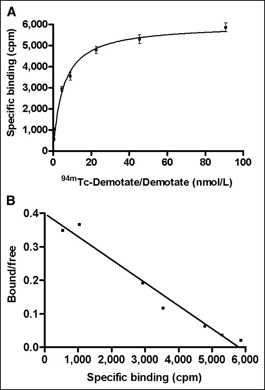
Saturation Binding Assays
A representative saturation binding curve and Scatchard transformation are shown in Figures 2A and 2B, respectively. The data show that 94mTc-Demotate 1 bound to a single class of binding sites with a Kd of 9.6 ± 2.8 nmol/L and a maximum binding capacity of 9,200 ± 2,700 fmol/mg of protein, as determined from 3 independent experiments. Because 94mTc-Demotate 1 consists of a mixture of radiolabeled Demotate 1 and unlabeled Demotate 1, the Kd represents a composite of these 2 species.

Representative plot of 94mTc-Demotate 1–Demotate 1 saturation binding curve (A) and Scatchard transformation (B) for membrane preparations from A-427 cells infected with AdHASSTR2 at 10 pfu per cell. It should be noted that 94mTc-Demotate 1 concentrations included unlabeled Demotate 1 and that saturation binding curve represented specific binding (nonspecifically bound subtracted from total bound). Each data point represents mean ± SEM of triplicate measurements.
Internalization Assays
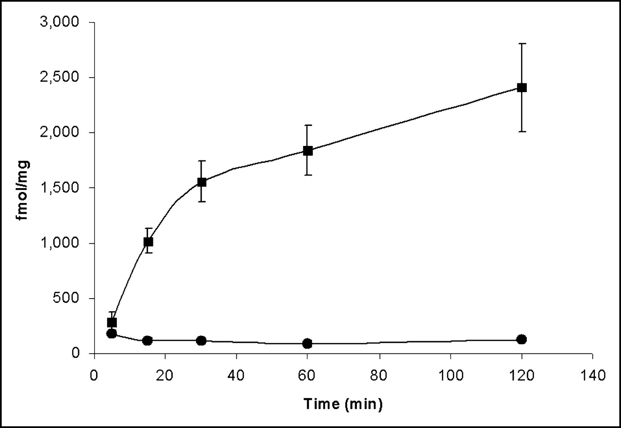
A-427 cells infected with AdHASSTR2 at 10 pfu per cell demonstrated time-dependent internalization of 94mTc-Demotate 1 (Fig. 3). The amount of specifically internalized 94mTc-Demotate 1 was 285 fmol/mg of protein at 5 min and increased to 1,022, 1,558, 1,842, and 2,407 fmol/mg at 15, 30, 60, and 120 min, respectively. The amount of internalized 94mTc-Demotate 1 was significantly greater (P < 0.0001) at all time points than the amount of surface-bound 94mTc-Demotate 1 (<182 fmol/mg at all time points), except for the 5-min time point.

Specific internalization at 37°C of 94mTc-Demotate 1 into A-427 cells infected with AdHASSTR2 at 10 pfu per cell. 94mTc-Demotate 1 (1 nmol/L) was incubated with cells for various times in presence or absence of inhibitor. Cells were acid washed to remove surface-bound radioactivity and then were harvested to determine internalized radioactivity. Specific internalized radioactivity (internalized with inhibitor subtracted from internalized without inhibitor) (▪) and specific surface-bound radioactivity (surface bound with inhibitor subtracted from surface bound without inhibitor) (•) are shown. Data for each time point are presented as mean ± SEM of 2–5 experiments each performed in triplicate.
Animal Biodistribution Studies
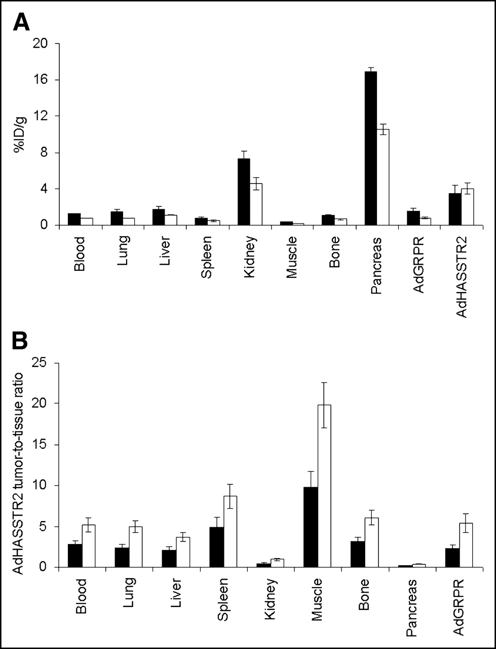
The tissue uptake of 94mTc-Demotate 1 in tumor-bearing mice receiving intratumoral injections of AdHASSTR2 is shown in Figure 4A. The uptake of 94mTc-Demotate 1 in tumors injected with AdHASSTR2 was 3.5 ± 0.8 %ID/g and 4.0 ± 0.6 %ID/g at 1 and 2 h after the administration of 94mTc-Demotate 1, respectively. This uptake was significantly higher (P < 0.03) than the uptake at both time points in all other tissues, except for the kidneys, the SSTR2-positive pancreas, and the liver at 1 h. There was rapid blood clearance of 94mTc-Demotate 1, with 1.2 ± 0.1 %ID/g remaining at 1 h and 0.8 ± 0.1 %ID/g remaining at 2 h. The uptake of 94mTc-Demotate 1 was <1.8 %ID/g in all other tissues, except for the pancreas, kidneys, and SSTR2-positive tumors at both time points. The ratios for the AdHASSTR2-injected tumors and other tissues at 1 and 2 h are shown in Figure 4B. The data show that all of the ratios were >2 at both time points, except for the kidney and pancreas ratios, which were both <1. The ratios also all increased from 1 h to 2 h in all tissues.

Biodistribution of 94mTc-Demotate 1 in mice bearing A-427 tumor xenografts. Tumors were injected directly with either AdHASSTR2 or AdGRPR as control, and 94mTc-Demotate 1 was injected via tail vein 2 d later. Mice were sacrificed 1 h (▪) and 2 h (□) later (n = 6 for each group). Data are presented as mean ± SEM %ID/g for each type of tissue (A) and as ratio of AdHASSTR2-injected tumor uptake to tissue uptake at both time points (B).
SSTR2-mediated uptake of 94mTc-Demotate 1 in tumors injected with AdHASSTR2 was demonstrated by coinjection of an excess of unlabeled Demotate 1 and by comparison with a tumor injected with AdGRPR as a control adenovirus (Table 1). The blocking study showed a significant decrease (P < 0.003) in the SSTR2-positive tumor uptake of 94mTc-Demotate 1 at 1 h (5.4 ± 0.8 %ID/g) with coinjection of the blocking agent (1.6 ± 0.3 %ID/g). The tumor injected with AdGRPR showed 94mTc-Demotate 1 uptake that ranged from 0.8 ± 0.1 %ID/g (Table 1) to 1.5 ± 0.3 %ID/g (Fig. 4A) at 1 h and was 0.8 ± 0.1 %ID/g at 2 h; these uptake values were significantly lower (P < 0.03) than the uptake values at those time points for the AdHASSTR2-injected tumors. The uptake of 94mTc-Demotate 1 also significantly decreased (P < 0.0001) from 21.3 ± 1.5 %ID/g without the blocking agent to 0.2 ± 0.03 %ID/g with the blocking agent in the pancreas. The only other tissues to show a significant decrease after administration of the blocking agent were the liver (P < 0.02) and the kidneys (P = 0.045). In addition, the radioactivity in the pancreas decreased 37% from 1 h to 2 h compared with no decrease in SSTR2-positive tumors. This finding likely was attributable to the high enzymatic activity in the pancreas, which led to higher tumor-to-pancreas uptake ratios at later time points. The only significant differences in the uptake of 94mTc-Demotate 1 between the 2 experiments conducted at 1 h (Fig. 4A; Table 1) were found in the blood, pancreas, and AdGRPR-injected tumor.
Biodistribution (n = 5) of 94mTc-Demotate 1 in Mice Bearing A-427 Tumor Xenografts Directly Injected with AdHASSTR2 or AdGRPR*
MicroPET Imaging
Tumor-bearing mice were imaged 1 and 2 h after the injection of 94mTc-Demotate 1, and 1-h coronal and transaxial microPET images are shown in Figures 5A and 5B, respectively. The data show clear uptake of 94mTc-Demotate 1 in the axillary tumor injected with AdHASSTR2 and background uptake in the tumor injected with AdGRPR. Also, as in the biodistribution studies, the clearance of radioactivity through the kidneys and excretion through the bladder were clearly visualized. The pancreas was not observed in Figure 5 because of the slice of the microPET image shown. SUVs also were determined for both tumors and the kidneys (Fig. 5C). The data show that there was significantly greater (P < 0.007) uptake of 94mTc-Demotate 1 in the AdHASSTR2-injected tumor than in the AdGRPR-injected tumor at both time points and that the uptake in the kidneys was significantly greater (P < 0.02) than the uptake in the AdHASSTR2-injected tumor at both time points. Comparison of the uptake in the AdHASSTR2-injected tumor with that in the AdGRPR-injected tumor and with that in the kidneys (by SUV and biodistribution analyses) is shown in Figure 5D. There was good agreement between the SUV analysis and the biodistribution analysis at 1 and 2 h for the ratio of the AdHASSTR2-injected tumor uptake to the AdGRPR-injected tumor uptake. These values differed by <12% at each time point. The differences between the SUV and biodistribution analyses were greater when the AdHASSTR2-injected tumor uptake was compared with the kidney uptake. A 35% difference between the values was observed at 1 h, whereas a 65% difference was observed at 2 h.

(A and B) Coronal (A) and transaxial (B) microPET projection images of A-427 tumor–bearing mice at 1 h after injection of 94mTc-Demotate 1. Mice carried axillary tumors in which left tumor was injected directly with AdHASSTR2 and right tumor was injected with AdGRPR. Coronal image shows uptake of 94mTc-Demotate 1 in AdHASSTR2-injected tumor and clearance through kidneys and bladder but background uptake in AdGRPR-injected tumor. (C) SUVs obtained from microPET images for AdGRPR-injected tumors, AdHASSTR2-injected tumors, and kidneys in 3 mice at 1 h (▪) and 2 h (□). (D) Ratios of uptake in AdHASSTR2-injected tumors to that in AdGRPR-injected tumors and of uptake in AdHASSTR2-injected tumors to that in kidneys, as determined by SUV analysis (red bars; n = 3) and biodistribution analysis (blue bars; n = 11 for 1 h and n = 6 for 2 h). Data in C and D are presented as mean ± SEM.
DISCUSSION
Initial studies of imaging of gene transfer with the SSTR2 gene as a reporter gene were carried out with probes radiolabeled with γ-emitters for γ-camera or SPECT analysis (14–17). These studies were carried out with 99mTc- or 111In-labeled probes for evaluating the tumor expression of SSTR2 after animals were administered a gene therapy vector carrying the sstr2 gene. SSTR2-specific uptake of these probes was observed in the tumors after direct injection of adenovirus vectors into the tumors or intraperitoneal injection of a vaccinia virus. In addition, Rogers et al. evaluated an adenovirus that contains SSTR2 with the 9-amino-acid HA sequence inserted in the N terminus of the receptor (27). That study demonstrated that insertion of the HA sequence into the N terminus of SSTR2 did not have an adverse effect on ligand binding to SSTR2 and that the HA sequence could serve as an alternative epitope for the binding of anti-HA antibodies. However, because PET has better sensitivity than SPECT at a given resolution, this system could be improved through the use of probes radiolabeled with positron emitters.
94mTc has the same chemistry as the γ-emitter 99mTc (the most important γ-emitting radionuclide) but is a positron emitter that can be used for PET studies. It is readily produced on a biomedical cyclotron at high specific activities for the radiolabeling of various biomolecules. Nickles et al. showed that 94mTc could be used in myocardial perfusion studies to obtain results that correlated well with those of standard PET perfusion studies (33). Griffiths et al. showed that an Fab′ fragment of an anti–carcinoembryonic antigen monoclonal antibody could be readily radiolabeled with 94mTc for potential PET of carcinoembryonic antigen–positive tumors (34). Luyt et al. used 94mTc to radiolabel an estrogen derivative to evaluate estrogen receptor expression in rats by microPET imaging (25). The advantages of using 94mTc over other positron emitters are that it has a relatively simple chemistry for the labeling of peptides and antibodies (compared with 18F, 76Br, or 124I), imaging studies must be conducted on the same day as radiopharmaceutical administration because of the short half-life (this is not necessarily true for longer-lived isotopes), and the short half-life should lead to radiation doses lower than those observed with longer-lived isotopes. However, 94mTc requires a biomedical cyclotron for production and has less-than-ideal physical properties for PET because of the high positron energy. This high positron energy leads to an intrinsic spatial resolution loss of 3.3 mm (35) but is still expected to provide resolution superior to that of corresponding studies with 99mTc and SPECT (34). Thus, 94mTc-labeled biomolecules can be used to model tracer kinetics, measure dosimetry, and determine structure–activity relationships by PET before their wider application with 99mTc and SPECT.
In this study, 94mTc was used to radiolabel the somatostatin analog Demotate 1. Demotate 1 was previously radiolabeled with 99mTc and has demonstrated good SSTR2-mediated tumor uptake in preclinical and clinical studies (26,36). These studies showed that tumor uptake could be observed within 15 min of 99mTc-Demotate 1 injection and that maximum tumor uptake and tumor/organ uptake ratios could be observed by 1 h after injection. These data are important for the radiolabeling of Demotate 1 with 94mTc because its 52-min half-life requires rapid target tissue uptake and clearance from nontarget organs. Radiolabeling of Demotate 1 with 94mTc was accomplished with high radiochemical purity in 15 min with an effective specific activity of 14,800 MBq/μmol (400 mCi/μmol). The effective specific activity was not optimized in our studies because we used 15 μg of Demotate 1 for all of the radiolabelings and never more than 273.8 MBq (7.4 mCi) of 94mTc. Therefore, it is expected that higher effective specific activities can be achieved by optimizing these parameters.
The saturation studies (Fig. 2) showed that 94mTc-Demotate 1 binds with a high affinity (9.6 nmol/L) to A-427 cells infected with AdHASSTR2 at 10 pfu per cell. Maina et al. showed that 99mTc-Demotate bound to SSTR2-expressing AR42J rat pancreatic carcinoma cells with a Kd that was approximately 100 times higher (70 pmol/L) (26). This result likely was attributable to 99mTc-Demotate having a higher affinity for SSTR2 than unlabeled Demotate. This finding is similar to the findings of studies showing that somatostatin analogs complexed with Tc/Re have a 20- to 30-fold higher affinity than noncomplexed analogs (37). In these saturation studies, Maina et al. ensured that all of the Demotate contained a Tc atom by using 99gTc as a carrier so that the final product was 99m/99gTc-Demotate. Because of the short half-life of 94mTc, we chose not to add 99gTc as a carrier and performed the studies with a mixture of 94mTc-Demotate 1 and unlabeled Demotate 1. This mixture is similar to many 99mTc “kit” formulations of radiopharmaceutical agents that contain an excess of the biomolecule. The expression of SSTR2 in A-427 cells infected with AdHASSTR2 at 10 pfu per cell in this study (∼9,200 fmol/mg) was similar to that previously described (∼33,000 fmol/mg) (27). These values for SSTR2 expression are 50–100 times higher than those reported with the commonly used AR42J cell line.
Rapid internalization of 94mTc-Demotate 1 was observed (Fig. 3), with 285 fmol/mg being internalized by 5 min. This internalization increased over the time course of the experiment, with 2,407 fmol/mg being internalized by 2 h. Nonlinear curve fitting showed that the curve reached a maximum at ∼3,300 fmol/mg. The initial velocity of internalization at this concentration of 94mTc-Demotate 1 was ∼49.7 fmol/mg/min, as determined by linear regression (r2 = 0.96) over the first 30 min. The amount internalized was greater than the amount surface bound, which remained constant over the course of the experiment. These results are similar to those reported previously for the internalization of 99mTc-Demotate 1 into AR42J cells, which showed rapid internalization that started to plateau by 15 min and reached its maximum by 30 min. Rapid internalization of radiolabeled peptides has been shown to be important for them to be used as diagnostic or therapeutic agents.
Biodistribution studies showed that there was good uptake of 94mTc-Demotate 1 in tumors injected with AdHASSTR2, with values of 3.5–5.4 %ID/g at 1 h after injection and 4.0 %ID/g at 2 h. This tumor uptake was comparable to that in other studies with the same A-427 tumor model and an intratumoral injection of adenovirus. Zinn et al. showed that at 48 h after the intratumoral injection of an adenovirus expressing SSTR2, tumor uptake at 3 h after the injection of 99mTc-P829 was 2.5 %ID/g (14). In another study by Zinn et al., 10.2 %ID/g was observed in tumors at 26 h after the injection of 99mTc-P2045 (15). Maina et al. (26) showed greater uptake of 99mTc-Demotate in AR42J cells at 1 h after injection (∼25 %ID/g) than was found in AdHASSTR2-infected A-427 cells in this study. This difference in uptake may be explained by the fact that only ∼10% of A-427 cells express SSTR2 after adenovirus infection compared with all natively expressing AR42J cells (14). Also, as discussed earlier, the specific activity of 94mTc-Demotate 1 was not optimized in our studies, a factor that may have led to lower tumor uptake, and was ∼2.5-fold lower than the specific activity described for 99mTc-Demotate 1. It is also clear from these studies that tumor uptake was SSTR2 mediated. The reduction in the liver and kidneys after coinjection of the blocking agent may have been attributable to low levels of expression of SSTR2 in these tissues. SSTR2 messenger RNA has been detected in mouse kidneys and liver, with SSTR2 protein being detected in the glomeruli of the kidneys (38,39).
As discussed above, several somatostatin analogs have been evaluated for γ-imaging of SSTR2-mediated gene transfer. This is the first study to use a somatostatin analog radiolabeled with a positron emitter to monitor SSTR2-mediated gene transfer by PET. Figures 5A and 5B clearly show the accumulation of 94mTc-Demotate 1 in tumors infected with AdHASSTR2 and background radioactivity in tumors infected with the control, AdGRPR. As in the biodistribution studies, the kidneys also were observed, with subsequent clearance through the bladder. The pancreas was not observed in these particular slices of the microPET images, but a strong pancreatic signal was observed in others (data not shown), a finding also consistent with the findings of the biodistribution studies. Analysis of the microPET images showed that there was good agreement between the SUVs obtained from microPET studies and the % ID/g values obtained from biodistribution studies. The SUV relationship between AdHASSTR2-injected tumor and the kidney is not as consistent with the biodistribution as the relationship with the control tumor, possibly because of the high levels of radioactivity in the kidneys and the high positron energy of 94mTc making the SUV measurements in the kidneys less accurate.
It is anticipated that these studies will be expanded to use somatostatin analogs radiolabeled with other positron emitters. Because 94mTc has a short half-life and is not commonly produced on most cyclotrons, other positron emitters will have more widespread applicability. The 18F-labeled somatostatin analogs could be used at most institutions with cyclotrons but still suffer from a relatively complex synthesis strategy (23,40). Somatostatin analogs radiolabeled with 86Y or 64Cu have the advantage of the long half-life of these isotopes for regional shipping and imaging at later time points, whereas 68Ga has the advantage of being generator produced for use at many institutions. Nevertheless, these studies demonstrate that 94mTc-Demotate 1 can be used for PET of SSTR2-mediated gene transfer and that 94mTc-Demotate 1 could be used to model gene transfer characteristics with PET before radiolabeling with 99mTc for additional SPECT studies.
CONCLUSION
This study shows that SSTR2-mediated gene transfer can be monitored by PET. Specific tumor localization of 94mTc-Demotate 1 in mice was visualized by microPET imaging after intratumoral injection of AdHASSTR2 but not after intratumoral injection of a control adenovirus. This investigation represents a first step in demonstrating that SSTR2 can serve as a reporter probe for PET gene transfer. The use of somatostatin analogs radiolabeled with other positron emitters and the development of better ligands that target the HA epitope may improve this system further.
Acknowledgments
This work was supported by Department of Energy grant DE-FG02-02ER63476 and the Department of Radiation Oncology, Washington University School of Medicine. The production of 94mTc at Washington University School of Medicine was supported by NCI grant R24 CA86307. Small-animal imaging at Washington University School of Medicine was supported by NIH grant 5 R24 CA83060. The authors gratefully acknowledge Dr. Petros Sotiriou for his contribution to the synthesis of Demotate 1, Deborah Sultan for the production of 94mTc, Nicole Fettig for the biodistribution studies, Ron Chen for tumor cell implantation, and Jerrel Rutlin and Lori Strong for the microPET imaging and data analysis. Heather Bigott is also thanked for helpful discussions on the production of 94mTc, and Carolyn Anderson is thanked for critical reading of the manuscript.
Footnotes
Received Jun. 8, 2005; revision accepted Aug. 11, 2005.
For correspondence or reprints contact: Buck E. Rogers, PhD, Department of Radiation Oncology, Washington University School of Medicine, 4511 Forest Park Blvd., Suite 411, St. Louis, MO 63108.
E-mail: rogers{at}radonc.wustl.edu
REFERENCES
In this issue


Jump to section
Related Articles
Cited By...
- Noninvasive Molecular Imaging Using Reporter Genes
- Imaging Expression of the Human Somatostatin Receptor Subtype-2 Reporter Gene with 68Ga-DOTATOC
- Multimodality Imaging of Gene Transfer with a Receptor-Based Reporter Gene
- Titration of Variant HSV1-tk Gene Expression to Determine the Sensitivity of 18F-FHBG PET Imaging in a Prostate Tumor