


Abstract
Radiolabeling of monoclonal antibodies (mAbs) with an intracellularly trapped form of 131I (residualizing 131I) involves radioiodinating a small molecular entity, conjugating it to the mAb, and purification. Column purifications are impractical during procedures involving multi-gigabecquerel levels of radioactivity. The goal of this study was to develop a simple, remote, “1-pot” method of radiolabeling and purification for the scaled-up radioiodination of a humanized anti–carcinoembryonic antigen (CEA) mAb, humanized MN-14 (hMN-14; labetuzumab), with an optimized residualizing 131I moiety, 131I-IMP-R4. IMP-R4 is MCC-Lys(MCC)-Lys(X)-d-Tyr-d-Lys(X)-OH, where MCC is 4-(N-maleimidomethyl)-cyclohexane-1-carbonyl and X is 1-((4-thiocarbonylamino)benzyl)-diethylenetriaminepentaacetic acid. Methods: An IODO-GEN-based remote labeling system was used. IMP-R4 was radioiodinated (0.13 μmol per 3.7 GBq of 131I) at a pH of 7.0–7.4 and conjugated to disulfide-reduced hMN-14 after quenching of unused reactive 131I. The product was purified by stirring for 5 min with a 20% (w/v) suspension of an anion-exchange resin and sterilely filtered into a sealed vial. Human serum albumin was added at a final concentration of 1%–2.5%. Immunoreactivity was determined by mixing with CEA and determining the complexation level by size-exclusion high-pressure liquid chromatography. Two control radiolabelings, either with unreduced hMN-14 or with IMP-R4 omitted, also were performed. Results: In 18 radiolabelings with 131I in the range of 2.04–4.81 GBq (55–130 mCi), yields of 59.9% ± 7.9% (mean ± SD) at specific activities of 200 ± 26 MBq/mg (5.4 ± 0.7 mCi/mg) were obtained, with ≥95% of the radioactivity being associated with hMN-14 and with ≤4% aggregation. Similar yields were obtained in a subset of radiolabelings (n = 7) with >3.7 GBq of 131I. The immunoreactivities of the preparations were typically >95%. Nonspecific incorporation in the absence of IMP-R4 was 0.5%, whereas that obtained with unreduced IgG was ∼8%, possibly because of conjugation of IMP-R4 at lysine sites. The process also removed >99% of the quenching reagent used. Radiolabelings performed with freshly prepared solutions or lyophilized preparations produced similar yields, a result that suggested the option for a single-use kit design. Conclusion: Efficient removal of 131I-IMP-R4 and quenched 131I by 5 min of stirring with anion-exchange resin renders a multi-gigabecquerel–level preparation of 131I-IMP-R4-hMN-14 safe, convenient, and practical.
Radioimmunotherapy (RAIT) is evolving into a conventional modality for cancer therapy (1), and there are now 2 U.S. Food and Drug Administration–approved products for non-Hodgkin’s lymphoma. Zevalin (Biogen IDEC) is a 90Y-labeled murine anti-CD20 antibody, and Bexxar (Corixa Pharmaceutical and GlaxoSmithKline) is a 131I-labeled murine anti-CD20 antibody. These products incorporate the 2 most commonly used radionuclides for RAIT, 90Y and 131I.
Directly radioiodinated monoclonal antibodies (mAbs) can lead to reduced retention of radioiodine in tumors because of intracellular antibody catabolism and the subsequent escape of 131I in the form of 131I-iodotyrosine (2,3). For example, the retention of the direct 125I label on an anti–epidermal growth factor 1 mAb, RS7, by a human lung adenocarcinoma cell line in vitro was found to be reduced to 4.7%, whereas that of the 111In radiolabel was reduced only to 42.7%, relative to the respective initial percentages of binding over a 69-h period (4). The phenomenon of reduced retention of radioiodine in tumors is severe with internalizing mAbs, such as RS7, and also has been shown to have an impact on slowly internalizing mAbs, such as an anti-epidermal growth factor 2 mAb, RS11 (4). In fact, mAbs that bind to the cell surface are gradually internalized, possibly through membrane turnover, and subsequently catabolized (5). There also have been reports of internalization of anti–carcinoembryonic antigen (CEA) mAbs (6,7).
Recognition of the factors contributing to reduced tumor retention of radioiodine from directly radioiodinated mAbs has spawned the development of several intracellularly trapped forms of 131I (residualizing 131I) (8–14). In all such examples, purification on a size-exclusion column was performed at the end of the process to furnish mAb incorporating residualizing 131I, and some of the methods also involved purification at the radioiodination stage (13,14). In many of these examples, there was also an activation step, after the radioiodination, to enable conjugation to antibodies. In our view, the single or multiple purification procedures associated with these methods are deterrents to further development for clinical RAIT applications.
Column purifications are generally cumbersome and have the added drawbacks of possible radiation exposure to personnel during procedures involving gigabecquerel levels of 131I, sterility and pyrogenicity issues, limitation on the volume that can be placed in a single column, the need to collect fractions, and potential column leakage and failure. A procedure involving remote transfers of reagents and a batch method of purification, similar to that routinely used for the direct radioiodination of mAbs (15–17), will address all of the issues related to radiolabeling and purification.
Here we describe such an approach for the labeling of an anti-CEA mAb, humanized MN-14 (hMN-14; labetuzumab) (18), with a residualizing 131I label, 131I-IMP-R4 (12), to safely and reliably generate multi-gigabecquerel levels of the pure product. IMP-R4 (Fig. 1) is MCC-Lys(MCC)-Lys(X)-d-Tyr-d-Lys(X)-OH, where MCC is 4-(N-maleimidomethyl)-cyclohexane-1-carbonyl and X is 1-((4-thiocarbonylamino)benzyl)-diethylenetriaminepentaacetic acid (DTPA). The therapeutic advantage of an 131I-IMP-R4-labeled mAb over a conventional 131I-labeled mAb has been documented in preclinical models of lung (12), breast (19), and colon (20) carcinomas.

Structure of the tetrapeptide IMP-R4. The peptide is also conveniently represented as MCC-Lys(MCC)-Lys(X)-d-Tyr-d-Lys(X)-OH, where MCC is 4-(N-maleimidomethyl)-cyclohexane-1-carbonyl and X is 1-((4-thiocarbonylamino)benzyl)-DTPA.
MATERIALS AND METHODS
hMN-14 and the peptide IMP-R4 were obtained from Immunomedics. 131I-Sodium iodide was purchased from Perkin-Elmer Life Science Products. CEA was obtained from Scripps Laboratories as lyophilized powder and was reconstituted with phosphate-buffered saline. Dithiothreitol (DTT) was purchased from Sigma-Aldrich Co. Human serum albumin (HSA) was purchased from Alpine Biologics. Pooled human serum from healthy volunteers was used in the stability study. IODO-GEN (1,3,4,6-tetrachloro-3a,6a-diphenylglycouril) was obtained from Pierce Chemical Co., and AG 1-X8 anion-exchange resin was obtained from Bio-Rad Laboratories.
The apparatus used for direct radioiodination (Fig. 2) was adapted for the 2-step radioiodination involving IMP-R4. This apparatus consisted of the radioiodide shipment vial, the IODO-GEN vial (defined below), and a product vial, all interconnected by syringe needles and plastic monitoring lines and placed in a glove box in a well-ventilated fume hood behind a lead shield. The vials also were connected to a manifold and a pump outside the fume hood for enabling remote transfers of reagents. The plastic monitoring lines and the unions were supplied by Cardio Medical Products, the approximately 30.5-cm (12-in.) butterfly adapter with a needle was obtained from Abbott Laboratories, and the 0.22-μm-pore-size Milli-Fil GS filter was obtained from Millipore Corp. Sterile vials were prefilled with buffers and resin suspension and used as needed. The IODO-GEN vial is a 10-mL-capacity septum-sealed vial provided with a stir bar and coated with 0.5 mg of IODO-GEN.
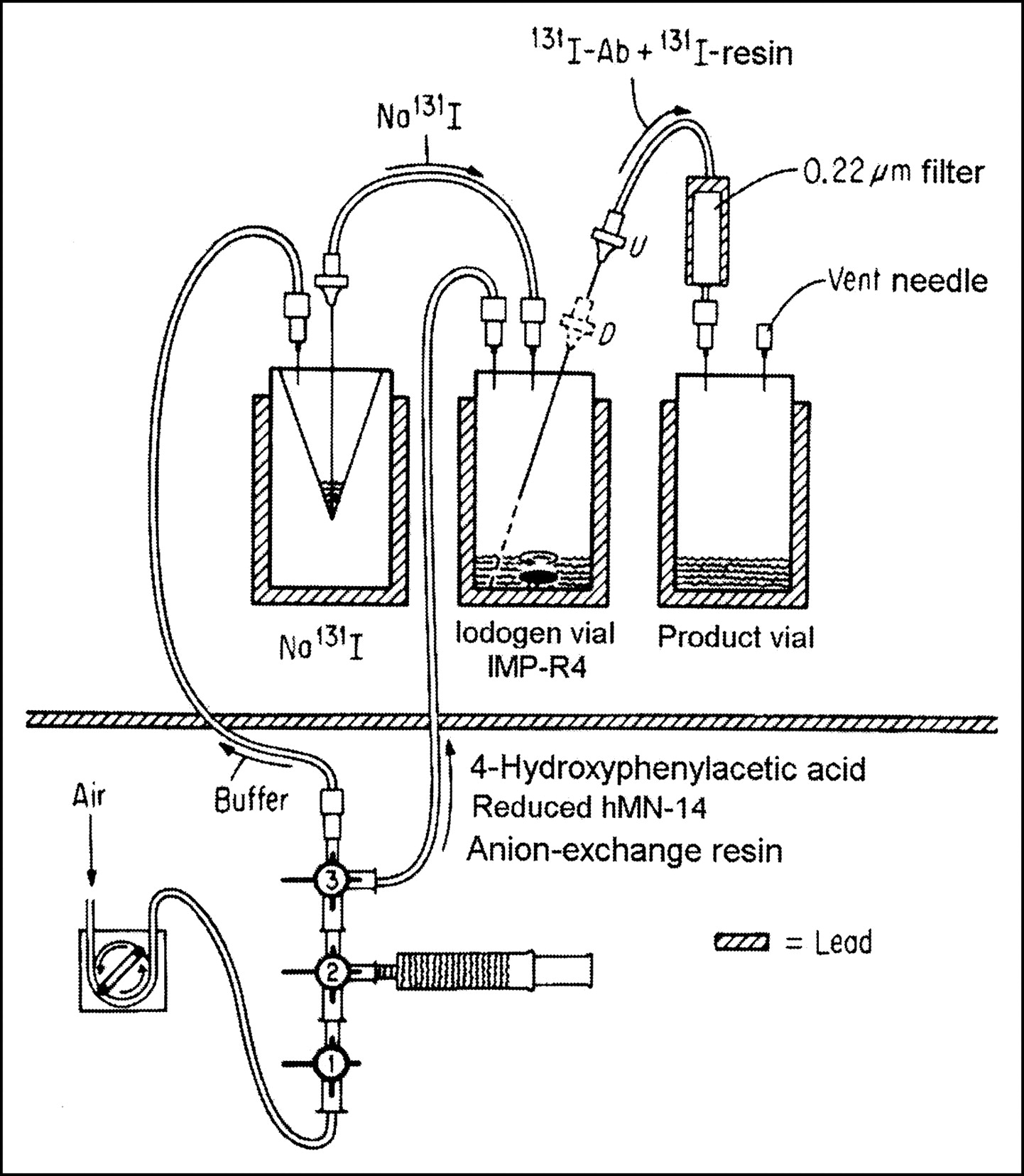
One-pot radioiodination and purification assembly. Ab = antibody.
High-pressure liquid chromatography (HPLC) analyses were performed with a Bio-Sil SEC250 (Bio-Rad Laboratories) analytic column (300 × 7.8 mm) fitted with a guard column (80 × 7.8 mm), with sodium phosphate (0.2 mol/L, pH 6.8) at a flow rate of 1 mL/min, and with in-line Bioscan FC-3300 radiomatic (radioactivity) and Gilson 155 UV-Vis detectors. A Gilson 234 autoinjector and a Gilson FC203B fraction collector also were used in these analyses.
Immunoreactivity was determined by mixing purified radiolabeled hMN-14 with a 20-fold molar excess of CEA at room temperature and determining the extent of the shift of the size-exclusion HPLC peak attributable to hMN-14 to the HPLC peak position corresponding to the antigen–antibody complex. The recovery of 131I radioactivity in HPLC analyses was routinely determined by counting the radioactivity of the entire eluate and comparing it with the injected radioactivity.
DTT Reduction of hMN-14 and Stability of Thiol Titer
A 3-mL quantity of mAb hMN-14 (8–10 mg/mL) was mixed with 0.18 mL of ethylenediaminetetraacetic acid (EDTA) (0.1 mol/L, pH 7) and 0.12 mL of aqueous DTT (0.08 mol/L) in a Vortex Mixer (Thermolyne). The solution was flushed briefly with argon and incubated at 37°C for 45 min. At least 2 successive purifications by centrifuged size-exclusion chromatography with Sephadex G50/80 (Sigma-Aldrich) and a buffer containing sodium phosphate (0.1 mol/L) and EDTA (5 mmol/L) (pH 6.5) and then filtration with a 0.22-μm-pore-size filter furnished the purified product. The concentration was determined by measuring the absorbance at 280 nm, and the thiol content was determined by Ellman’s assay. The material was stored frozen at −80°C. Alternatively, the material was lyophilized in 3- to 5-mg lots and stored in septum-sealed vials under vacuum at 2°C–8°C.
Single determinations of the stability of the thiol content under different conditions were performed as follows. The frozen material was thawed after 6 mo; material was prepared under radioiodination conditions without radioiodine and IMP-R4; and evaluated after being stirred with IODO-GEN for 30 min or as is after 18 h at room temperature and assayed after 5 min of incubation with sodium tetrathionate (STT; 5 mmol/L).
Radioiodination
131I radioiodination with 131I-IMP-R4 was performed first by radioiodinating IMP-R4; this step was followed by conjugation with reduced hMN-14 and in situ purification by stirring with AG 1-X8 anion-exchange resin. An aqueous solution of IMP-R4 at 1.0 mg/mL (0.48 mmol/L) was prepared at a pH of 5–5.5, divided into aliquots, placed in vials, and stored at −80°C. Alternatively, a 1.0-mg/mL solution in sodium phosphate (0.1 mol/L, pH 6.1) was prepared, lyophilized in 0.25- to 0.4-mg lots in vials sealed under vacuum, and stored at −20°C. The IODO-GEN vial containing a preadded volume of IMP-R4 solution (1.0 mg/mL) was assembled with the product vial and the transfer lines in a ventilated glove box behind a lead shield, and the 131I shipment vial was connected last. In Figure 2, the spinal syringe needle in the IODO-GEN vial was in the elevated position during radiolabeling, conjugation, and resin treatment but was placed at the bottom of the vial for transfer of the purified product through a 0.22-μm-pore-size filter into the product vial. Stopcock 3 was suitably positioned for adding either buffer to the isotope vial or other reagents to the IODO-GEN vial. In early experiments, radioiodide was buffered with 7 times its volume of sodium phosphate (0.5 mol/L, pH 7.4) and transferred with 1.4 mL of sodium phosphate (0.04 mol/L, pH 7.4) into the IODO-GEN vial. The labeling and conjugation pH was ∼7.4. This protocol was later changed to buffering with 7 times its volume of sodium phosphate (0.3 mol/L, pH 6) and transferring with 1.4 mL of sodium phosphate (0.1 mol/L, pH 6.1). With these conditions, the labeling and conjugation pH was ∼7.0. After the mixture was stirred for a specified period (12 min for labelings at >3.7 GBq and 5 min for labelings at <1.85 GBq), an excess of 4-hydroxyphenylacetic acid was added to capture unreacted radioiodine, with stirring for an additional 3 min. This quenching step was followed by the addition of reduced hMN-14 and incubation without stirring for 20–30 min. In the initial 8 radiolabelings, STT also was added to a final concentration of 1.6–3.1 mmol/L, but this step was omitted in the subsequent runs. Finally, 3 mL of a 20% (w/v) suspension of AG 1-X8 anion-exchange resin in sodium phosphate (40 mmol/L, pH 7.4) was added, and the mixture was stirred for 5 min. The solution and the resin were passed through a 0.22-μm-pore-size Milli-Fil GS filter into a septum-sealed product vial (or bottle). The IODO-GEN vial was rinsed with sodium phosphate buffer (40 mmol/L, pH 7.4), and the rinse was transferred into the product vial. HSA was added to a final concentration of 1%–2.5%. A total of 18 radiolabelings with 2.04–4.81 GBq (55–130 mCi) of 131I were conducted. Two control runs involved a 2.07-GBq (56-mCi) labeling with unreduced hMN-14 and a 2.3-GBq (62-mCi) labeling with reduced hMN-14 but without IMP-R4. The labeling and purification protocol was otherwise unchanged.
In adapting the apparatus used for direct radioiodination, materials were added remotely when the volumes were generally >1 mL. This process typically involved transfer of buffer into the 131I shipment vial, antibody with volumes of >1 mL, 3 mL of anion-exchange resin suspension, and the final rinse buffer. The quenching reagent, 4-hydroxyphenylacetic acid solution, was added directly to the IODO-GEN vial because of the small volume involved. Further, instead of the pump, the liquids were pushed through the lines by use of air from an empty 10-mL syringe.
Typically, for 3.7-GBq (100-mCi)-scale labeling, 0.27 mg (0.13 μmol) of the peptide IMP-R4 and 12 mg (0.08 μmol) of reduced hMN-14 were used. The specific activity of commercial 131I is ∼370–740 GBq (10–20 Ci)/mg, with the shipment specification stated as >185 GBq/mg. At a specific activity of 131I of 370 GBq/mg, the peptide-to-iodide molar ratio was ∼1.9:1, and the antibody-to-peptide ratio was 0.6:1. The quenching reagent was used in a 60-fold molar excess with respect to iodide.
Stability studies were performed as follows. The product was left at room temperature and examined after 20 h, the product solution was made 2.5% in HSA at a concentration of 0.87 mCi/mL and a mAb concentration of 0.2 mg/mL and was reexamined after 20 h, and 0.3 mL of the product solution was mixed with 0.97 mL of human serum to yield a final mAb concentration of 39 nmol/L and was incubated at 37°C for 20 h.
RESULTS
Reduced hMN-14 and Its Stability
DTT-reduced hMN-14 was obtained at a concentration (mean ± SD) of 6.8 ± 0.4 mg/mL and a thiol titer of 7.2 ± 0.4 thiol groups per IgG molecule (n = 20). When the material was stored frozen and examined after 6 mo, the thiol content was unchanged. When stirred for 30 min with IODO-GEN under conditions mimicking radiolabeling but without the addition of IMP-R4, the material lost 40% of the thiol titer. The thiol content was unchanged when the material was prepared under radiolabeling conditions but without IODO-GEN and IMP-R4 and was left at room temperature for 18 h. The material did not aggregate during storage, as determined by size-exclusion HPLC with absorbance detection (data not shown). Finally, the thiol content was reduced to ∼4% of the initial value after 5 min of incubation with STT at 5 mmol/L, a result that confirmed reoxidation, as expected.
Both the liquid product and the lyophilized material produced similar overall yields in the radioiodination procedure.
Radioiodination
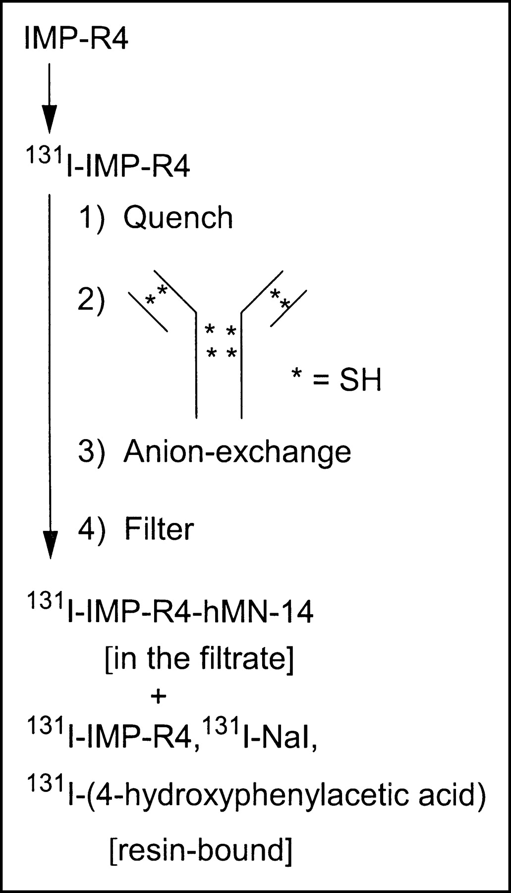
Figure 3 shows the flow chart for the “1-pot” radiolabeling and purification process described here. Thawed liquid aliquots and reconstituted lyophilized peptide IMP-R4 resulted in similar radiolabeling yields.

Flow chart for radioiodination of IMP-R4 and conjugation to reduced hMN-14. The anion-exchange resin was AG 1-X8. SH = thiol group.
Yields were determined by measuring the percentage recovery of radioactivity in the final product vial relative to the initial activity in the shipment vial, and these were 59.9% ± 7.9% at specific activities of 200 ± 26 MBq/mg (5.4 ± 0.7 mCi/mg) in 18 large-scale radiolabelings with 2.04–4.81 GBq (55–130 mCi) of 131I. The immunoreactivity of the radiolabeled hMN-14 preparations, as determined by CEA complexation and HPLC analyses, was typically ≥95%; this value was similar to the value obtained for directly radioiodinated hMN-14 prepared by the conventional chloramine-T procedure.
Table 1 shows the labeling efficiencies, specific activities, and immunoreactivities obtained in these labelings. Incorporation into IgG reached >95%, as determined by HPLC analyses, with radioactivities recovered from the HPLC column being mostly >95%. Aggregation, frequently observed only as a shoulder, was estimated to be ≤4%. In a subset of 7 radiolabelings with >3.7 GBq (100 mCi) of 131I, the yields were 57.9% ± 7.6% at specific activities of 207 ± 26 MBq/mg; these yields were similar to the yields for all of the radiolabelings.
Data for 2-Step Radioiodination with 2.04–4.81 GBq (55–130 mCi) of 131I
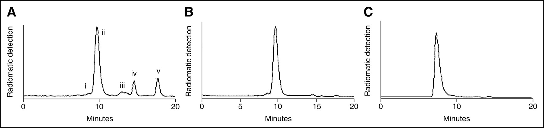
Representative HPLC traces are shown in Figure 4. The HPLC trace of the crude labeled product, before treatment with anion-exchange resin, is shown in Figure 4A. Monomeric 131I-IMP-R4-hMN-14 centered at 9.7 min, along with an aggregate appearing as a shoulder, accounted for 70%; 131I-IMP-R4 appeared as a broad peak at 13.0–13.5 min (5%); and 2 other low-molecular-weight (low-MW) radioactive peaks appeared at 14.6 min (9.4%) and ∼17.7 min (12.6%). In the HPLC trace of the purified product (Fig. 4B), low-MW peaks accounted for 2.3% and the main monomeric IgG peak, together with an aggregate appearing as a shoulder, accounted for the rest. Complexation with CEA and the consequent shift of the peak to near the voided volume at 7.3 min are depicted by the HPLC trace in Figure 4C.

(A) Size-exclusion HPLC analysis of the crude product before anion-exchange treatment, showing aggregate (i), 131I-IMP-R4-hMN-14 (ii), 131I-IMP-R4 (iii), and 131I-iodo-4-hydroxyphenylacetic acid (iv and v). (B) Purified product. (C) Antibody–antigen complex after mixing of the purified product with CEA.
Variations in the duration of the conjugation reaction also were examined. In labelings with ∼2 GBq of the radionuclide, a 10-min conjugation furnished a 59% yield, compared with a 66.7% yield in a 15-min conjugation. In subsequent runs, the mAb-coupling duration was 20–30 min.
In the absence of IMP-R4, the radioactivity recovered with the anion-exchange process was only 1.2%, and HPLC analysis of the product revealed 41.7% of the radioactivity in IgG and 55.4% in low-MW species. In another control, involving unreduced hMN-14, HPLC analysis of the crude product revealed ∼8% incorporation into IgG.
In the absence of HSA, radiolytic damage caused the loss of ∼19% of the label in a 20-h period. Although the nature of the low-MW material was unknown, its appearance as a relatively sharp HPLC peak suggested that it was probably not the labeled peptide. The addition of 2.5% HSA greatly enhanced the label integrity in terms of the percentages of radioactivity in hMN-14 (2.6% loss) and immunoreactivity (92.9%) after a 20-h period. The product maintained stability (1% loss as low-MW material) and immunoreactivity (96.7%) after incubation with serum for 20 h at 37°C.
DISCUSSION
Govindan et al. documented the advantage of residualizing 131I-IMP-R4-hMN-14 over directly labeled 131I-hMN-14 in a preclinical model of human colon carcinoma (20). The promising clinical results obtained with directly radioiodinated hMN-14 (21,22) prompted us to develop a residualizing 131I-based approach to further increase the achievable tumor dose. In this context, devising a simple method for generating gigabecquerel levels of 131I-IMP-R4-hMN-14 became imperative.
Rationale for Anion-Exchange Method
The built-in maleimide group on the IMP-R4 reagent makes conjugation to thiol-containing mAbs facile under neutral-pH conditions. Thus, both radioiodination and conjugation reactions are expected to occur quickly. With water-insoluble IODO-GEN removable by filtration, one has to remove only unused radioiodine, captured with the quenching reagent, and unconjugated 131I-IMP-R4 in a manner that renders the entire radiolabeling and purification method simple and rapid. We hypothesized that, because this peptide contains a phenolic group and multiple carboxyl groups, the unconjugated peptide and the quenched 131I may be amenable to removal by simple stirring with an anion-exchange resin and that the mAb–peptide conjugate may be recoverable by filtration because of the large size of the protein. That is, the same set-up that is used for direct radioiodination of mAbs may be suitable for purification after the 2-step procedure of peptide labeling and mAb conjugation.
AG 1-X8 is a quaternary ammonium–based strong anion-exchange resin (23) with phosphate as the counterion in our experiments. The selectivity for anion exchange on this resin increases from hydroxide at an arbitrary value of 1 to acetate at 3.2, phosphate at 5, phenolate at 110, and iodide at 175 (23). IMP-R4 with multiple acetate groups of DTPA and the phenolic group in the form of d-tyrosine would appear to be ideally suited to be exchanged on the resin. Li et al. (24) reported that a 1,4,7,10-tetraazacyclododecane-N,N′,N″,N‴-tetraacetic acid-based bifunctional chelating agent could be exchanged on a diethylaminoethyl cellulose anion-exchange resin in a process designed to separate the charged chelating agent from the corresponding neutral metal chelate. With IMP-R4, it is likely that the DTPA and the tyrosine portions of the molecule contribute to various degrees, resulting in efficient retention by the resin. Unoxidized iodide, if any, and 4-hydroxyphenylacetic acid are removed almost quantitatively (>99%), reflecting the high anion-exchange selectivities for phenolate and iodide ions. These data suggest that the present method should be generally applicable to the removal of other types of bifunctional tyrosine- or phenol-containing residualizing components by anion exchange after IODO-GEN-based radioiodination and coupling to mAbs. Antibody-bound IMP-R4 is excluded because of the large size of the protein and can be isolated by filtration.
DTT-Reduced mAb
The thiol titer in disulfide-reduced hMN-14 is stable under a variety of storage conditions when stored in the presence of EDTA. Oxidation of thiol groups of the reduced mAb by IODO-GEN, consistent with the reported capability of IODO-GEN to react with thiol groups (25), may be less of a concern, because it is the rate of thiol–maleimide reaction relative to that of thiol oxidation that is important, with the former reaction, leading to the formation of the conjugate, being facile.
Radioiodination
Peptides of the IMP-R4 type usually elute as a broad peak from size-exclusion HPLC columns, and this pattern also was seen in the HPLC analysis of the crude radioiodinated product (Fig. 4A), in which unconjugated 131I-IMP-R4 appears as a broad peak. Low-MW peaks at 14.6 and 17.7 min in the HPLC analysis of crude radiolabeled hMN-14 are both attributable to radioiodinated 4-hydroxyphenylacetic acid, which is used to capture unused radioiodine. 125I-radioiodinated 4-hydroxyphenylacetic acid itself shows 2 HPLC peaks in an ∼1:1 ratio at these retention times; these peaks correspond to mono- and diiodinated species (data not shown). The alternative explanation of the peak at 14.6 min being attributable to radioiodide seems to be contraindicated by the lack of a sharp peak attributable to radioiodide in the HPLC analysis of the 131I-IMP-R4 preparation without the use of the quenching reagent (data not shown). Besides, a reductant is not used to convert unused reactive radioiodine back to radioiodide in these labelings.
Initially, STT was used to reoxidize unreacted thiol groups on the antibody, but this step was omitted in later radiolabelings to render the process simpler. Omitting this step did not have any practical effect on yields, level of aggregation, immunoreactivity, or performance in preclinical evaluations (20).
Attempts to increase the specific activity of the final product were guided by the fact that the molar excess of IMP-R4 used was probably not sufficient to capture all of the reactive radioiodine generated, as inferred from the presence of 131I-iodo-4-hydroxyphenylacetic acid in the unpurified product (Fig. 4A). Accordingly, in one run, when the IMP-R4 content was increased by 40%, with a concurrent decrease in the reduced hMN-14 content by 20%, the final specific activity improved from an average of 200 MBq/mg to 248 MBq/mg (6.71 mCi/mg).
In the absence of IMP-R4, nonspecific radiolabeling on IgG was 0.5% (1.2% × 0.417); almost all (>99%) of the radioiodinated 4-hydroxyphenylacetic acid was scavenged by the anion-exchange resin because, overall, only 0.66% (1.2% × 0.554) of the same could be accounted for by actual recovery and HPLC analyses. In the control run involving unreduced hMN-14, the observation of ∼8% incorporation into IgG suggested that some conjugation might have occurred at lysine sites. This control labeling was performed with sodium phosphate buffer at 0.5 mol/L and pH 7.4 for buffering radioiodide in the shipment vial. Subsequently, the labeling pH was lowered with phosphate buffer at 0.3 mol/L and pH 6, with a view to further reducing conjugation at lysine sites; however, in the presence of reduced IgG in the actual labeling, the facile thiol–maleimide reaction would probably render the issue of conjugation at lysine sites moot. Yields were not affected by lowering of the labeling pH.
The overall yield of the purified product in these radiolabelings was ∼60%, compared with the yield of ∼85% for direct radioiodination of hMN-14 by the conventional chloramine-T method. The overall yield in the 2-step procedure is the combination of yields at the radioiodination and conjugation steps. Further optimization of the peptide-to-radioiodide and peptide-to-mAb molar ratios to further increase the overall yield must also take into account the final specific activity of the product. One of the unknown factors with respect to such optimization is the variability in the specific activity of commercial 131I. An option in this regard is to use higher-specific-activity 131I, which could help increase the overall yield and specific activity, because the same level of IMP-R4 would represent a much higher molar excess with respect to the radionuclide. 131I with a specific activity of 1,850 GBq/mg (50 Ci/mg) may be commercially available soon (personal communication from MDS Nordion, Kanata, Ontario, Canada; conversation on April 20, 2004). The specific activity of the product can be improved further by lowering the amounts of both IMP-R4 and reduced hMN-14. However, a specific activity higher than that achieved currently may not be necessary clinically, because unlabeled antibody frequently has been added to radiolabeled material to maintain a certain protein dose.
In patients, the maximum tolerated dose with directly labeled 131I-hMN-14 was reached at 2.22 GBq/m2 (21), with myelotoxicity being dose limiting. From preclinical data, the maximum tolerated dose with 131I-IMP-R4-hMN-14 was determined to be in the same range as that with conventionally labeled 131I-hMN-14 (20). Thus, the preparation of 3–4 GBq of purified labeled material per patient would appear to be adequate for initial clinical studies evaluating 131I-IMP-R4-hMN-14. We have so far demonstrated that 3 GBq of the label can be readily prepared by the simplified 1-pot approach.
The present format of radiolabeling leads to practically pure material, with <1% of extraneous reagents and <5% of low-MW radioactive species being present in the final product. Further simplification is achieved by use of lyophilized preparations of IMP-R4 and reduced hMN-14, with reconstitution with water being all that is needed at the time of radiolabeling.
CONCLUSION
This is the first report of the production of a residualizing 131I-labeled mAb at levels needed for clinical therapy, exemplified with the anti-CEA mAb hMN-14 and the residualizing 131I label 131I-IMP-R4. We have shown that all unincorporated 131I radioactivity can be removed readily by a simple, rapid, batch-type anion-exchange procedure. An overall yield of 60% ± 8% (n = 18) of the purified product, with the preservation of immunoreactivity, was documented with 131I in the range of 2.04–4.81 GBq (55–130 mCi). Further yield improvement is contingent on the use of higher-specific-activity 131I-sodium iodide, anticipated to be commercially available soon, and subsequent optimization of the relative molar ratios of the reagents. Because the radiolabeling results are unchanged when lyophilized preparations are used, additional process simplification may be possible with the development of a single-use kit design.
Acknowledgments
This work was supported in part by National Institutes of Health grant CA103312. We thank Nicholas Kumburis and Nino Velasco for HPLC analyses.
Footnotes
Received May 11, 2004; revision accepted Aug. 12, 2004.
For correspondence or reprints contact: Serengulam V. Govindan, PhD, Immunomedics, Inc., 300 American Rd., Morris Plains, NJ 07950.
E-mail: sgovindan{at}immunomedics.com
REFERENCES
In this issue


Jump to section
Related Articles
Cited By...
- Immuno-PET Quantitation of de2-7 Epidermal Growth Factor Receptor Expression in Glioma Using 124I-IMP-R4-Labeled Antibody ch806
- Immunization with Mimotopes Prevents Growth of Carcinoembryonic Antigen Positive Tumors in BALB/c Mice
- Advantage of a Residualizing Iodine Radiolabel in the Therapy of a Colon Cancer Xenograft Targeted with an Anticarcinoembryonic Antigen Monoclonal Antibody