

FIGURE 7.
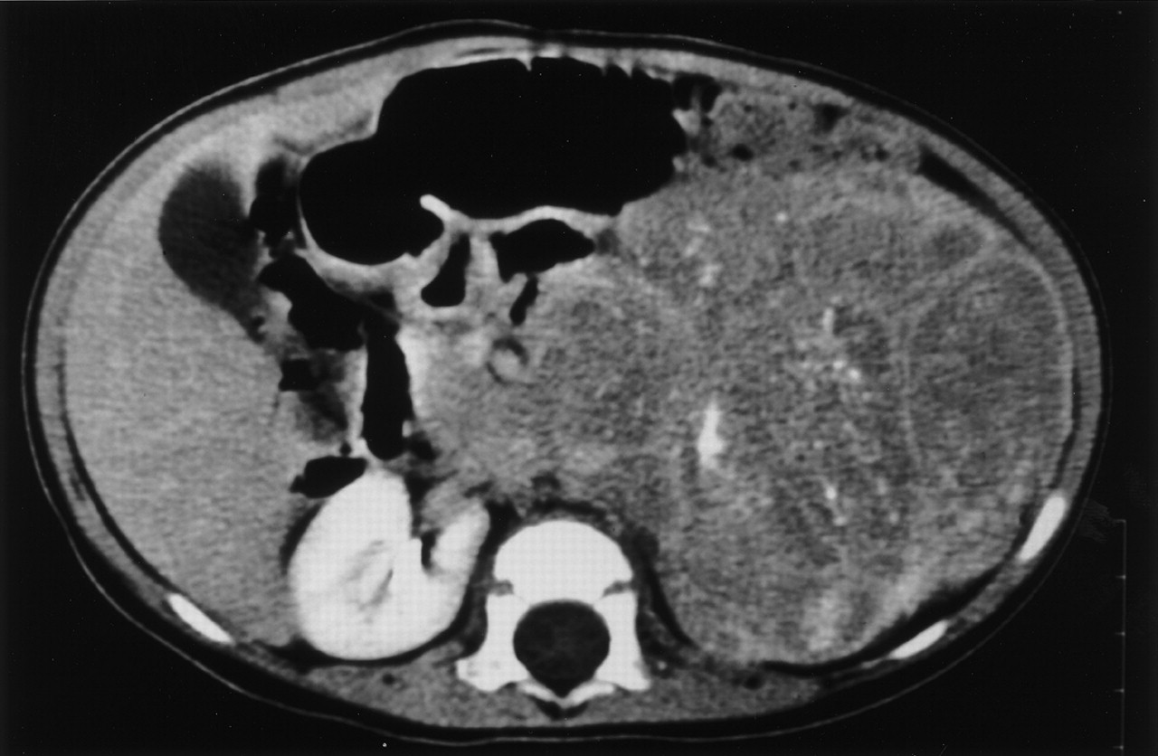
FIGURE 7.
CT image of large abdominal neuroblastoma in 13-mo-old child. Mass crossed midline and encased major blood vessels—findings typical for high-risk neuroblastoma. However, mass (and regional lymph nodes) proved to be resectable, making this stage 2B, as previously described (29). Patient remains well 9+ y later without having received any cytotoxic therapy.
In this issue


Related Articles
Cited By...
- IGF2BP1 induces high-risk neuroblastoma and forms a druggable feedforward loop with MYCN promoting 17q oncogene expression
- Congenital Neuroblastoma
- Positron Emission Tomography Detects In Vivo Expression of Disialoganglioside GD2 in Mouse Models of Primary and Metastatic Osteosarcoma
- Arsenic Trioxide as a Radiation Sensitizer for 131I-Metaiodobenzylguanidine Therapy: Results of a Phase II Study
- Characterization of Neuroblastic Tumors Using 18F-FDOPA PET
- 64Cu-p-NH2-Bn-DOTA-hu14.18K322A, a PET Radiotracer Targeting Neuroblastoma and Melanoma
- Complicated appearance of an abdominal mass in the I-131 MIBG and Tc-99m bone scintigraphy of a patient with neuroblastoma
- 18F-FDG PET/CT and 123I-Metaiodobenzylguanidine Imaging in High-Risk Neuroblastoma: Diagnostic Comparison and Survival Analysis
- Neem Leaf Extract Induces Radiosensitization in Human Neuroblastoma Xenograft Through Modulation of Apoptotic Pathway
- Neuroblastoma: contemporary management
- Comparison of Iodine-123 Metaiodobenzylguanidine (MIBG) Scan and [18F]Fluorodeoxyglucose Positron Emission Tomography to Evaluate Response After Iodine-131 MIBG Therapy for Relapsed Neuroblastoma
- Sensitivity of Surveillance Studies for Detecting Asymptomatic and Unsuspected Relapse of High-Risk Neuroblastoma
- In vivo Imaging and Quantitation of Adoptively Transferred Human Antigen-Specific T Cells Transduced to Express a Human Norepinephrine Transporter Gene
- Whole-Body PET/CT with 11C-Meta-Hydroxyephedrine in Tumors of the Sympathetic Nervous System: Feasibility Study and Comparison with 123I-MIBG SPECT/CT
- Surgical Risk Factors in Primary Surgery for Localized Neuroblastoma: The LNESG1 Study of the European International Society of Pediatric Oncology Neuroblastoma Group