


Abstract
The aim of this study was to develop a scintigraphic test to measure gastric emptying and accommodation simultaneously. Methods: Gastric emptying and accommodation were measured in healthy subjects. To determine gastric accommodation, the stomach was imaged with SPECT 20 min after intravenous administration of 185 MBq (5 mCi) 99mTc-pertechnetate. After ingestion of 11 MBq (300 μCi) 111In-diethylenetriaminepentaacertic acid in a liquid nutrient drink or an 111In-oxine–labeled egg sandwich, dual-isotope imaging assessed SPECT gastric dimensions and gastric emptying every 20 min up to 240 min. Gastric accommodation was calculated as the percentage change in planar (2-dimensional) gastric cross-sectional area (CSA) using a left anterior oblique planar projection and the percentage change in total SPECT gastric voxel counts (3-dimensional) compared with the baseline image. Results: With the liquid nutrient drink (9 subjects), maximal mean CSA (158% ± 12% of baseline; P < 0.05) occurred 40 min after meal ingestion, when only 69% ± 3% of the radiolabeled liquid nutrient drink remained in the stomach. At 120 min, mean CSA was 125% ± 8% of baseline, but only 35% ± 3% of the liquid nutrient drink remained in the stomach. Using SPECT to measure 3-dimensional volumes, maximal gastric volume occurred 20 min after meal ingestion (189% ± 25% of baseline). With the solid egg meal (10 subjects), maximal total CSA (159% ± 13% of baseline) occurred immediately after meal ingestion; total CSA remained significantly increased above baseline for the first 3 h after ingestion of the egg meal, despite only 12% ± 4% gastric retention at 3 h. Using SPECT to measure 3-dimensional volumes, maximal gastric volume occurred immediately after the meal (184% ± 19% of baseline). Conclusion: This method permits simultaneous measurement of gastric emptying and accommodation. In healthy subjects, the gastric accommodation response is prolonged and persists despite nearly complete emptying of a liquid or solid meal.
Normally with food ingestion, the proximal stomach (fundus) relaxes, with an associated increase in volume, allowing the stomach to accommodate the meal (1). In the antrum, regular peristaltic contractions grind down (triturate) solid food so that it can be passed from the stomach.
Dyspepsia refers to symptoms originating in the upper gastrointestinal (GI) tract and includes upper abdominal pain or discomfort, early satiety, postprandial abdominal bloating, and nausea (2,3). Impaired gastric accommodation to a meal, antral hypomotility leading to delayed gastric emptying, and visceral hypersensitivity have been proposed as important pathophysiologic factors in the genesis of functional dyspepsia. Delayed gastric emptying is present in 25%–40% of patients but does not correlate with dyspeptic symptoms in most patients (4). Impaired fundic accommodation promotes rapid transit from the proximal stomach into the antrum, producing antral distension, which seems to correlate with dyspeptic symptoms in some patients (5).
Gastric volume has been traditionally measured with a barostat, a balloon placed into the stomach through oral intubation (5). Impaired fundic accommodation to a meal in functional dyspepsia has been described (5–7). However, the gastric barostat is invasive and uncomfortable since the patient needs to swallow a compliant balloon attached to a tube. The procedure is not tolerated by many patients, often making it impractical for individual patient evaluation.
Noninvasive techniques have been proposed to measure gastric accommodation (8–10). Radionuclide imaging of the gastric wall can be performed after the intravenous injection of 99mTc-pertechnetate, which is selectively taken up and localizes in the gastric mucosa (10–12). This property is widely used to identify ectopic gastric mucosa in patients with suspected Meckel’s diverticulum (11). Recently, intravenous administration of 99mTc-pertechnetate to label the gastric mucosa with SPECT imaging has been found to be a noninvasive method that provides a 3-dimensional (3D) representation of the stomach wall from which gastric volume can be calculated (10). 99mTc labeling with SPECT has been shown to provide a noninvasive measurement of the effect of a meal on total gastric volume that is comparable to changes in balloon volume as observed with the gastric barostat (13). However, in the previous studies, gastric emptying of solids was assessed on a different study day (14).
The aim of this study was to develop a noninvasive, clinically applicable scintigraphic test that could measure simultaneously both gastric emptying and accommodation after ingestion of either a solid or a liquid test meal. This combined gastric emptying/accommodation test should be useful to define the pathophysiology of symptoms in patients with functional dyspepsia. Some of the results of this study were presented at the 2003 Digestive Disease Week Meeting in Orlando, FL (15), and the 2003 Society of Nuclear Medicine Annual Meeting in New Orleans, LA (16).
MATERIALS AND METHODS
Overview
In this study, gastric emptying and accommodation were measured simultaneously in healthy subjects. This study was approved by the Institutional Review Board of Temple University. A healthy subject was defined as a person with no GI symptoms; no prior history of peptic ulcer disease, functional dyspepsia, or irritable bowel syndrome; no prior surgery on the esophagus, stomach, or small intestine; a normal physical examination; and no medications for GI disorders. The study was performed in 2 parts with different healthy subjects in each part. In the first part, gastric emptying and SPECT 3D imaging were measured after ingestion of a radiolabeled liquid nutrient drink (Ensure; Ross Productions Division, Abbott Laboratories). In the second part, gastric emptying and 3D volume were assessed after ingestion of a radiolabeled egg sandwich. The accommodation response analysis was performed comparing 2 methods: 3D SPECT for gastric volume and a potentially simpler left anterior oblique (LAO) 2-dimensional (2D) planar imaging to determine whether the 2D measurement could be used in place of SPECT.
Study Protocol 1
Gastric emptying and accommodation were measured simultaneously in 9 healthy subjects (4 females, 5 males; average age, 36 ± 5 y; body mass index [BMI], 25.6 ± 1.0) after a liquid nutrient meal (liquid nutrient drink, 300 mL; 1.05 calories/mL; 7.6 g fat, 50.6 g carbohydrate, 11.4 g protein). Each study was performed with the subject fasting for at least 6 h. To determine baseline gastric dimensions, the stomach was imaged 20 min after intravenous injection of 185 MBq (5 mCi) 99mTc-pertechnetate. SPECT was performed using a dual-head MAXXUS Camera (General Electric Medical Systems) connected to a General Electric Starcam 4000i computer. For SPECT, a 64 × 64 word mode matrix and a 360° rotation around the subject was obtained using a circular orbit (32 stops at 15 s per stop). The subjects then ingested 300 mL liquid nutrient drink containing 11 MBq (300 μCi) 111In-diethylenetriaminepentaacetic acid (111In-DTPA). Dual-isotope imaging of 99mTc and 111In began immediately after meal ingestion (time 0). Both anterior and posterior images for 111In counts were used to measure geometric mean solid-phase gastric emptying. This was followed immediately by 3D SPECT. This imaging sequence (anterior and posterior planar images for 111In gastric emptying, SPECT for 99mTc activity) was repeated every 20 min for 120 min after liquid nutrient drink ingestion. The 111In imaging was performed using a 20% window centered on the 274-keV photopeak of 111In with a medium-energy, parallel-hole collimator. The 99mTc imaging was performed using a 20% window on the 140-keV photopeak of 99mTc with the same collimator.
Study Protocol 2
The scintigraphic test used to measure gastric emptying and accommodation with the liquid nutrient drink was modified to simultaneously measure gastric emptying and volume changes (accommodation) in 10 healthy subjects (3 females, 7 males; average age, 36 ± 3 y; BMI, 26.9 ± 2.0) using a 111In-oxine–labeled egg sandwich, a conventional meal used to measure gastric emptying (17,18). For these studies, additional images were obtained at 150, 180, 210, and 240 min after the meal ingestion, since our prior studies have shown that imaging to 4 h after meal ingestion is helpful to assess gastric emptying (19).
Image Processing
Gastric Emptying.
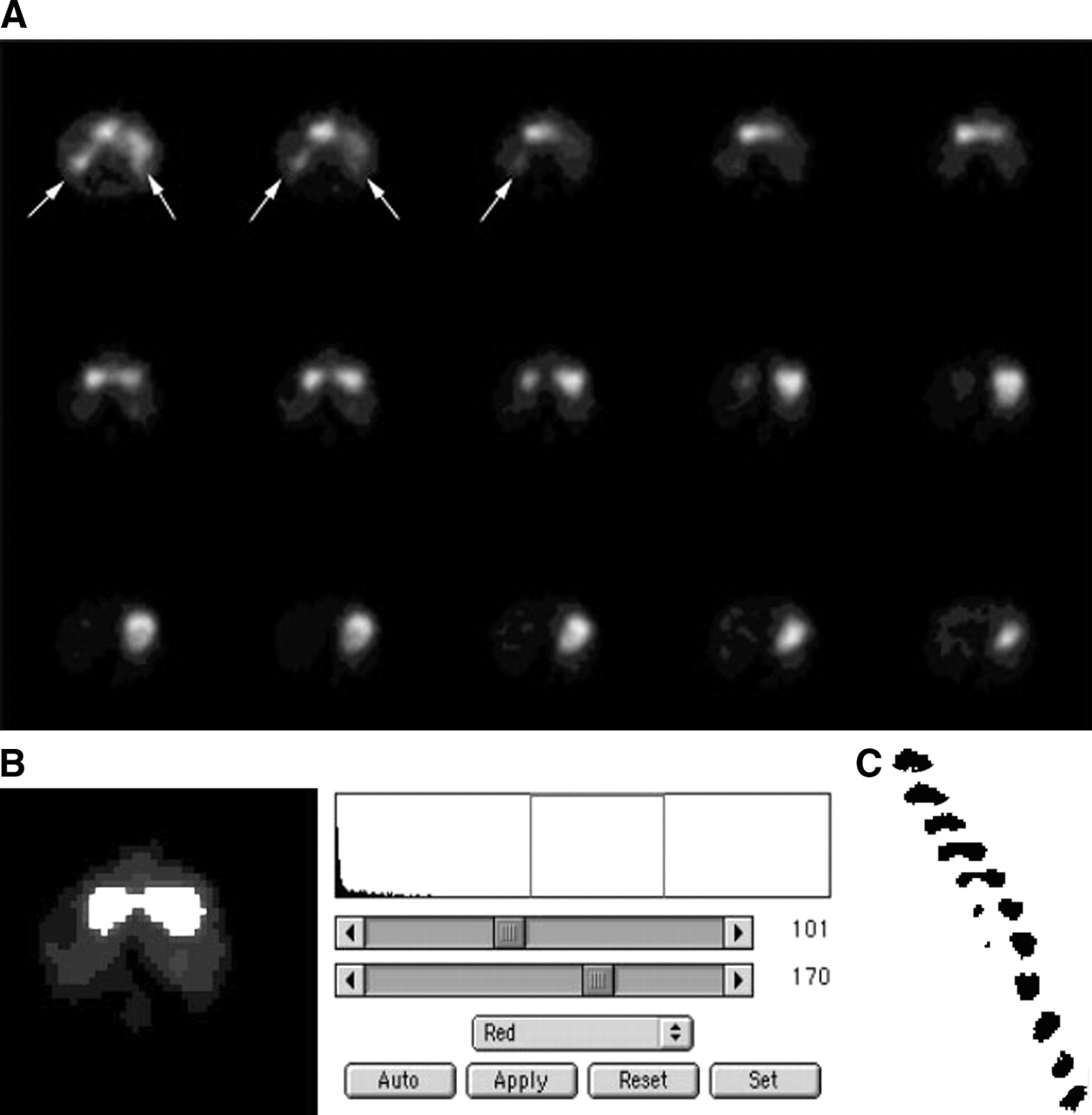
Gastric emptying of the liquid nutrient drink labeled with 111In-DTPA and the egg sandwich labeled with 111In-oxine was calculated by measuring total gastric counts obtained from manual regions of interest (ROIs) drawn around the stomach (Fig. 1A) and calculating the percentage retention in the stomach as a function of time using the decay-corrected, geometric mean of the anterior and posterior 111In counts (18,19).

Gastric images obtained during simultaneous assessment of gastric emptying and accommodation. (A) Gastric emptying of 111In-labeled egg sandwich meal. Typical images of the 111In-labeled egg meal are shown (anterior views only; posterior images not shown). (B) Selected LAO planar images from serial SPECT images of stomach used to measure planar cross-sectional area of stomach after 99mTc-pertechnetate labeling of gastric mucosa. (C) Three-dimensional volume rendered images from SPECT reconstruction shows early gastric dilatation that occurs after meal.
2-Dimensional Cross-Sectional Area (CSA).
The 2D CSA of the stomach (17) was measured to determine whether changes in this measurement could be used as an index of gastric accommodation. The raw tomographic SPECT data were rotated and visually examined to find the projection that gave the maximal CSA of the stomach (approximately the LAO projection image) (Fig. 1B). The CSA of the stomach in this image was determined by drawing a manual ROI to outline the entire stomach. In addition, regional changes in gastric CSA were assessed by measuring changes in the proximal gastric (fundus) and distal gastric (antrum) CSA defined as the area of the stomach above and below the incisura, respectively.
3-Dimensional Gastric Volume.
SPECT was used for the 3D volume analysis (Fig. 1C). The image of the stomach with the longest fundus-to-antrum dimension was used to determine the upper and lower limits of the stomach. The reconstructed transaxial stomach images were converted to TIFF format for SPECT volume reconstructions and analysis (G4 computer; Macintosh). Total gastric volumes were calculated from the SPECT images by summing total gastric voxels for each transaxial slice (Fig. 2). Total gastric voxel counts were obtained from a semiautomated thresholding method with visual confirmation using National Institutes of Health Image J software. Image J is image processing and analysis software in Java language (20), which is in the public domain (http://rsb.info.nih.gov/ij/index.html). The Image J analysis used existing subroutines that permit masking extragastric activity (renal), automated thresholding, and voxel counting. Gastric accommodation was calculated for each time period as the percentage change in gastric voxels compared with the baseline.

Image J analysis of gastric volume. (A) Series of reconstructed transaxial images from top of stomach (fundus) (bottom right corner) down to bottom of antrum (top left corner) are shown. Renal activity (arrows) is masked out of image before performing thresholding to detect gastric boundaries. (B) Image J software permits threshold levels to be adjusted to mark all pixels within gastric ROI. Sample histogram is shown before thresholding (right). After thresholding, all voxels within gastric outlines are marked in white (left). (C) To obtain total number of voxels in stomach, all thresholded voxels are summed for all slices from top of stomach (bottom right) to bottom of stomach (top left).
Phantom Studies for Scatter and Cross Talk
Phantom studies were performed to assess scatter and cross talk between the different radionuclides and to standardize the volume measurements from the SPECT-derived voxel counts. A phantom was constructed from different volume bags of normal saline (100, 250, 500, and 1,000 mL) into which was introduced either 99mTc or 111In or both. The phantom was imaged using the same γ-camera and collimators as those used for the clinical studies. 111In imaging was performed using a 20% window on the upper photopeak of 274 keV. 99mTc imaging was performed using a 20% window on the 140-keV photopeak.
Data and Statistical Analysis
Gastric accommodation was calculated both as the percentage change in gastric CSA in the largest LAO projection (2D) and from the total 3D SPECT gastric voxels compared with the baseline image. Total, proximal, and distal gastric CSA and gastric emptying were measured serially.
The 2D (LAO planar) approach calculating gastric dimensions was compared with the 3D SPECT gastric volume analysis with the Pearson correlation coefficient.
Results are expressed as mean ± SEM. Statistical significance was determined with P < 0.05 (21).
RESULTS
Phantom Studies
Phantom studies demonstrated negligible scatter between the 99m Tc and 111In imaging windows. The 99mTc counts in the 111In window showed only a 0.1% contribution. The 111In counts contributed 15% of counts in the 99mTc window; however, since these contributed to intragastric activity, they did not affect the edge detection used to outline the stomach to detect gastric volume.
Using Image J analysis, the voxel count of a known 1,000-mL volume standard radiolabeled with 99mTc was 1,881 voxels. Repeated imaging of this 1,000-mL standard was within 3% on 3 independent measurements by one observer. The correlation between voxel counts and known volumes of 100-, 250-, 500-, and 1,000-mL standard volumes was linear with r = 0.9995 and the equation of the best fit line: Volume in milliliters = (0.55 × voxel count) − 50.
Liquid Meal (Nutrient Drink) Studies
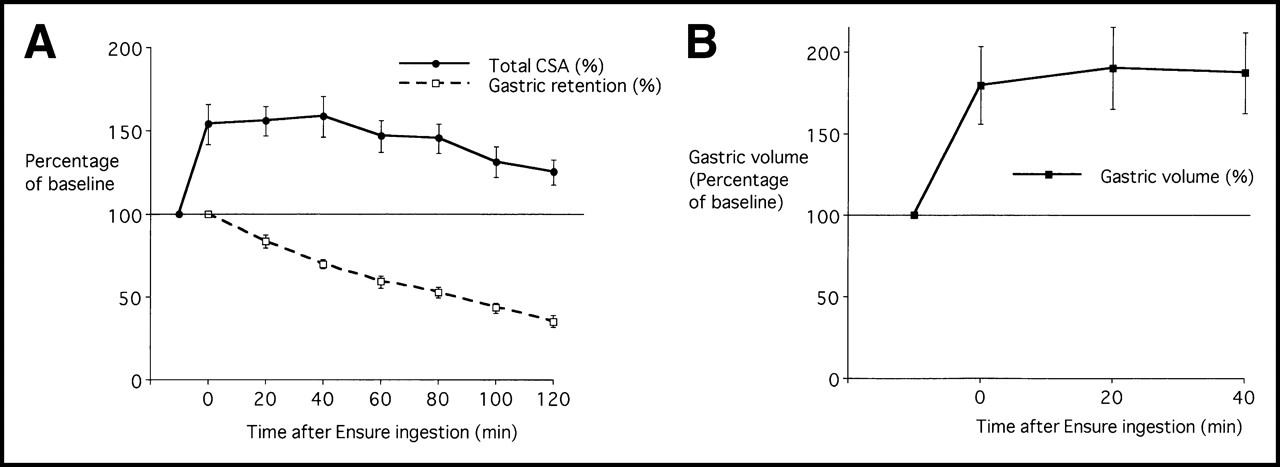
Gastric emptying and accommodation were measured simultaneously in 9 healthy subjects after a liquid nutrient drink (300 mL; 316 calories). The maximal CSA (158% ± 12% of baseline; P < 0.05) occurred 40 min after meal ingestion, a time when only 70% ± 3% of the liquid nutrient drink remained in the stomach (Fig. 3A). At 120 min after ingestion of the liquid nutrient drink, CSA was 125% ± 8% of baseline, but only 35% ± 4% of the liquid nutrient drink remained in the stomach (Fig. 3A). Proximal CSA increased to a maximum of 160% ± 9% at 20 min after ingestion; distal CSA increased to a maximum of 161% ± 16% at 40 min. Using SPECT to measure 3D volume, gastric volume increased immediately after meal ingestion (Fig. 3B; Table 1), being maximal at 20 min after meal ingestion (189% ± 25% of baseline).

Gastric emptying and accommodation curves after liquid nutrient meal in healthy subjects. (A) Total gastric CSA determined from LAO projection and gastric retention after liquid nutrient drink ingestion are shown in relation to time after meal ingestion. (B) Gastric accommodation after liquid nutrient meal. Data shown were obtained from SPECT and Image J analysis for 3D volume. Results are expressed as percentage of baseline total gastric volume before meal ingestion. Results are expressed as mean ± SEM in 9 healthy subjects.
Gastric CSA and Volume After Liquid Nutrient Drink Meal or Solid Egg Sandwich Meal
Solid Meal (Egg Sandwich) Studies
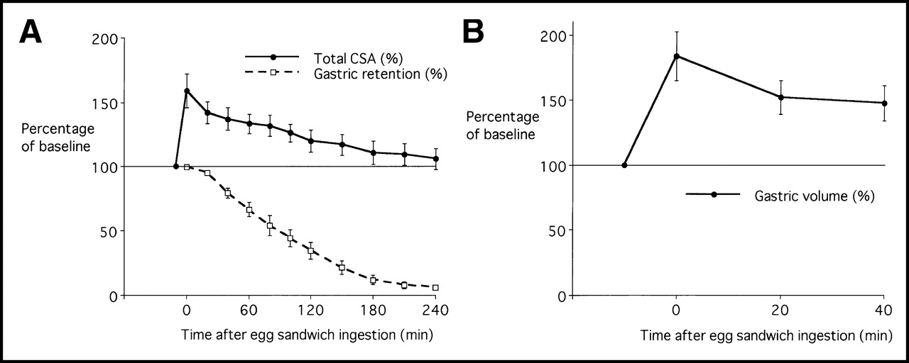
Gastric emptying and gastric accommodation were measured simultaneously in 10 healthy subjects after ingesting a radiolabeled egg sandwich. Total gastric CSA (2D) increased after meal ingestion, with the maximum (159% ± 13% of baseline; P < 0.05) occurring immediately after meal ingestion (Fig. 4A; Table 1). Total CSA remained significantly increased above baseline for the first 2.5 h after ingestion of the egg meal, despite 78% ± 5% gastric emptying at 2.5 h. Maximal proximal CSA (159% ± 10% of baseline) occurred immediately after meal ingestion. Distal CSA also increased after meal ingestion and remained elevated at 134% of control from 0 to 80 min after meal ingestion.

Gastric emptying and accommodation after solid egg sandwich meal in healthy subjects. Results are expressed as mean ± SEM in 10 healthy subjects. (A) Total gastric CSA as determined from LAO projection and gastric retention after egg meal are shown in relation to time after meal ingestion. (B) Gastric accommodation after egg sandwich meal. Data shown were obtained from SPECT and Image J analysis for 3D volume. Results are expressed as percentage of baseline total gastric volume before meal ingestion.
Maximal SPECT 3D gastric volume (mean ± SEM) occurred immediately after the meal (184% ± 19% of baseline; P < 0.05 vs. baseline) compared with the volume at 20 min (152% ± 13% of baseline; P < 0.05) and at 40 min (147% ± 14% of baseline; P < 0.05) (Fig. 4B).
One observer performed both manual ROI determinations for the gastric outlines on the SPECT transaxial slices and semiautomated edge detection using the Image J software. There was excellent correlation between the manual ROI method versus the semiautomated edge detection using Image J software (slope = 0.869; r = 0.95). Using the SPECT 3D volume measurements and the equation derived from the volume standards (volume in mL = [voxel count × 0.55] − 50), the mean gastric dimensions were calculated at baseline to be 397 ± 55 mL, which increased 730 ± 92 mL after meal ingestion—an average increase of 333 mL.
There was, however, a poor correlation of the gastric accommodation measurement with the planar CSA LAO view (r = 0.622; P = 0.055) for the immediate postprandial period. This was worse in the 20-min postprandial period (r = 0.445) and for the 40-min postprandial period (r = 0.431). These findings showed that the 2D LAO approximation did not correlate well with the 3D SPECT measurements.
DISCUSSION
Noninvasive techniques have been proposed to measure gastric accommodation, the relaxation of the proximal stomach with food ingestion. In this study, radionuclide imaging of the gastric wall was performed after the intravenous injection of 99mTc-pertechnetate, which is taken up selectively and localizes in the gastric mucosa. Three-dimensional calculation of gastric volume from SPECT imaging was used as a noninvasive measure of gastric accommodation (10, 13). Simultaneous measurement of gastric emptying of either a liquid or a solid meal using a different radionuclide (111In) was unique to our study. Thus, this study describes a noninvasive, clinically applicable scintigraphic test that measures simultaneously both gastric emptying and accommodation after ingestion of either a solid or liquid test meal.
99mTc labeling of the stomach with SPECT, as performed in this study, has been shown previously to provide a noninvasive estimate of the effect of a meal on total gastric volume that is comparable with changes in balloon volume as observed with the gastric barostat (13). In 32 patients with functional dyspepsia referred to a tertiary medical center, SPECT after 99mTc-pertechnetate infusion showed that impaired gastric accommodation was present in 41% of dyspeptic patients and was more common than abnormal gastric emptying (22%) (14). Further studies have shown that pharmacologic modulation of human gastric volumes can be demonstrated using SPECT (22–24). For example, sublingal isosorbide increases fasting volume, whereas erythromycin lowers postprandial volume (22). In those studies, gastric accommodation assessment was performed on a different day than the conventional gastric emptying test. In addition, the gastric accommodation test was performed with a different meal (liquid rather than solid) and only measured accommodation at very early times after liquid meal ingestion.
Several interesting aspects of gastric postprandial volume and gastric emptying were found in this study in healthy subjects. First, this study shows that both gastric volume and emptying can be measured simultaneously in a single test using dual-isotope imaging. Second, the gastric accommodation response is immediate and prolonged, lasting several hours, persisting despite nearly complete emptying of a liquid or solid meal. Third, both the proximal and distal stomach undergoes an accommodation response; the fundic relaxation response occurs temporally before the antral response.
It had been hoped that the measurement of the gastric accommodation response could be performed using a simple 2D planar image of the stomach. For this, the LAO projection, which maximizes the gastric CSA, was evaluated (17). However, there was a poor correlation between the planar (2D) and SPECT (3D) measurements. Other studies have shown a good correlation between gastric volumes measured by SPECT and the gastric barostat (13). Thus, true 3D SPECT gastric volume determination appears to be required to accurately measure total gastric volume. This is not surprising given the complex and variable shape of the stomach, which is J shaped in the vertical direction and with varying cross-sectional diameters along its major axis.
This scintigraphic method can be used to measure gastric emptying and accommodation simultaneously after ingestion of either a liquid or a mixed solid/liquid meal. This test will improve and extend the current methodology for noninvasive imaging by using a scintigraphic technique to quantitate 2 physical parameters of gastric motility: gastric emptying of solid food and gastric accommodation. A combined gastric emptying/accommodation technique is expected to be of clinical value to study patients with a range of upper GI symptoms, including functional dyspepsia. This is expected to help differentiate dyspeptic patients with impaired fundic accommodation or delayed stomach emptying. Better understanding of the underlying pathophysiology may help direct medical therapy.
CONCLUSION
This study has demonstrated the feasibility and standardization of a new scintigraphic test for simultaneous measurement of gastric emptying and accommodation. The results show that this dual-isotope technique allowed gastric accommodation and emptying to be successfully measured in a single test. In healthy subjects, gastric accommodation was maximal soon after the meal ingestion and was prolonged, persisting for several hours despite nearly complete emptying of a liquid or solid meal. Future applications of this method are expected to improve evaluation and treatment of patients with upper GI symptoms.
Acknowledgments
This study was supported in part by funding through a National Institutes of Health (NIH) Midcareer Investigator Award in Patient-Oriented Research (NIH DK02921) and by the NIH General Clinical Research Center Program of the National Center for Research Resources (RR00349).
Footnotes
Received Aug. 28, 2003; revision accepted Apr. 6, 2004.
For correspondence or reprints contact: Henry P. Parkman, MD, Gastroenterology Section, Department of Medicine, Temple University School of Medicine, Parkinson Pavilion, 8th floor, 3401 N. Broad St., Philadelphia, PA 19140.
E-mail: henry.parkman{at}temple.edu
REFERENCES
In this issue


Jump to section
Related Articles
Cited By...
- Gastric Emptying Scintigraphy
- Gastrointestinal Motility, Part 1: Esophageal Transit and Gastric Emptying
- Gastrointestinal Motility, Part 1: Esophageal Transit and Gastric Emptying
- Diabetic gastroparesis: what we have learned and had to unlearn in the past 5 years
- Impaired gastric accommodation and its role in dyspepsia