
Main menu

  • Home
  • Content
    • Current
    • Ahead of print
    • Past Issues
    • JNM Supplement
    • SNMMI Annual Meeting Abstracts
    • Continuing Education
    • JNM Podcasts
  • Subscriptions
    • Subscribers
    • Institutional and Non-member
    • Rates
    • Journal Claims
    • Corporate & Special Sales
  • Authors
    • Submit to JNM
    • Information for Authors
    • Assignment of Copyright
    • AQARA requirements
  • Info
    • Reviewers
    • Permissions
    • Advertisers
  • About
    • About Us
    • Editorial Board
    • Contact Information
  • More
    • Alerts
    • Feedback
    • Help
    • SNMMI Journals
  • SNMMI
    • JNM
    • JNMT
    • SNMMI Journals
    • SNMMI

User menu

  • Subscribe
  • My alerts
  • Log in
  • Log out
  • My Cart

Search

  • Advanced search
Journal of Nuclear Medicine
  • SNMMI
    • JNM
    • JNMT
    • SNMMI Journals
    • SNMMI
  • Subscribe
  • My alerts
  • Log in
  • Log out
  • My Cart
Journal of Nuclear Medicine

Advanced Search

  • Home
  • Content
    • Current
    • Ahead of print
    • Past Issues
    • JNM Supplement
    • SNMMI Annual Meeting Abstracts
    • Continuing Education
    • JNM Podcasts
  • Subscriptions
    • Subscribers
    • Institutional and Non-member
    • Rates
    • Journal Claims
    • Corporate & Special Sales
  • Authors
    • Submit to JNM
    • Information for Authors
    • Assignment of Copyright
    • AQARA requirements
  • Info
    • Reviewers
    • Permissions
    • Advertisers
  • About
    • About Us
    • Editorial Board
    • Contact Information
  • More
    • Alerts
    • Feedback
    • Help
    • SNMMI Journals
  • View or Listen to JNM Podcast
  • Visit JNM on Facebook
  • Join JNM on LinkedIn
  • Follow JNM on Twitter
  • Subscribe to our RSS feeds
OtherInvited Perspectives

Imaging the Vulnerable Plaque: A Scintillating Light at the End of the Tunnel?

H. William Strauss, Mark Dunphy and Naoki Tokita
Journal of Nuclear Medicine July 2004, 45 (7) 1106-1107;
H. William Strauss
  • Find this author on Google Scholar
  • Find this author on PubMed
  • Search for this author on this site
Mark Dunphy
  • Find this author on Google Scholar
  • Find this author on PubMed
  • Search for this author on this site
Naoki Tokita
  • Find this author on Google Scholar
  • Find this author on PubMed
  • Search for this author on this site
  • Article
  • Figures & Data
  • Info & Metrics
  • PDF
Loading

Atheromas are very prevalent in humans at autopsy. Most of the lesions are found in the aorta and in the carotid, iliac, and coronary arteries. Histologic studies have demonstrated that atheromas go through a cycle of lipid deposition, inflammatory cell infiltration, increased vasa vasorum, plaque disruption—either by erosion (sudden loss of endothelial cells leading to exposure of underlying thrombogenic collagen) or rupture (loss of integrity of the cap of the plaque by weakening at the shoulders of the lesion or sudden increase in pressure within the plaque due to hemorrhage, exposing thrombogenic contents)—platelet deposition, thrombus formation, and reendothelialization, followed by quiescence (Fig. 1). This cycle is repeated multiple times for each lesion, leading to the conclusion that most plaque disruptions are clinically silent (1). This concept is supported by the observation in autopsy studies, as well as during angioscopy of patients with acute coronary syndromes, that patients with an acute thrombus have multiple vulnerable lesions (2–4).

There are 2 potential clinical consequences of thrombus formation on plaque. The first is that emboli arising from the plaque thrombus occlude a critical vessel downstream. The second is that the thrombus occludes a major portion of the vessel lumen at the site of the lesion. Lesions in the aorta are associated with embolism, lesions in the coronaries are associated with occlusion, and lesions in the carotids are associated with both. Because plaques are common and patients can survive for many years with flow-limiting lesions (when the underlying atheroma is stable, such as in chronic stable angina), only a small subset of all atheromas requires immediate treatment. These are the lesions most likely to cause a clinical event.

Libby (5) suggests “… most myocardial infarctions result not from critical blockages but from lesions that produce stenoses that do not limit flow. For example, in individuals who have undergone coronary arteriography in the months preceding myocardial infarction, the culprit lesion most often shows less than 50% stenosis. In a compilation of 4 such serial angiographic studies, only approximately 15% of acute myocardial infarctions arose from lesions with degrees of stenosis greater than 60% on an antecedent angiogram (6,7).”

Atheromas causing a clinical event are often called vulnerable plaque, a term coined by Muller in 1985 (8) in an article describing the circadian variation in the frequency of myocardial infarction (a 3-fold higher incidence of myocardial infarction in the morning [peak at 9 am] than in the evening [trough at 11 pm]). Although vulnerable plaques are a small fraction of the total burden of fibroatheroma (9), these lesions are numerous in patients with clinical events. Histologically, the broad category of vulnerable plaques is characterized by a fibrous cap < 65 μm thick (10); a lipid core occupying at least 50% of the plaque volume; numerous lipid-filled macrophages near the necrotic core and in the cap; abundant lymphocytes in the adventitia (11); and other inflammatory cells, such as mast cells, in the lesion. Histologic reconstruction of lesions that were found at autopsy demonstrated that 70% of lesions had ruptured and healed without causing a clinical event (9). However, compared with calcified fibroatheroma, thin-capped lipid-filled lesions with intense inflammation have a higher likelihood of rupture leading to a major acute coronary event. Recent studies suggest, however, that the composition of atheromas leading to acute events in young patients is different from that seen in patients in their 70s and 80s. In the latter group, the majority of culprit atheromas are calcified, contain limited amounts of lipid and relatively few inflammatory cells, and often cause clinical events through erosion rather than rupture. These age-related differences have a major impact on the potential approaches available to detect patients at high risk of a clinical event.

Although ultrasound (12), CT (13,14), and MRI (15) can identify atheromas on the basis of degree of stenosis, intima-to-media thickness ratio, presence and extent of calcium, or appearance of lipid content, these technologies cannot see a key component of the lesion: inflammation. 18F-FDG PET, on the other hand, can readily identify the metabolic activity of macrophages and other hypermetabolic cells in inflammatory lesions (16). Localization of 18F-FDG in atheromas has been documented for the carotid arteries (17), aorta (18), and other major vessels (19) of humans.

On pages 1245–1250 of this issue of The Journal of Nuclear Medicine, Ogawa and colleagues describe the results of 18F-FDG imaging in rabbits with hereditable hypercholesterolemia (20). The Watanabe rabbit (WR) possesses a heritable trait of hyperlipidemia, with an arterial system lacking functional low-density lipoprotein receptors. Aortic atherosclerosis develops spontaneously by the age of 5 mo (21). A few weeks after birth, fatty streaks develop in the aortae of young WRs; macrophages (i.e., lipid-laden foam cells) are the most prevalent cell type in these streaks (22,23). As young WRs grow older, fatty streaks develop in the intramyocardial and carotid arteries (24). WR atherosclerosis predominates in the arch of the aorta, with diffuse thickening of the vascular wall. Descending thoracic atherosclerosis in WRs varies from a patchwork of atheromatous plaques to more diffuse disease.

Ogawa et al. (20) correlated 18F-FDG uptake determined on ex vivo well-counting studies of 6-mm segments of the aorta with histologic quantitation of the number of macrophages based on the macrophage-specific stain, RAM-11. The investigators found an r value of 0.81 between macrophage number and 18F-FDG uptake.

There is a cautionary note in their results, however. The investigators compared uptake in the hyperlipidemic WRs with that in control animals. 18F-FDG localization was almost 3-fold greater in hyperlipidemic vessels than in control vessels. The in vivo images demonstrated a modest degree of 18F-FDG uptake, which was difficult to identify in images. The clinical challenge is not identifying the patient with atheroma (which can be done through blood lipid measurement and assays of inflammatory factors) but recognizing specific lesions likely to cause clinical events. To fulfill this role, the imaging agent must have a high concentration only in these critical lesions, with a minimal concentration in other atheromas. 18F-FDG may concentrate in too broad a range of lesions to provide unambiguous clinical data. Other markers, such as 99mTc-labeled annexin V, a marker of apoptosis (25), have also been suggested for these studies (26). The pathophysiologic basis for using annexin is the presence of large numbers of apoptotic cells in unstable lesions. At this point it is not clear whether 18F-FDG or annexin (27) or perhaps some other marker will provide the specific localization required to make the technique clinically useful.

Regardless of which tracer proves most useful, however, the relationship between tracer localization and macrophage activity in the plaque is likely to be a key to identifying lesions likely to cause clinical events.

  FIGURE 1.
  • Download figure
  • Open in new tab
  • Download powerpoint
FIGURE 1.

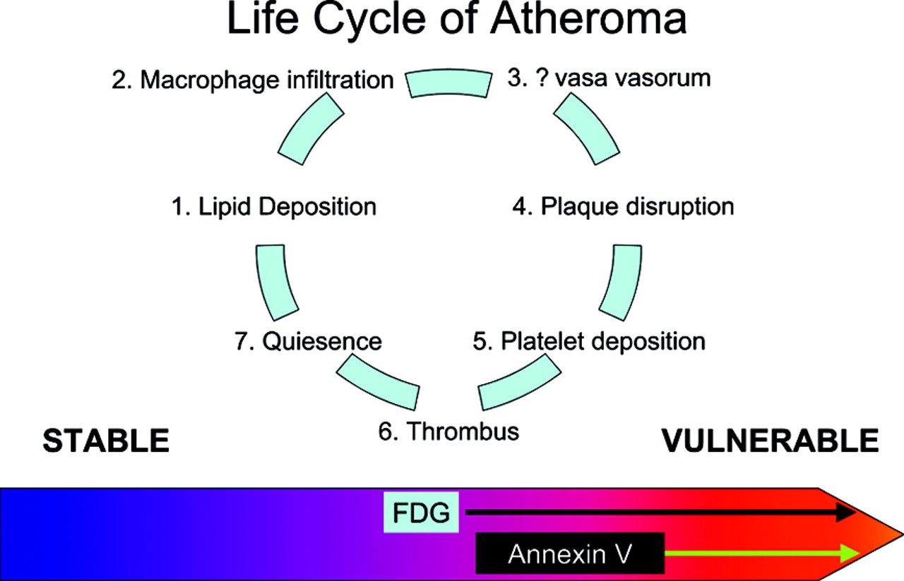
Diagram depicting the life cycle of an atheroma. The lesion goes through several cycles of endothelial injury, lipid deposition, macrophage attraction, increased vasa vasorum, and quiescence. The plaque may progress to the point where its cap is disrupted, leading to platelet deposition and thrombus formation. Most of these episodes are clinically silent. The lesion heals, undergoes reendothelialization, and may remain quiescent for decades. The major goal is to identify lesions likely to cause a clinical event in the near term. Several tracers can accomplish this task. The leading contenders appear to be 18F-FDG (metabolic activity of the inflammatory cells) and annexin V (apoptosis of cells in the lesion). 18F-FDG, however, may identify a broader range of lesions than does annexin and, as such, may be less helpful as a marker identifying patients for intervention and intense local therapy.

Footnotes

  • Received May 21, 2004; revision accepted May 27, 2004.

    For correspondence or reprints contact: H. William Strauss, MD, Nuclear Medicine Service, Memorial Sloan Kettering Cancer Center, Room S212, 1275 York Ave., New York, NY 10021.

    E-mail: straussH{at}mskcc.org

REFERENCES

  1. ↵
    Davies MJ. Stability and instability: two faces of coronary atherosclerosis—The Paul Dudley White Lecture 1995. Circulation. 1996;94:2013–2020.
    OpenUrlFREE Full Text
  2. ↵
    Asakura M, Ueda Y, Yamaguchi O, et al. Extensive development of vulnerable plaques as a pan-coronary process in patients with myocardial infarction: an angioscopic study. J Am Coll Cardiol. 2001;37:1284–1288.
    OpenUrlCrossRefPubMed
  3. Spagnoli LG, Bonanno E, Mauriello A, et al. Multicentric inflammation in epicardial coronary arteries of patients dying of acute myocardial infarction. J Am Coll Cardiol. 2002;40:1579–1588.
    OpenUrlCrossRefPubMed
  4. ↵
    Davies MJ. Atlas of Coronary Artery Disease. Philadelphia, PA: Lippincott Raven; 1999.
  5. ↵
    Libby PF. The vascular biology of atherosclerosis. In: Braunwald EB, Zipes DP, Libby P, eds. Heart Disease: A Textbook of Cardiovascular Medicine. 6th ed. Philadelphia, PA: WB Saunders and Co.; 2001:chap 30.
  6. ↵
    Falk E, Shah P, Fuster V. Coronary plaque disruption. Circulation. 1995;92:657–671.
    OpenUrlFREE Full Text
  7. ↵
    Smith S Jr. Risk-reduction therapy: the challenge to change. Circulation. 1996;93:2205–2211.
    OpenUrlFREE Full Text
  8. ↵
    Muller JE, Stone PH, Turi ZG, et al. Circadian variation in the frequency of onset of acute myocardial infarction. N Engl J Med. 1985;313:1315–1322.
    OpenUrlCrossRefPubMed
  9. ↵
    Davies MJ. Acute coronary thrombosis: the role of plaque disruption and its initiation and prevention. Eur Heart J. 1995;16(suppl L):3–7.
  10. ↵
    Virmani R, Burke AP, Kolodgie FD, Farb A. Vulnerable plaque: the pathology of unstable coronary lesions. J Interv Cardiol. 2002;15:439–446.
    OpenUrlCrossRefPubMed
  11. ↵
    Katsuda S, Kaji T. Atherosclerosis and extracellular matrix. J Atheroscler Thromb. 2003;10:267–274.
    OpenUrlPubMed
  12. ↵
    Bard RL, Kalsi H, Rubenfire M, et al. Effect of carotid atherosclerosis screening on risk stratification during primary cardiovascular disease prevention. Am J Cardiol. 2004;93:1030–1032.
    OpenUrlCrossRefPubMed
  13. ↵
    Agatston AS, Janowitz WR, Hildner FJ, Zusmer NR, Viamonte M, Detrano R. Quantification of coronary artery calcium using ultrafast computed tomography. J Am Coll Cardiol. 1990;15:827–832.
    OpenUrlCrossRefPubMed
  14. ↵
    Stanford W, Thompson BH, Burns TL, Heery SD, Burr MC. Coronary artery calcium quantification at multi-detector row helical CT versus electron-beam CT. Radiology. 2004;230:397–402.
    OpenUrlPubMed
  15. ↵
    Luo Y, Polissar N, Han C, et al. Accuracy and uniqueness of three in vivo measurements of atherosclerotic carotid plaque morphology with black blood MRI. Magn Reson Med. 2003;50:75–82.
    OpenUrlCrossRefPubMed
  16. ↵
    Kubota R, Yamada S, Kubota K, Ishiwata K, Tamahashi N, Ido T. Intratumoral distribution of fluorine-18-fluorodeoxyglucose in vivo: high accumulation in macrophages and granulation tissues studied by microautoradiography. J Nucl Med. 1992;33:1972–1980.
    OpenUrlAbstract/FREE Full Text
  17. ↵
    Rudd JH, Warburton EA, Fryer TD, et al. Imaging atherosclerotic plaque inflammation with [18F]-fluorodeoxyglucose positron emission tomography. Circulation. 2002;105:2708–2711.
    OpenUrlAbstract/FREE Full Text
  18. ↵
    Tatsumi M, Cohade C, Nakamoto Y, Wahl RL. Fluorodeoxyglucose uptake in the aortic wall at PET/CT: possible finding for active atherosclerosis. Radiology. 2003;229:831–837.
    OpenUrlPubMed
  19. ↵
    Yun M, Jang S, Cucchiara A, Newberg AB, Alavi A. 18F FDG uptake in the large arteries: a correlation study with the atherogenic risk factors. Semin Nucl Med. 2002;32:70–76.
    OpenUrlCrossRefPubMed
  20. ↵
    Ogawa M, Ishino S, Mukai, et al. 18F-FDG accumulation in atherosclerotic plaques: immunohistochemical and PET imaging study. J Nucl Med. 2004;45:1245–1250.
    OpenUrlAbstract/FREE Full Text
  21. ↵
    Tanzawa K, Shimada Y, Kuroda M, Tsujita Y, Arai M, Watanabe H. WHHL-rabbit: a low density lipoprotein receptor-deficient animal model for familial hypercholesterolemia. FEBS Lett. 1980;118:81–84.
    OpenUrlCrossRefPubMed
  22. ↵
    Rosenfeld ME, Tsukada T, Gown AM, Ross R. Fatty streak initiation in Watanabe Heritable Hyperlipemic and comparably hypercholesterolemic fat-fed rabbits. Arteriosclerosis. 1987;7:9–23.
    OpenUrlAbstract
  23. ↵
    Rosenfeld ME, Tsukada T, Chait A, Bierman EL, Gown AM, Ross R. Fatty streak expansion and maturation in Watanabe Heritable Hyperlipemic and comparably hypercholesterolemic fat-fed rabbits. Arteriosclerosis. 1987;7:24–34.
    OpenUrlAbstract
  24. ↵
    Spagnoli LG, Orlandi A, Mauriello A, et al. Aging and atherosclerosis in the rabbit. 1. Distribution, prevalence and morphology of atherosclerotic lesions. Atherosclerosis. 1991;89:11–24.
    OpenUrlCrossRefPubMed
  25. ↵
    Blankenberg FG, Katsikis PD, Tait JF, et al. In vivo detection and imaging of phosphatidylserine expression during programmed cell death. Proc Natl Acad Sci USA. 1998;95:6349–6354.
    OpenUrlAbstract/FREE Full Text
  26. ↵
    Kietselaer BLJH, Reutelingsperger CPM, Heidendal GAK, et al. Noninvasive detection of plaque instability with use of radiolabeled annexin A5 in patients with carotid-artery atherosclerosis. N Engl J Med. 2004;350:1472–1473.
    OpenUrlCrossRefPubMed
  27. ↵
    Mari C, Blankenberg F, Narula Z, et al. 99mTc-annexin V versus 18F-FDG in the identification of atherosclerotic plaques in apoE −/− mice [abstract]. J Nucl Med. 2002;43(suppl):1P.
    OpenUrl
PreviousNext
Back to top

In this issue

Journal of Nuclear Medicine: 45 (7)
Journal of Nuclear Medicine
Vol. 45, Issue 7
July 1, 2004
  • Table of Contents
  • About the Cover
  • Index by author
Print
Download PDF
Article Alerts
Sign In to Email Alerts with your Email Address
Email Article

Thank you for your interest in spreading the word on Journal of Nuclear Medicine.

NOTE: We only request your email address so that the person you are recommending the page to knows that you wanted them to see it, and that it is not junk mail. We do not capture any email address.

Enter multiple addresses on separate lines or separate them with commas.
Imaging the Vulnerable Plaque: A Scintillating Light at the End of the Tunnel?
(Your Name) has sent you a message from Journal of Nuclear Medicine
(Your Name) thought you would like to see the Journal of Nuclear Medicine web site.
Citation Tools
Imaging the Vulnerable Plaque: A Scintillating Light at the End of the Tunnel?
H. William Strauss, Mark Dunphy, Naoki Tokita
Journal of Nuclear Medicine Jul 2004, 45 (7) 1106-1107;

Citation Manager Formats

  • BibTeX
  • Bookends
  • EasyBib
  • EndNote (tagged)
  • EndNote 8 (xml)
  • Medlars
  • Mendeley
  • Papers
  • RefWorks Tagged
  • Ref Manager
  • RIS
  • Zotero
Share
Imaging the Vulnerable Plaque: A Scintillating Light at the End of the Tunnel?
H. William Strauss, Mark Dunphy, Naoki Tokita
Journal of Nuclear Medicine Jul 2004, 45 (7) 1106-1107;
Twitter logo Facebook logo LinkedIn logo Mendeley logo
  • Tweet Widget
  • Facebook Like
  • Google Plus One
Bookmark this article

Jump to section

  • Article
    • Footnotes
    • REFERENCES
  • Figures & Data
  • Info & Metrics
  • PDF

Related Articles

  • This Month in JNM
  • PubMed
  • Google Scholar

Cited By...

  • Imaging Atherosclerotic Plaque Inflammation by Fluorodeoxyglucose With Positron Emission Tomography: Ready for Prime Time?
  • Multimodality Imaging of Carotid Artery Plaques: 18F-Fluoro-2-Deoxyglucose Positron Emission Tomography, Computed Tomography, and Magnetic Resonance Imaging
  • Detection and Treatment of Vulnerable Plaques and Vulnerable Patients: Novel Approaches to Prevention of Coronary Events
  • Google Scholar

More in this TOC Section

  • Radiomics in PET/CT: More Than Meets the Eye?
  • Metabolic Tumor Volume: We Still Need a Platinum-Standard Metric
  • Citius, Altius, Fortius: An Olympian Dream for Theranostics
Show more Invited Perspectives

Similar Articles

SNMMI

© 2025 SNMMI

Powered by HighWire