


Abstract
Conventional nuclear medicine imaging with large radiolabeled molecules such as antitumor antibodies suffers from slow localization and clearance. Pretargeting is under active investigation as an alternative using either (strept)avidin/biotin, bispecific antibodies, or oligomers. However, only the use of oligomers such as phosphorodiamidate morpholinos (MORFs) in pretargeting offers the potential of signal amplification at the target. Amplification targeting is a multistep procedure with the potential to greatly improve target localization of radioactivity (and eventually drugs) through the intermediate use of polymers conjugated with multiple copies of oligomers. Objective: This study was conducted to prove the concept in vivo in tumored mice of amplfication targeting. Methods: Nude mice bearing LS174T tumors received in order: the anti-CEA antibody MN14 conjugated with MORF, a polymer conjugated with multiple copies of complementary MORFs (cMORFs), and, finally, 99mTc-MORF. Results: In tumored animals, dual radiolabels (99mTc and 111In) were used to demonstrate that, after 18 h, about 25% of antibody MORFs in tumor were targeted with polymeric cMORFs and, after 3 h, about 12% of the polymeric cMORFs in tumor were targeted with 99mTc-MORF. Therefore, hybridization in tumor in both cases (i.e., polymeric cMORF to antibody MORF and radiolabeled MORF to polymeric cMORF) was surprisingly efficient given the barriers to targeting in vivo and the competition between targeting and clearance. Moles of radiolabeled MORF accumulating in tumor were more than tripled for study animals receiving all 3 injections compared with control animals not receiving the antibody or the polymer. Furthermore, MORF expression (on antibody) and cMORF expression (on polymer) were rapidly lost in normal organs such as liver, spleen, and kidneys but not in tumor, thus improving the target-to-nontarget ratios. Conclusion: Although signal amplification has not yet been convincingly demonstrated and amplification targeting will require further studies for optimization, the concept has now been shown to be feasible.
This investigation was designed to provide proof that amplification targeting is feasible in vivo and may potentially increase the accumulation of radioactivity in tumor as target tissue. In one of its many possible forms, pretargeting relies on oligomers such as phosphodiester and phosphorothioate DNAs, peptide nucleic acids (PNAs), or phosphordiamidate morpholinos (MORFs). In the approach to amplification targeting described herein, the first administration is an antitumor antibody conjugated with MORF, the second administration is a polymer conjugated with multiple copies of the complementary MORF (cMORF), followed by a final administration of radiolabeled MORF. Thus, amplification targeting bears some similarities to pretargeting (1–6), which, in this case, would involve the administration of the MORF-conjugated antitumor antibody, followed only by the administration of radiolabeled cMORF. Amplification targeting therefore adds the intermediate administration of a polymer conjugated with multiple copies of cMORF. If accessibility and pharmacokinetics factors are favorable, signal amplification will result from the presence of multiple cMORF targets accessible to the radioactive MORF in the tumor provided by the polymer. Obviously, should amplification targeting become practical, its usefulness will extend beyond radioactivity to the increased localization of drugs in tumor and other lesions as well. This investigation extends preliminary work from this laboratory on amplification targeting in which PNAs instead of MORFs served as the oligomer and tumor accumulation was by nonspecific diffusion rather than through the use of an antitumor antibody as is now the case (7).
Described herein are the first studies of amplification targeting in tumored mice. The availability of the antibody MORF to polymeric cMORF as well as the availability of polymeric cMORF to radiolabeled MORF was established in dual-labeling studies using 111In as well as 99mTc. Thus, the first animal study consisted of 111In-labeled antibody and 99mTc-labeled polymer and the second consisted of unlabeled antibody, 111In-labeled polymer, and 99mTc-MORF. The last animal study tested the concept of amplification targeting and consisted of unlabeled antibody, unlabeled polymer, and 99mTc-MORF. By using the dual radioactivity labels in this manner it was possible to measure separately in tumor the absolute accumulation of the antibody, the polymer, and the radiolabeled MORF. Using these values along with the number of MORFs and cMORFs in each injection, it was possible to calculate the percentage of polymeric cMORFs targeting the antibody MORFs localized in tumor and the percentage of radiolabeled MORF that was subsequently able to target the cMORFs on the polymer localized in the tumor. Control studies were also performed and the results were used to establish the extent of nonspecific accumulation of both the polymer in tumor and the radiolabeled MORF in tumor for use in these calculations.
MATERIALS AND METHODS
The 25 mer MORF and cMORF were purchased (Gene-Tools) with a 3′-amine via a 9-member succinylated piperidine linker and were identical to that used by us previously (8). Each MORF was analyzed by size-exclusion high-performance liquid chromatography (HPLC) and MALDI-TOF to ensure quality. The base sequence and molecular weight were as follows: MORF: 5′-TGGTGGTGGGTGTACGTCACAACTA-linker-amine, 8,701 Da; cMORF: 5′-TAGTTGTGACGTACACCCACCACCA-linker-amine, 8,519 Da. The high-affinity murine anti-CEA– carcinoembryonic antigen (CEA) antibody (MN14, IgG 1 subtype; molecular weight, 160 kDa) was a gift from Immunomedics. Polylysine, uniformly succinylated (PL) with an average molecular weight of about 30 kDa, was purchased (Sigma-Aldrich) as was 1-ethyl-3-(3-dimethylaminopropyl) carbodiimide hydrochloride (EDC) (Pierce). N-Hydroxysuccinimidyl S-acetylmercaptoacetyltriglycine (NHS-MAG3) was synthesized in house (9), and the structure was confirmed by elemental analysis, proton nuclear magnetic resonance, and mass spectroscopy. Reagent grade diethylenetriaminepentaacetic acid (DTPA) cyclic anhydride and carbonyldiimidazole were from Sigma-Aldrich and were used as received. The P-4 resin (Bio-Gel P-4 Gel, medium) was purchased (Bio-Rad Laboratories) as was the Sephadex G-100 resin (Pharmacia Biotech). The 99mTc-pertechnetate was eluted from a 99Mo-99mTc generator (Bristol-Myers Squibb Medical Imaging). The 111In was purchased as the chloride (PerkinElmer Life Science Inc.). All other chemicals were reagent grade and were used without further purification.
Preparation of MN14-MORF, MN14-DTPA, MORF-MAG3, MORF-DTPA, and Radiolabelings
The conjugation of MN14 with MORF was accomplished by reacting amine-derivitized MORF with the native antibody using EDC, followed by purification on Sephadex G-100 with 0.05 mol/L phosphate buffer, pH 7.2, as previously described (6). The antibody conjugated with MORFs was characterized by HPLC for concentration and for the average number of MORFs per antibody molecule (groups per molecule) using a differential ultraviolet (UV) method at 265 and 280 nm (8). A measure of immunoreactive fraction for MN14-MORF was obtained by adding an excess of CEA to the MN14 radiolabeled with trace 99mTc-cMORF to shift the peak. That the shift was quantitative indicates that the immunoreactive fraction remained close to unity despite the conjugation (data not presented). MN14-DTPA and MORF-DTPA were prepared using DTPA cyclic anhydride as previously described (10). The average number of DTPA groups per MN14 was determined by labeling the mixture with 111In before purification, assuming the identical accessibility of 111In to both conjugated and free DTPA. 99mTc-MORF-MAG3 was prepared and analyzed as described previously (6). Radiolabeling was achieved by first adding 99mTc-pertechnetate generator eluant to a solution of 5–10 μL of either MORF-MAG3 or cMORF-MAG3 (concentrations >0.1 μg/μL), 25 μL 0.25 mol/L ammonium acetate buffer, pH 5.2, 10 μL pH 9.2 tartrate solution (50 μg sodium tartrate dihydrate per μL), and 4 μL stannous chloride solution (1 μg stannous chloride dihydrate and 1 μg sodium ascorbate per μL in 10 mmol/L HCl), followed by heating in boiling water for 20 min. The product was purified on a P-4 column with 0.05 mol/L phosphate buffer, pH 7.2, as eluant. 111In-MORF-DTPA was prepared by incubating MORF-DTPA with 111In for 1 h at room temperature and was followed by purification as described for 99mTc-MORF. Both labeled (c)MORFs were routinely analyzed by size-exclusion HPLC and found to provide essentially identical chromatograms both with UV and radioactivity detection.
Preparation of PL-cMORF and Radiolabelings
Ten milligrams of uniformly succinylated polylysine polymer (PL) with an initial molecular weight of 30 kDa was dissolved in 1.0 mL of the aprotic solvent N-methyl pyrrolidinone (NMP) and to this was added 20.4 mg of 1,1′-carbonyldiimidazole and 3.0 μL of diisopropylethylamine (DIEA). The mixture was incubated at room temperature for 2 h. To 500 μL of a 1.2 mg/mL solution of cMORF in NMP, a designated amount of the activated PL mixture and equivalent moles of DIEA were added to reach a 100:1 molar ratio of cMORF to PL. The solution was incubated overnight at room temperature.
An aliquot of the solution before purification was analyzed by size-exclusion HPLC with UV detection at 265 nm to estimate the average number of cMORF groups bonded to each PL molecule. Since PL does not absorb appreciably at 265 nm, the peak areas of free, nonconjugated cMORF and PL-coupled cMORF were compared. As an alternative method of estimating the average number of groups per molecule, radiolabeled MORF at tracer concentrations was also added to another aliquot of the PL-cMORF solution and the radioactivity of 99mTc-MORF hybridized to free, nonconjugated cMORF was compared with that hybridized to PL-coupled cMORF. The PL-cMORF conjugates were then purified by open-column gel-filtration chromatography on a 1 cm × 30 cm Sephadex G-100 column using water as eluant. The concentration of PL-cMORFs with respect to cMORFs in the recovered fraction(s) was estimated by UV absorbency using the molar absorbency value of cMORFs provided by the manufacturer.
PL-cMORFs were radiolabeled by incubation with a trace amount of 99mTc-MORF or 111In-MORF for 30 min at room temperature such that, on average, only about 1 cMORF on each polymer was hybridized with MORF. Quality assurance was routinely performed based on size-exclusion HPLC chromatography in which radioactivity recovery was routinely monitored.
Animal Studies
All animal studies were performed with the approval of the University of Massachusetts Medical School Institutional Animal Care and Use Committee. The LS174T cells were grown in Minimum Essential Medium (MEM; Gibco BRL Products) with 2 mmol/L l-glutamine, 1.5 mg/L sodium bicarbonate, 0.1 mmol/L nonessential amino acids, and 1.0 mmol/L sodium pyruvate supplemented with 10% fetal bovine serum (FBS) and 100 mg/mL of penicillin-streptomycin (GIBCO). Cells were maintained as monolayers in a humidified 5% CO2 atmosphere, normally in T75 flasks (Falcon, Becton Dickinson). The cells at 80%–90% confluence were trypsinated in the T75 flasks using 0.05% trypsin/0.02% ethylenediaminetetraacetic acid and were then suspended in MEM with 10% FBS. Nude mice (NIH Swiss, 30–40 g; Taconic Farms) were each injected subcutaneously in the left thigh with a 0.1 mL suspension containing 106 LS174T colon tumor cells. Animals were used after 14 d when the tumors were no more than 1.5 cm in any dimension.
To estimate the optimum dosage of the MN14-MORF antibody and 30 kDa PL-cMORF polymer, in preliminary studies, tumored animals were administered 60 μg (as an initial guess) of MN14 conjugated with an average of 0.2 MORF per molecule and 3 μg of MN14-DTPA radiolabeled with about 7.4 × 10⁴ Bq (2.0 μCi) of 111In 2 d before the administration of 99mTc-labeled polymer at 3 dosages from 24 to 76 μg per animal. On the basis of these results, a 25-μg dosage of MN14-MORF and a 15 μg dosage of polymer were selected for subsequent animal studies.
Figure 1 presents a schematic of timing and dosages for the following 3 animal studies of amplification targeting. In the first amplification targeting study, nude mice received simultaneously 25 μg of MN14-MORF mixed with 3 μg (7.4 × 10⁴ Bq [2.0 μCi]) of 111In-MN14. About 30 h later, the animals received 15 μg (9.25 MBq [250 μCi]) of the 30 kDa PL-cMORF polymer labeled by hybridization with 99mTc-MORF occupying an average of only about 1 of the 12–15 cMORFs on the polymer. Control animals did not receive either the antibody or the polymer. Animals were killed by heart puncture under anesthesia at 18 h after administration of the polymer. Organs and blood were harvested for simultaneously counting of 111In and 99mTc in an automatic γ-counter (Cobra II; Packard Instrument Co.). All counts were corrected for physical decay and for the small contribution of 111In activity in the 99mTc window. Results are presented as percentage of injected dosage per gram (%ID/g).

Schematic diagrams show timing and dosages for 3 amplification targeting animal studies.
In a second amplification targeting study, targeting was performed in a similar manner with 25 μg of the MN14-MORF (now unlabeled) administered to LS174T tumored mice 30 h before the administration of 15 μg of the PL-cMORF polymer (now radiolabeled with trace 0.11 MBq [3 μCi] 111In-MORF). Animals received 1.5 μg (about 11.1 MBq [300 μCi]) of 99mTc-MORF at 41 h after administration of the polymer and were killed 3 h later. The labeled MORF dosage was selected to be reasonably low yet capable of carrying sufficient radioactivity. For controls, the antibody was eliminated in one set of animals while both the antibody and the polymer were eliminated in another set.
A third animal study was designed specifically to evaluate targeting amplification. Nude mice first received 25 μg of the unlabeled MN14-MORF antibody 30 h before administration of 15 μg of the unlabeled 30 kDa PL-cMORF polymer. Animals received 1.5 μg of 99mTc-MORF at either 18 or 40 h after administration of the polymer and were killed 3 h later. Control animals either did not receive the antibody or received neither the antibody nor the polymer. After carefully voiding the bladder with a syringe, the animals were imaged on an Elscint APEX 409 M large-view γ-camera. After imaging, animals were killed and dissected as described. Statistical significance was established by the Student paired t test based on Microsoft Excel with 2-tailed distribution.
RESULTS
Conjugations and Radiolabeling
Using the UV HPLC chromatograms and the slopes of the standard curves (8), the average MORFs per MN14 molecule for the MN14-MORF preparation used in this investigation was calculated as 0.20. The HPLC radiochromatograms of the MN14-MORFs showed one prominent peak when freshly prepared and purified but, on storage for 1 mo at refrigerator temperatures, showed evidence of <10% of a free MORF peak. However, only freshly prepared conjugated antibodies were used in this investigation. The HPLC radiochromatograph of 111In-MN14-DTPA before purification was used to calculate that an average of 0.7 DTPA groups was conjugated to each MN14 antibody.
The 99mTc labeling procedure used in this investigation always provided a labeled MORF with >90% radiochemical purity after purification as demonstrated by routine HPLC analysis with >90% recovery in all analyses. The radiolabeled cMORFs were routinely shown to be capable of hybridizing to MORF conjugated on antibodies or polymers or immobilized on magnetic beads (6,11).
The average number of cMORF groups attached to each PL polymer molecule was determined by size-exclusion HPLC analysis, both by UV absorbance of the conjugated but unpurified PL-cMORF polymers and by radioactivity before and after the addition of trace 99mTc-MORF. The 30 kDa PL polymer was estimated to have been conjugated with an average of 12–15 cMORF groups per molecule. The 2 methods of measuring groups per molecule showed 80%–90% agreement. The final molecular weight was estimated as 130–160 kDa. HPLC analysis of the purified polymer showed a single peak by HPLC with >90% recovery.
Animal Studies
Before animal studies of amplification targeting, the influence of antibody and polymer dosage on tumor accumulation was investigated. Tumored animals were administered 60 μg (as an initial guess) of 111In-MORF antibody 2 d before the administration of 99mTc-labeled 30 kDa PL polymer at 3 dosages from 24 to 76 μg per animal. Each polymer control animal received 24 μg of polymer but not the antibody. The 99mTc biodistribution results are presented in Table 1 and show an increasing tumor accumulation of polymer in %ID/g with decreasing dosage of polymer at a fixed antibody dosage of 60 μg. The 111In results (not presented) show an accumulation in tumor of MN14-MORF antibody at 60 μg that is statistically identical to that shown in Table 1 for 24 μg of antibody. Therefore, a lower antibody dosage of 25 μg MN14-MORF was selected to conserve reagent as was a polymer dosage of 15 μg. These dosages were then used in all subsequent animals studies of amplification targeting. In this and subsequent tables, statistical significance was established by the Student paired t test based on Microsoft Excel with 2-tailed distribution. Statistically significant values (P ≤ 0.05) are underlined. The results have been corrected for radionuclide physical half-life.
Biodistribution in Tumored Mice 18 Hours After Administration of 99mTc-cMORF Polymer
The first animal study related to amplification targeting was designed to evaluate the degree to which antibody MORF in tumor can be targeted by polymeric cMORF. Thus, nude mice first received radiolabeled antibody (i.e., MN14-MORF along with 111In-MN14). Two days later, the animals received the radiolabeled PL-cMORF polymer (i.e., PL-cMORF along with 99mTc-MORF). Animals were killed at 18 h after administration of the polymer (Fig. 1). Control animals did not receive the antibody and received only the labeled polymer 18 h earlier.
From the biodistribution results in Table 2, the number of antibody MORFs per gram of tumor may be calculated using the 111In values, while the 99mTc values in tumor for the study minus the control may be used to calculate the number of polymeric cMORFs per gram of tumor localized specifically due to the antibody. These calculations show that under the conditions of this study, 25% of the antibody MORFs in tumor were targeted with polymeric cMORFs. This calculation assumes that 1 polymer molecule can bind to no more than 1 antibody molecule. If this assumption is incorrect and a polymer can crosslink antibodies on the tumor, the above value would then be >25%. Although the polymer may be capable of crosslinking multiple antibody molecules in tumor, in a separate study, hybridization of the antibody MORFs in tumor by free (i.e., not conjugated to a polymer and therefore monovalent) labeled cMORF was found to be only 50% (10). Since it is unlikely that the polymeric cMORFs target antibody MORFs more effectively than free cMORF, crosslinking may be judged as unlikely and, if it occurs at all, probably results in crosslinking no more than 2 antibody molecules per polymer molecule.
Biodistribution in Tumored Mice (Study 1)
In connection with a previous pretargeting investigation, this laboratory had reported on similar 99mTc and 111In dual-labeling studies in animals with LS174T tumors to estimate accessibility as a function of time of radiolabeled cMORF to MN14-MORF in tumor, liver, and spleen (10). The results showed that access to MORF in liver and spleen by radiolabeled cMORF was limited to about ≤6% after 24 h following administration of the antibody. Apparently the MORF on MN14 quickly becomes largely “invisible” once the antibody is localized in these organs. Fortunately this was not the case in tumor, where >50% was accessible throughout.
The second animal study related to amplification targeting was designed to evaluate the degree to which polymeric cMORF in tumor can be targeted by radiolabeled MORF. Thus, nude mice first received 25 μg of unlabeled antibody and, 30 h later, received 15 μg of the 30 kDa PL-cMORF polymer labeled by hybridization with trace 111In-MORF. Forty-one hours later, animals received 1.5 μg of 99mTc-MORF and were killed 3 h later (i.e., 74 h after administration of antibody, 44 h after administration of 111In-polymer) (Fig. 1). The labeled MORF dosage was selected to be reasonably low yet capable of carrying sufficient radioactivity for imaging. For controls, the antibody was eliminated in one set of animals (control I), whereas both the antibody and the polymer were eliminated in another set (control II). Biodistribution results are presented in Table 3.
Biodistribution in Tumored Mice 3 Hours After Administration of 99mTc-MORF (Study 2)
That the 99mTc values in tumor are significantly higher with versus without antibody administration (study vs. control I) illustrates the specific nature of amplification targeting. The number of polymeric cMORFs per gram of tumor may be calculated from the 111In values and used along with the 99mTc values in tumor for animals receiving the antibody minus that of animals not receiving the antibody to calculate that 12% of the polymeric cMORFs in tumor were targeted with 99mTc-MORF.
The 111In and 99mTc results may also be used to calculate that after 44 h following administration of the PL-cMORF polymer, <1% of the polymeric cMORFs in liver, spleen, and kidneys were targeted by the radiolabeled MORF. Just as MORF on antibody carried into liver and spleen was shown to become invisible rapidly to the radiolabeled cMORF (10), these latest results show that polymeric cMORF also rapidly become invisible to radiolabeled MORF. Fortunately, once again this phenomenon of disappearing expression is much less evident in tumor. Even after >40 h, 12% of polymeric cMORFs were targeted in tumor. The above calculation may also be used on the blood values to show that about 30% of the polymeric cMORFs were targeted in circulation with the labeled MORF.
That the polymer in normal tissues is rapidly sequestered may also be seen by comparison in Table 3 of 99mTc-MORF values with and without the polymer (control I vs. control II). With the exception of spleen and blood, the differences are not significant in the normal tissues. Thus, once again, the evidence is that cMORF expression carried by the polymer into these tissues is sequestered within the 41 h and becomes effectively invisible to the radiolabeled MORF. In the case of MORF expression on antibody and cMORF expression on polymer, the result is a lowering in normal tissue of the radioactivity levels. Fortunately, in both cases, accessibility in tumor was largely maintained over this period.
The third animal study was designed specifically to evaluate amplification targeting. Nude mice first received 25 μg of the unlabeled MN14-MORF antibody 30 h before the administration of 15 μg of the unlabeled 30 kDa PL-cMORF polymer. Animals then received 1.5 μg of 99mTc-MORF and were killed 3 h later at either 21 h (Table 4) or 43 h (Table 5) after administration of the polymer. Control animals either did not receive the antibody (control I) or received neither the antibody nor the polymer (control II). Animals were imaged before they were killed (Figs. 2 and 3).

Whole-body images obtained simultaneously. (Left) Image of mouse with tumor obtained 3 h after administration of 99mTc-MORF, 43 h after administration of PL-cMORF, and 73 h after administration of MORF-MN14. (Right) Image of control animal receiving polymer, but not antibody. Tumors are in thigh on right.
Biodistribution in Tumored Mice 3 Hours After Administration of 99mTc-MORF (Study 3)
Biodistribution in Tumored Mice 3 Hours After Administration of 99mTc-MORF (Study 3)
In summary, these results provide evidence that in vivo amplification targeting is feasible and has been achieved. Under the conditions of these studies, about 25% of the MORFs localized in tumor via the MN14 antibody are accessible and can be targeted with the PL-cMORF polymer. Furthermore, about 12% of the cMORFs localized in tumor in this way can be successfully targeted with radiolabeled MORF. Proof of the concept for amplification targeting is apparent in that the tumor accumulation in each study is consistently statistically higher than that of the controls. Equally important with the absolute accumulation in the target are the target-to-normal tissue ratios. The calculation of these ratios for each tissue for each of the 3 animal studies shows that amplification targeting provided higher tumor-to-nontarget ratios in 33 of the 35 evaluations compared with the polymer-only and radiolabeled MORF-only controls. Finally, as shown in Table 6, the number of picomoles of radiolabeled MORF in tumor is 3- to 4-fold greater in the study animals receiving all 3 injectates compared with any of the control animals.
Tumor Accumulations of Radiolabeled MORF from s 4 and 5
Animal Imaging
Figure 2 presents whole-body images obtained simultaneously of 2 nude mice each bearing LS174T tumors in the right thigh. Both animals received 99mTc-MORF (3 h before imaging) and both received cMORF-polymer (43 h before imaging). Only the study (amplification targeting) animal on the left received the MN14-MORF antibody (73 h before imaging).
Figure 3 presents whole-body images obtained simultaneously of 3 nude mice each bearing LS174T tumors in the right thigh under conditions identical to those of Figure 2. Thus, the animal on the left received only the 99mTc-MORF (3 h before imaging), the animal in the center received the 99mTc-MORF and cMORF-polymer (21 h before imaging), and the study animal (amplification targeting) on the right received the 99mTc-MORF, cMORF-polymer, and MN14-MORF (51 h before imaging). The images presented in Figures 2 and 3 clearly show the increased accumulation in tumor in the study animals receiving both the antibody and the polymer.
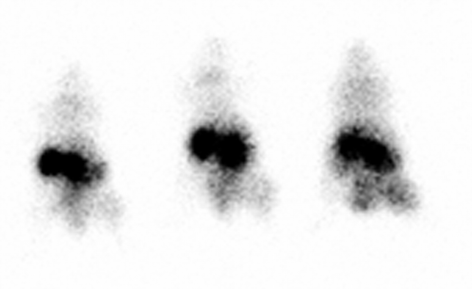
Whole-body images obtained simultaneously. (Right) Image of mouse with tumor obtained 3 h after administration of 99mTc-MORF, 21 h after administration of PL-cMORF, and 51 h after administration of MORF-MN14. (Center) Control animal receiving polymer, but not antibody. (Left) Control animal receiving neither polymer nor antibody. Tumors are in thigh on right.
DISCUSSION
Coming from an interest in oligomers for radiopharmaceutical development, this laboratory has examined phosphodiester and phosphorothioate DNAs, PNAs, and MORF oligomers for a variety of applications. The native phosphodiester DNA differs from the phosphorothioate by the substitution of a nonbonding oxygen with a sulfur atom. In the case of PNA, the phosphate backbone of DNA has been replaced with a (2-aminoethyl) glycine polypeptide linkage to which the nitrogenous bases are attached via methylenecarbonyl groups, whereas the phosphodiester backbone in MORFs has been substituted with a phosphorodiamidate group and the ribose sugar has been replaced with a morpholino ring. Like the DNAs, MORFs and PNAs are commercially available but, unlike DNAs, they are both uncharged and (unlike phosphodiester DNAs) stable to nucleases and (unlike phosphorothioate DNAs) nonchiral.
The goal of this investigation was to provide evidence that amplification targeting may be successfully achieved in a mouse tumor model. By introducing a multivalent cMORF polymer, it should ultimately be possible to greatly increase the accumulation of radioactivity in tumor and, at the same time, improve the tumor-to-normal tissue ratios. One alternative to amplification targeting is pretargeting using either (strept)avidin/biotin, bispecific antibodies, or oligomer pairs. Recently, encouraging results have been reported, especially using bispecific antibodies (5) and oligomers (6). However, since pretargeting does not involve a multivalent polymer, amplification is not possible (other than a factor of up to 4 in the case of certain streptavidin/biotin protocols (12)). Another alternative to the amplification strategy described herein is to conjugate the multiple copies of MORF directly to the antibody, thus avoiding the second administration. This would first require the preparation of a MORF polymer that would ultimately be conjugated to the antibody. Only in this way is there any hope of avoiding significant denaturation of the antibody. There have been numerous reports of antitumor antibodies conjugated directly with multiple copies of low-molecular-weight species such as boron for boron neutron capture (13) or metal-chelating agents for a variety of applications (14). The obvious disadvantage is the risk of denaturing the antibody as a result of the conjugation, especially when the aim is to attach multiple copies of a relatively high-molecular-weight MORF. For example, to carry 50 MORFs per molecule, the molecular weight of an IgG antibody must be raised from about 150 kDa to least 600 kDa. Under these conditions, it may not be possible to preserve immunoreactivity. Furthermore, amplification targeting may eventually be useful with tissue-specific agents other than antibodies such as antitumor and antitissue peptides, which, because of their low molecular weight, could not tolerate conjugation with multiple MORFs.
This laboratory began consideration of oligomers for amplification targeting and other radiopharmaceutical applications by examining DNA and concluded that neither the phosphodiester nor the phosphoromonothioate would be ideal for the applications of interest—the former being too unstable to nucleases in vivo and the latter showing too high an affinity for serum and tissue proteins (15). We subsequently showed that PNAs are stable and exhibit minimal protein binding affinities (16). Accordingly, our first amplification targeting study used PNAs (7). However, difficulties were encountered due to a low aqueous solubility of PNAs with the selected base sequence. Because of difficulties in conjugation, the PNA study did not use an antitumor antibody (7). Possibly because of higher aqueous solubility, the amine-derivatized MORFs were successfully conjugated both to the antitumor antibody MN14 and to the chelator MAG3 to permit radiolabeling with 99mTc. The radiolabel and the MORFs were found to be “stable” in vitro and in vivo, to show little affinity for proteins, and, like the labeled PNAs, are capable of rapid hybridization (11).
One application of radiolabeled MORFs is in pretargeting (6). Amplification targeting shares some similarities with pretargeting (as described herein) in the use of both a MORF-antibody and a radiolabeled MORF (cMORF in the case of pretargeting) but differs in the intermediate use of the polymer in amplification targeting. Amplification targeting is obviously more complicated than pretargeting but with potential for signal amplification. Should it be possible to significantly improve localization of radioactivity in targets such as tumor by this strategy, the approach would obviously be of value in the targeting of drugs, MRI contrast agents, and so forth to tumor and other targets.
One important aspect of amplification targeting is in situ accessibility. To achieve high tumor radioactivity levels, the MORFs on antibody in tumor must be accessible to the polymeric cMORFs and, in turn, the cMORF on the polymer in tumor must be accessible to the radiolabeled MORF. Another important aspect results from accumulation of the polymeric cMORF in liver, spleen, kidneys, and other normal organs. To lower background radiation levels in these normal organs, the polymeric cMORF expression should rapidly become inaccessible to the radiolabeled MORF.
The polymer may be the most important ingredient in amplification targeting. It must be fairly large to carry sufficient numbers of oligomers; the conjugated polymer should be water soluble and stable in both saline and serum; the pharmacokinetic properties must be favorable and the oligomers must be arranged on the polymer such that binding is not unduly hindered sterically. The polymer used in the earlier PNA study from this laboratory was a polymethylvinylether maleic acid (PA) with an initial molecular weight of 80 kDa and approximately 900 carboxyl groups per molecule (7). Each molecule was modified with an average of 80 PNAs (each with a 19 member polyetherpolyamide linker). Because of the limited aqueous solubility of PNA, the PA polymer showed unfavorable pharmacokinetics (i.e., high liver and low blood levels) unless the polymer was also conjugated with an average of 200 polyethylene glycol (PEG) groups. As a consequence, the molecular weight of the final polymer was raised to 1.4 MDa with >70% due to PEG. Increasing the molecular weight to this extent is expected to limit diffusion and penetration into tumor. Nevertheless, the most encouraging aspect of the PNA study was accessibilty. Although 75% of the PNAs on PA were accessible to the radiolabeled cPNA in solution and even when immobilized (on beads), between 35% and 58% of the PNAs on PA were still accessible (7).
The use of MORFs over PNAs simplified the synthesis of the polymers and provided a larger variety of polymer choices. In a parallel investigation, 3 PLs, completely succinylated to provide the required carboxyl groups and of different initial molecular weights, were synthesized and tested (17). In addition, 1 PA similar to that used previously with PNAs was also studied but without the PEG such that the final molecular weight of the PA polymer was now about 0.4 MDa compared with 1.4 MDa for only slightly fewer accessible oligomers per molecule. The biodistribution in normal mice showed the advantage of MORF over PNA since PA shows about half the liver and 4 times the blood levels when conjugated with MORFs compared with PNAs under nearly identical conditions. The results also suggested that a good choice for in vivo study is the 30 kDa PL, which shows higher blood levels and lower liver levels than the other 3 polymers tested. It was for this reason that the 30 kDa PL polymer was used in the studies described herein. However, this polymer had only 12–15 cMORFs per molecule.
Animal studies obviously provide a more realistic and more stringent test of amplification targeting compared with tissue culture studies. The first 2 animal studies were designed to quantitate antibody and polymer in tumor. By using dual radioactivity labels, it was possible to calculate that under the conditions of this investigation, about 25% of the antibody MORFs in tumor were targeted with polymeric cMORF in about 2 d and that 12% of these polymeric cMORFs in tumor could be targeted with radiolabeled MORF after 3 h. Because the previous study from this laboratory with PNAs did not use the antitumor antibody, no comparison with the 25% value is possible. However, the 12% accessibility of polymeric oligomer in tumor achieved under the conditions of this investigation is lower than the 35%–60% targeting found earlier (7). There are many possible explanations for this difference, including the use of a different polymer backbone (i.e., PA vs. PL), different-size polymers (initial molecular weight of 80 vs. 30 kDa), different oligomer (i.e., PNA vs. MORF), different linkers (i.e., 19 member polyetherpolyamide vs. 9 member succinylated piperidine), different tumor models (i.e., ACHN vs. LS174T), and different times between administration of the polymer and the radiolabel (i.e., 3 vs. 18 h). Perhaps the most likely explanation is the use of a different mechanism of localization. Whereas the PNA polymer localized in tumor by nonspecific diffusion and therefore was presumably free in the interstitial fluid, the MORF polymer was tethered to its antibody target presumably on the tumor cell surface. Some support for this possibility comes from closer agreement in accessibility of both polymers in solution of 70% for the cPNA polymer (7) compared with 50% for the cMORF polymer (17).
Over all animal studies, the only tissue consistently showing statistically significant higher values between study and control animals was tumor. Blood was often significantly higher for the study animals but this is expected as antibody and polymer remaining in circulation (and in tissues) at the time of the 99mTc-MORF administration will hybridize and retain radiolabeled MORF. Use of a clearing agent would likely be helpful (18). Kidney levels are elevated in this investigation since the labeled MORF localizes is this organ. We have recently shown that radiolabeled MORFs with cytosine bases such as that used in this research show elevated kidney levels that may be reduced by eliminating the cytosines (8). Future studies will use these modified MORFs.
CONCLUSION
Proof of the concept of amplification pretargeting has been demonstrated in that it was possible to target efficiently both the MORF on MN14 in tumor with the cMORF polymer and the cMORF on polymer in tumor with the radiolabeled MORF despite the barriers to accumulation that exist in vivo and the competition between clearance and targeting. Though it cannot yet be claimed that amplification itself has been convincingly demonstrated, accumulation in tumor of radiolabeled MORF was more than tripled over that of controls. Furthermore, MORF expression (on antibody) and cMORF expression (on polymer) was rapidly lost in normal organs such as liver, spleen, and kidneys but not in tumor, thus improving the target-to-nontarget ratios. These results were achieved despite an antibody conjugated with an average of only 0.2 MORF and with what is probably a less-than-ideal polymer with no more than 15 cMORFs per molecule. Furthermore, no clearing agent was used. Thus, there is considerable room for improvement.
Acknowledgments
The authors are grateful to Dr. Gary Griffiths of Immunomedics (Morris Plains, NJ) for providing the MN14 in this investigation. Financial support for this investigation was provided, in part, by the National Institutes of Health (grants CA79507 and CA94994).
Footnotes
Received Sep. 23, 2003; revision accepted Jan. 14, 2004.
For correspondence or reprints contact: Donald J. Hnatowich, PhD, Department of Radiology, University of Massachusetts Medical School, Worcester, MA 01655.
E-mail: Donald.hnatowich{at}umassmed.edu
REFERENCES
In this issue


Jump to section
Related Articles
Cited By...
- An experimental and theoretical evaluation of the influence of pretargeting antibody on the tumor accumulation of effector
- Successful Radiotherapy of Tumor in Pretargeted Mice by 188Re-Radiolabeled Phosphorodiamidate Morpholino Oligomer, a Synthetic DNA Analogue.
- Antibody Pretargeting Advances Cancer Radioimmunodetection and Radioimmunotherapy
- The Progress and Promise of Molecular Imaging Probes in Oncologic Drug Development
- Improving the Delivery of Radionuclides for Imaging and Therapy of Cancer Using Pretargeting Methods