


Abstract
99mTc-Mercaptoacetyltriglycine (99mTc-MAG3) and 99mTc-l,l-ethylenedicysteine (99mTc-ll-EC) are useful renal radiopharmaceuticals; however, both agents have renal clearances less than that of 131I-orthoiodohippurate (131I-OIH), and 99mTc-ll-EC exists in dianionic and monoanionic forms at physiologic pH. In an effort to develop a superior 99mTc agent with a rapid clearance comparable with that of 131I-OIH, we have designed a new ligand system, mercaptoacetamide-ethylene-cysteine (MAEC), which combines important structural features of both MAG3 and EC. Methods: Biodistribution and clearance studies were performed on Sprague–Dawley rats using syn- and anti-99mTc-l- and -d-MAEC coinjected with 131I-OIH. Studies were also performed by coinjecting each isomer (∼74 MBq [∼2 mCi]) and 7.4–11.1 MBq (200–300 μCi) of 131I-OIH in 3 volunteers with dual-isotope imaging performed using a camera system fitted with a high-energy collimator. Blood samples were obtained from 3 to 90 min after injection and urine samples were obtained at 30, 90, and 180 min. Results: In the rats, <10% of the injected dose remained in the blood at 10 min after injection for all isomers, and the urine dose at 60 min ranged from 84% to 99% that of 131I-OIH. The clearances of syn- and anti-99mTc-l-MAEC in the rats were higher than the clearances for the d-isomers (P ≤ 0.02) and were 102% and 105% that of 131I-OIH, respectively. In humans, the plasma protein binding of the 99mTc-MAEC complexes ranged from 82% to 89%. All 4 complexes provided excellent renal images. The 99mTc-MAEC complex/131I-OIH plasma clearance ratio in humans ranged from 45% (anti-99mTc-l-MAEC) to 74% (syn-99mTc-d-MAEC) with the 180-min urine excretion equivalent to that of 131I-OIH for all 4 complexes. Conclusion: Initial data in humans suggest that syn-99mTc-d-MAEC has a higher clearance than that of 99mTc-MAG3; however, none of the 99mTc-MAEC tracers have a clearance equivalent to that of 131I-OIH and further ligand design is needed.
- 99mTc-mercaptoacetamide-ethylene-cysteine
- 99mTc-ethylenedicysteine
- 99mTc-mercaptoacetyltriglycine
- renal radiopharmaceuticals
During the past 20 y several 99mTc complexes have been synthesized and tested as potential alternatives to 131I- or 123I-orthoiodohippurate (OIH) (1–3). The most widely used agent is 99mTc-mercaptoacetyltriglycine (99mTc-MAG3), which has been extensively studied and considered by many to be the 99mTc renal agent of choice (4–6). Nevertheless, 99mTc-MAG3 is still not the ideal replacement for 131I-OIH because its clearance is only 50%–60% that of 131I-OIH and it does not provide a direct measurement of effective renal plasma flow (ERPF) (1). Furthermore, a small percentage of 99mTc-MAG3 is transported into the small intestine via the hepatobiliary system in healthy volunteers; this percentage increases in patients with renal failure and can lead to problems in image interpretation (7–9). Increased hepatobiliary activity can also occur with suboptimal kit preparation (10).
In 1990, Verbruggen et al. (11) observed that 99mTc-l,l-ethylenedicysteine (99mTc-ll-EC), the polar metabolite of the brain agent 99mTc-l,l-ethylenedicysteine diethyl ester (99mTc-ll-ECD), was rapidly and efficiently excreted into the urine in mice. Subsequent studies have shown that the clearance of 99mTc-ll-EC is higher than that of 99mTc-MAG3 and ranges from 70% to 76% that of OIH (12–15). Nevertheless, the clearance of 99mTc-ll-EC is significantly less than that of 131I-OIH. Moreover, 99mTc-ll-EC exists in dianionic (80%) and monoanionic (20%) forms at physiologic pH, and it is highly unlikely that these 2 forms have the same clearance or protein binding affinity (15). The pH-dependent distribution between monoanionic and dianionic forms may lead to greater clearance variability if 99mTc-ll-EC is used to monitor a patient’s renal function over time.
The limitations of 99mTc-MAG3 and 99mTc-ll-EC coupled with the need for a 99mTc tracer that can directly measure ERPF have prompted a continuing search for an improved 99mTc renal imaging agent that has a clearance comparable to that of 131I-OIH. Our previous successful speciation studies with Re-EC complexes led directly to our design of a new series of ligands, mercaptoacetamide-ethylene-cysteine (MAEC), which combined important structural features of EC (cysteine moiety) and MAG3 (mercaptoacetamide moiety) (Fig. 1) (16–22). This study compares the pharmacokinetics of syn- and anti-99mTc-d- and l-MAEC with those of 131I-OIH in rats and healthy volunteers.

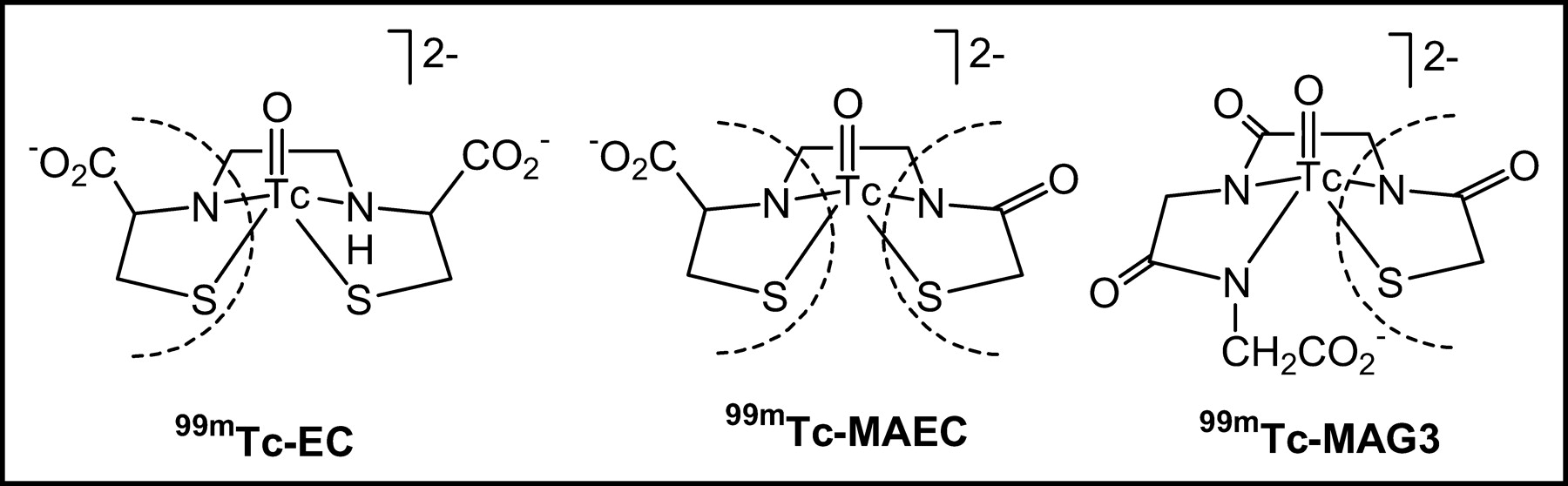
99mTc-MAEC combines structural characteristics of 99mTc-MAG3 and 99mTc-EC.
MATERIALS AND METHODS
All chemicals and solvents were of reagent grade and were used without further purification. N-(S-Benzoylmercaptoacetamide)ethylene-l-cysteine (l-MAEC) was prepared as described previously (23). The synthesis of N-(S-benzoylmercaptoacetamide)ethylene-d-cysteine (d-MAEC) was performed in a similar manner as that of l-MAEC except that d-cysteine was used instead of l-cysteine. 99mTc-Sodium pertechnetate (Na[99mTcO4]) was eluted from a 99Mo/99mTc generator (Amersham Health) using 0.9% saline. High-performance liquid chromatography (HPLC) analyses were performed on a Beckman System Gold Nouveau equipped with model 170 Radioisotope Detector, 166 UV Detector, and 32 Karat workstation software.
99mTc Radiolabeling
Each ligand (1 mg) was dissolved in EtOH (0.1 mL) and glycine buffer (0.1 mL; 50 mg of glycine sodium salt in 10 mL of 0.9% saline). Freshly prepared stannous tartrate solution (4 mmol/L in H2O, 0.1 mL) was added. The final pH of the solution was ∼9. Na[99mTcO4] in generator saline (0.25 mL) was added to the solution and the mixture was heated at 100°C for 10 min. After cooling to room temperature, the pH was adjusted to 6 with 1N HCl. The 99mTc-MAEC complexes were isolated by reverse-phase HPLC on a Beckman Ultrasphere ODS 5-μm column (4.6 × 250 mm); flow rate, 1 mL/min; mobile phase, 8% EtOH, 0.01 mol/L sodium phosphate buffer, pH 6.5. Stannous reduction of 99mTcO4− at pH 9 in the presence of either l-MAEC or d-MAEC ligands produced a mixture of two 99mTc-MAEC products. The two 99mTc-MAEC products were resolved by HPLC in an approximate ratio of 2:3. The radiochemical purity of the HPLC-separated 99mTc-MAEC complexes was >99%. The complexes were buffered to pH 7.4 and tested for stability by HPLC up to 6 h; no measurable decomposition was observed.
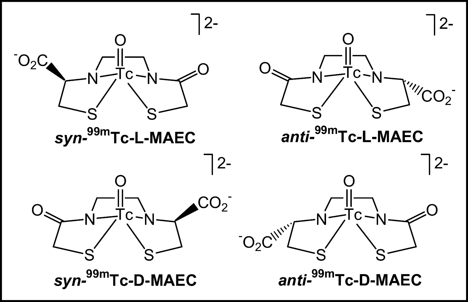
The first eluting peak was assigned as anti-99mTc-MAEC and the second peak as syn-99mTc-MAEC. We assigned the syn configuration to the 99mTc-MAEC complex because it was favored at high pH, a characteristic found in related systems and confirmed by chemical methods (21,23,24). Although the anti isomer formed at pH 12, it converted to the syn isomer on heating, a result similar to that obtained for Re-MAEC complex (23); a lower pH (∼9) must be used to slow the anti-to-syn conversion rate. At high pH, the syn isomer is thermodynamically preferred (Re, 94%; 99mTc, 94%) and the rate of anti-to-syn equilibration is fast (reaction completion, <30 min) (3,24).
Rat Studies
Biodistribution Studies.
The animal experiments followed the principles of laboratory animal care and were approved by the Institutional Animal Care and Use Committee of Emory University. syn- and anti-99mTc-l- and d-MAEC were each evaluated in 5 Sprague–Dawley rats at 10 and 60 min, respectively; data for 2 rats studied with anti-99mTc-l-MAEC at 10 min and 1 rat studied at 60 min had to be excluded due to problems with injection, standard preparation, or hypotension. A solution of each 99mTc complex (3.7 MBq/mL [100 μCi/mL]) and 131I-OIH (925 kBq/mL [25 μCi/mL]) was prepared and six 0.2-mL aliquots were drawn with insulin syringes. Five aliquots were used for doses; the sixth aliquot was diluted to 100 mL, and three 1-mL portions of the resulting solution were used as standards. Each rat was anesthetized with ketamine/xylazine (2 mg/kg body weight) injected intramuscularly. The bladder was catheterized using heat-flared PE-50 tubing for urine collection.
The radiopharmaceutical solution was injected intravenously via a tail vein, and 5 animals were killed at 10 min and 5 were killed at 60 min after injection. A blood sample was obtained and the heart, lung, spleen, liver, intestine, stomach, and kidney were removed. The whole liver was weighed and random sections were obtained for counting. Blood, whole organs, and tissue samples were placed in tubes, and each sample was weighed. Radioactivity of the sample and standards was measured in a double-channel well counter with 20% windows centered on the photo peaks of 99mTc (140 keV) and 131I (360 keV). Counts were corrected for background radiation, physical decay, and spillover of 131I counts into the 99mTc window. The percentage dose found in each tissue or organ was calculated by dividing the counts in each tissue or organ by the total injected counts. The value given for bowel represents combined stomach and intestine activity. The percentage injected dose in whole blood was estimated assuming a blood volume of 6.5% of total body weight.
Metabolism Studies.
Rats were prepared according to the procedure described for the biodistribution studies. A bolus injection of the radiopharmaceutical (∼37 MBq [∼1 mCi]) was given, and the urine was collected for 30 min and analyzed by HPLC alone and with purified complex added. Each 99mTc-MAEC complex was tested in 2 rats.
Renal Clearance, Extraction Fraction (EF), and Plasma Protein Binding (PPB).
Each of the 99mTc-MAEC isomers was compared with 131I-OIH in 6 Sprague–Dawley rats to determine steady-state plasma clearance, EF, and PPB. Each rat was anesthetized with ketamine/xylazine (2 mg/kg body weight) injected intramuscularly and placed on a heated surgical table. After tracheotomy, the left jugular vein was cannulated with 2 pieces of PE-50 tubing (one for infusion of the radiopharmaceuticals and one for infusion of normal saline [5.2 mL/h] to maintain hydration and additional anesthetic [up to 4 mg/h] as necessary). The right carotid artery was cannulated for blood sampling and the bladder was catheterized using heat-flared PE-50 tubing for urine collection. The core temperature of each animal was continually monitored using a rectal temperature probe. Each 99mTc complex (370 kBq/mL [10 μCi/mL]) was coinfused with 131I-OIH (185 kBq/mL [5 μCi/mL]) as an internal control at a flow rate of 1.5 mL/h through the left jugular vein for 45–60 min to establish steady-state blood levels. Urine was collected for three 10-min clearance periods, and midpoint blood samples (0.3 mL) were obtained. The following equation was used to calculate renal clearance: Cl (mL/min) = (urine volume/min × urine concentration)/plasma concentration. The average of the three 10-min clearance periods was used as the clearance value.
To measure EF, a left renal venous blood sample (0.5 mL) followed by a carotid artery sample (3 mL) was obtained at the conclusion of the study. The venous sample was centrifuged within 10 min of collection. EF was calculated by the following equation: EF = (arterial concentration-venous concentration)/arterial concentration; there was no correction for leakage of any of the tracers out of the red cells into the plasma. PPB was determined by ultracentrifugation (Centrifree micropartition system; Amicon Inc.) of 1 mL of arterial plasma: PPB = (1.0 –[ultrafiltrate concentration/plasma concentration]) × 100. A Beckman γ-counter system was used to determine the concentration of radioactivity in plasma, in red blood cells, and in urine samples with correction for 131I scatter into the 99mTc window.
Healthy Volunteer Studies
All studies were performed with the approval of the Human Investigations Committee, and a signed consent form was obtained. syn- and anti-99mTc-d- and l-MAEC complexes were each evaluated in 3 healthy volunteers. HPLC-purified complexes and phosphate-buffered saline (pH 7.4) were passed through a Sep-Pak Plus C18 cartridge (Waters Co.) (primed with 1 mL of ethanol) and a sterile Millex-GS 22-μm filter (Millipore Co.) into a sterile, pyrogen-free empty vial. The final concentration was 37 MBq/mL (∼1 mCi/mL) and the final pH was 7.4. Test samples of each complex were analyzed and determined to be sterile and pyrogen free. Approximately 74 MBq (∼2 mCi) of each 99mTc-MAEC complex were coinjected with 7.4–11.1 MBq (200–300 μCi) 131I-OIH, and imaging was performed using a General Electric camera with a 0.953 cm (0.375 in.) crystal fitted with a high-energy collimator; a 20% window was centered over the 365-keV photopeak of 131I, and a second 20% window was centered over the 140-keV photopeak of 99mTc. Data were acquired in a 128 × 128 matrix using a 3-phase dynamic acquisition and processed on a General Electric StarCam computer using QuantEM renal software. Blood samples were obtained at 3, 5, 10, 20, 30, 45, 60, and 90 min after injection and plasma clearances for 131I-OIH and each 99mTc-MAEC complex were determined using the single-injection, 2-compartment model of Sapirstein et al. (25). The volunteers voided at 30 and 180 min after injection to determine the percentage dose in the urine at each time period. For metabolism studies, a urine sample from the 30-min urine collection was obtained from each volunteer and analyzed by HPLC alone and with purified complex added.
Statistical Analysis
The statistical analysis was based on a 1-way ANOVA and paired t test. P ≤ 0.05 was considered to be significant. Datasets with n ≤ 3 were not analyzed.
RESULTS
Rat Studies
Biodistribution Studies.
All 4 99mTc-MAEC isomers (Fig. 2) showed a rapid blood clearance in rats with <10% of the injected dose remaining in the blood at 10 min after injection (Table 1). There was rapid urine excretion at 10 min as well as high specificity for renal excretion, with the dose in the urine at 60 min ranging from 84% to 99% that of 131I-OIH (Table 1). anti-99mTc-d-MAEC and anti-99mTc-l-MAEC showed more hepatic uptake than 131I-OIH at 10 min after injection and significantly more bowel activity at 60 min than 131I-OIH (P < 0.05), indicating hepatobiliary transport. Uptake in the spleen, heart, and lungs was ≤ 0.4% of the injected dose for all isomers at both time periods.

Isomers of 99mTc-MAEC.
Percentage Injected Dose in Rats of syn- and anti-99mTc-d- and l-MAEC Isomers in Blood, Urine, and Selected Organs at 10 and 60 Minutes Compared with 131I-OIH
Renal Clearance, EF, and PPB.
The renal clearances, EFs, and PPB studies of the 4 complexes in rats are summarized in Table 2; historical 99mTc-MAG3 data are shown for comparison (26). To minimize the effect of the experimental conditions on the results, the clearance and EFs for each complex were normalized to the corresponding 131I-OIH value in each rat. The 99mTc-MAEC/131I-OIH clearance ratio for syn- and anti-99mTc-l-MAEC was significantly higher than that of syn- and anti-99mTc-d-MAEC (P ≤ 0.02); similarly, the EF ratio of syn- and anti-99mTc-l-MAEC/131I-OIH was also higher than that of the d-complexes (P ≤ 0.02). PPB was moderately high (68%–96%) for all 99mTc-MAEC complexes. On the basis of the animal data, syn-99mTc-l-MAEC was the most promising complex, with a 78% protein binding, minimal bowel activity, and a clearance and extraction ratio comparable with those of 131I-OIH.
Renal Clearance (mL/min/100 g), EF, and PPB of 99mTc-MAEC Isomers in Rats Compared with Simultaneously Injected 131I-OIH (n = 6)
Metabolism Studies in Rats.
The urine was analyzed by HPLC to determine if the complexes were excreted intact. The 99mTc-l-MAEC complexes demonstrated syn-to-anti interconversion that was catalyzed in vivo. Approximately 20% of each injected agent converted to the corresponding isomer (syn to anti and anti to syn), giving the ratio of isomers in urine of 80% injected agent/20% corresponding isomer. The 99mTc-d-MAEC complexes did not demonstrate syn-to-anti conversion and each isomer was excreted unchanged in the urine.
Healthy Volunteer Studies
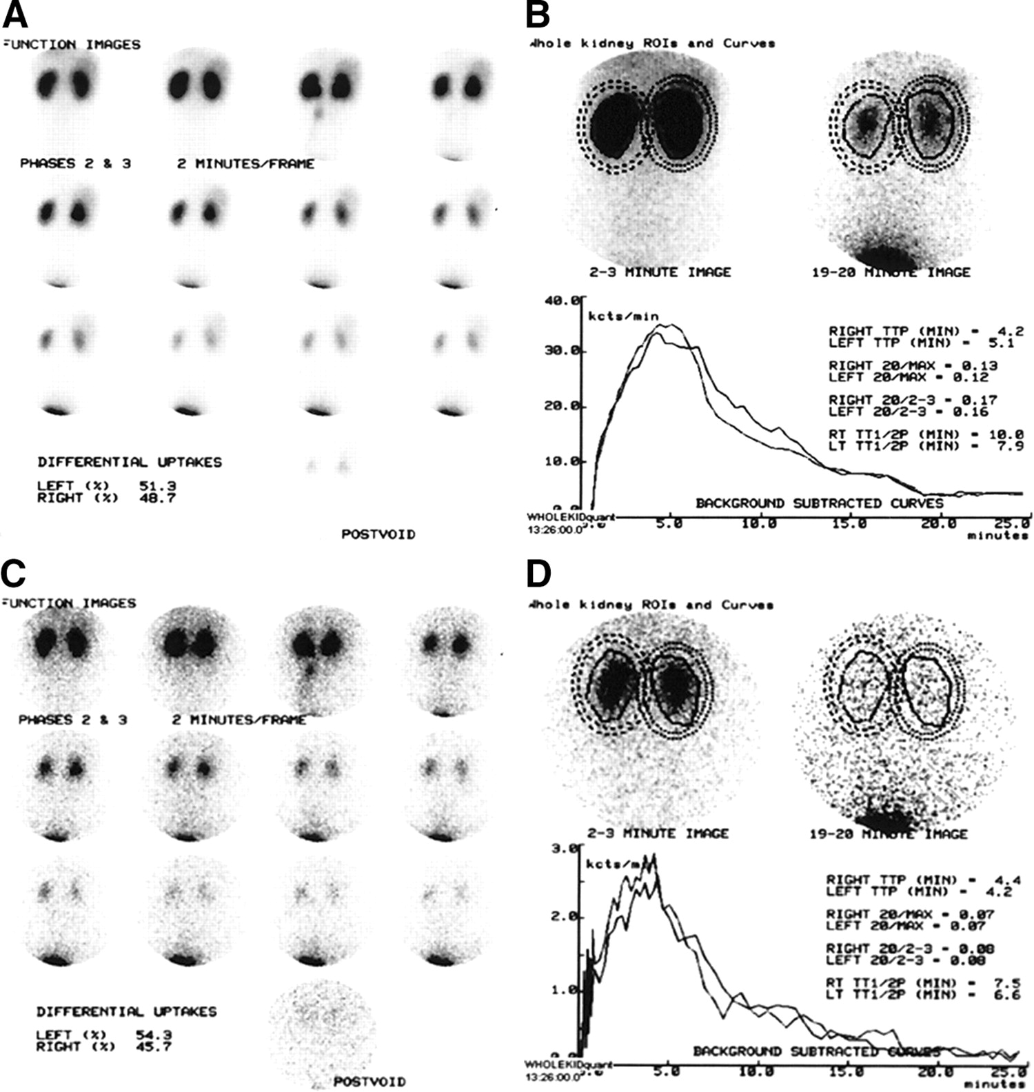
The PPB of the 4 99mTc-MAEC isomers ranged from 82% to 89%; red cell binding ranged from 19% to 23% (Table 3). With the exception of anti-99mTc-l-MAEC, almost all the injected dose was recovered in the urine at 3 h. The clearance of syn-99mTc-d-MAEC averaged 74% that of 131I-OIH compared with 49% for syn-99mTc-l-MAEC and 45% and 59% for anti-99mTc-l- and d-MAEC, respectively (Table 3), and is higher than the 50%–60% ratio reported for 99mTc-MAG3 (1). Image quality was excellent with all agents. Relative function measured with the 4 99mTc-MAEC complexes was equivalent to relative function measured with 131I-OIH; renogram parameters (time to peak [TTP] and the 20 min/max ratio for whole kidney regions of interest [ROIs]) were slightly more prolonged with the 99mTc-MAEC complexes than with 131I-OIH (Table 4). Representative syn-99mTc-l-MAEC images, renogram curves, and simultaneous 131I-OIH images and curves are shown in Fig. 3.

Data were acquired in 1 volunteer after simultaneous injection of 69.93 MBq (1.89 mCi) syn-99mTc-d-MAEC and 11.47 MBq (0.31 mCi) 131I-OIH; postvoid images were acquired ∼30 min after injection. The 2-min syn-99mTc-d-MAEC images (A) and renogram curves (B) can be compared with simultaneously acquired 131I-OIH images (C) and renogram curves (D). (A) Sequential 2-min images after injection of 69.93 MBq (1.89 mCi) syn-99mTc-d-MAEC; differential (relative) uptake is 51.3% in left kidney and 48.7% in right kidney. Final image is postvoid image obtained ∼30 min after injection. (B) Renogram curves and curve parameters based on whole kidney ROIs assigned over kidneys in A. Whole kidney and background ROIs are superimposed over the 2- to 3- and 19- to 20-min images at top. TTP refers to time-to-peak height of renogram curve and values are expressed in minutes; 20/max expression refers to ratio of counts at 20 min divided by maximum counts; 20/2–3 expression refers to ratio of counts at 19–20 min divided by counts at 2–3 min; TT1/2P refers to time (minutes) for counts in renogram curve to fall to half of peak value. RT = right; LT = left. (C) Sequential 2-min images after injection of 11.47 MBq (0.31 mCi) 131I-OIH. 131I-OIH images were acquired simultaneously as syn-99mTc-d-MAEC images in A. Differential (relative) uptake is 54.3% in left kidney and 45.7% in right kidney. Final image is postvoid image obtained ∼30 min after injection. (D) Renogram curves and curve parameters based on whole kidney ROIs assigned over kidneys in A and applied to 131I-OIH image data (C). Whole kidney and background ROIs are superimposed over 2- to 3- and 19- to 20-min images at top. TTP, 20/max expression, 20/2–3 expression, and TT1/2P are as described in B.
Clearance, Protein Binding, Red Cell Binding, and Urine Excretion of 99mTc-MAEC Complexes in Humans Compared with Simultaneously Injected 131I-OIH (n = 3)
Renogram Parameters of 99mTc-MAEC Complexes in Humans Compared with Simultaneously Injected 131I-OIH Using Whole Kidney ROIs (n = 3)
Metabolism Studies in Humans.
A urine sample from the 30-min urine collection for each complex was obtained for HPLC analysis. The results were similar to those of the metabolism studies performed in rats. As observed in the rat, the syn-to-anti conversion in vivo occurred only for 99mTc-l-MAEC complexes and was present in a ratio of 80% injected isomer to 20% of the corresponding isomer. 99mTc-d-MAEC complexes showed no syn-to-anti conversion and each injected isomer was excreted unchanged in the urine.
DISCUSSION
131I-OIH is typically used as the benchmark for new 99mTc agents because it provides a measure of ERPF and it is easy to measure in plasma and urine; however, the clearance of 131I-OIH is about 10% less than the clearance of p-aminohippuric acid (PAH) and it is no longer commercially available in the United States. A new 99mTc tubular agent with a clearance comparable with that of 131I-OIH would optimize renal imaging and allow a direct measurement of ERPF.
Wide clinical applicability would be enhanced by a radiopharmaceutical that contains only one isomer and exists as a single species under physiologic conditions. This goal is complicated by the observation that optimal tubular transport appears to require a pendant carboxyl group; this carboxyl group is often attached via a chiral carbon, and the coordination of a ligand bearing such pendant carboxyl group to the metal produces 2 isomers. (The carboxyl group can wrap close to the oxo group, syn, or away from the oxo group, anti.) (Fig. 2).
Quadridentate N2S2 and N3S ligands used for chelation of the metals Tc and Re are linear and form stable M(V)=O complexes with the N and S donor atoms normally coordinating in the equatorial plane with the oxo ligand in an axial position. The S donors are terminal; the 2 N donors are interior and each anchors 2 chelate rings on complexation to the metal (in the N3S system the third N is terminal). The N donors can be amide groups, amines, or a combination of the 2 donor types. Complexes with 2 secondary amine donors such as 99mTc-ll-EC and 99mTc-dd-EC have higher clearances than those of 99mTc(N2S2) and 99mTc(N3S) renal agents that contain exclusively amide N donors (1–3,15,27). However, solution studies of Re-ll-EC and its tetramethyl analog, Re-dd-TMEC, have shown that the complexes exist as a mixture of monoanionic CO2− ligated and dianionic CO2− deligated forms at physiologic pH (16,17). These ligated and deligated forms differ in charge, denticity, and structure; because of these differences, the 2 forms are highly unlikely to have comparable protein binding affinities, nor are they likely to be cleared at the same rate. The apparent protein binding and clearance of 99mTc-ll-EC reflects an average of the 2 species. Because the partition between the 2 species is pH dependent in the physiologic range, changes in renal clearance could reflect changes in pH rather than changes in renal function.
Each (ll, dd, or dl) EC ligand forms Re- and Tc-complexes with 2 geometric forms (3). In contrast, Re- and Tc-MAG3 have only one geometric form but, because they lack C2 symmetry, they have 2 chiral forms. These forms have been separated and evaluated independently, but there are only small differences in plasma clearance and renal transit (28). The search for new agents led us to investigate analogs of these species with the goal of identifying the underlying chemistry that would permit us to develop agents that exist in only one form at physiologic pH.
We designed and investigated Re-MAEC because it has a combination of amide and amine donors and only one noncoordinating carboxyl group (23). Because the carboxyl group is electron withdrawing and separated by only 2 bonds from the NH group, the acidity of the NH group is enhanced. Moreover, the amide donor bears a negative charge and is deprotonated; consequently, the metal–MAEC complex has only one dissociable NH group, and it was reasonable to expect that dissociation of the NH would occur outside the physiologic pH range. This is, in fact, what occurs. We found that the pKa of the single amine in the Re-MAEC complexes was near pH 6 and the dianionic form predominates (96%) at physiologic pH (23,24).
In general, the rat has been a good model for predicting the behavior of 99mTc renal radiopharmaceuticals; however, interspecies differences do occur and there is no substitute for human testing. For example, syn-99mTc-l-MAEC is the best 99mTc-MAEC complex in rats, whereas syn-99mTc-d-MAEC is clearly the best isomer in humans. The fact that the clearance of the 99mTc-MAEC complexes exceeds the glomerular filtration rate indicates that these complexes must be transported by the renal tubules and, as anionic tracers, they likely share the same tubular transport process as for 131I-OIH, 99mTc-MAG3, and 99mTc-EC. 99mTc-MAG3, 99mTc-ll- and -dd-EC, and syn-99mTc-CO2DADS (syn-bismercaptoacetamidopropanoate) are all cleared rapidly by the renal tubules and, like the best 99mTc-MAEC complex in humans (syn-99mTc-d-MAEC), all contain an oxo-technetium-glycyl sequence (O=Tc-N-C-CO2−) (1–3,27) with, except for 99mTc-MAG3, a CO2− group syn to the oxo ligand. However, for 99mTc-MAG3, the terminal glycyl residue is conformationally flexible and conformers are possible with a syn relationship of the carboxyl to the oxo ligand. The anti-99mTc-CO2DADS isomer is not rapidly excreted in the urine (27) and the anti-99mTc-MAEC complexes have prolonged half-time values compared with the syn-complexes. These structure–distribution relationships strongly suggest that the combination of the oxo group and the carboxyl moiety is responsible for receptor recognition, with the syn relationship being the preferred structure in humans.
The fact that syn- and anti-99mTc-l-MAEC complexes convert to the corresponding isomers (syn to anti and anti to syn) in vivo is an interesting observation. Since this conversion does not occur in vitro at physiologic pH, the conversion must be catalyzed by enzymatic activity. Furthermore, the enzyme only recognizes the l configuration since syn-to-anti interconversion does not occur for the 99mTc-d-MAEC isomers. The enzyme is probably located in the proximal tubular cell, although further testing must be performed to confirm this hypothesis. If the enzyme is identified and confirmed to reside in the cells of the renal tubules, syn-99mTc-l-MAEC could potentially be used as a probe to monitor enzyme activity in disease states.
CONCLUSION
In summary, these data provide a better understanding of the structure–function relationships of the 99mTc-MAEC complexes and will be helpful in the design of future renal radiopharmaceuticals. syn-99mTc-d-MAEC can be prepared as a single species (94% syn) when labeled at high pH. Initial data in humans suggest that its clearance is greater than that of 99mTc-MAG3 and is comparable to the clearance reported for 99mTc-ll-EC. Nevertheless, the clearance of 99mTc-MAEC is still less than those of 131I-OIH and PAH, and further ligand design and testing are required to develop a 99mTc renal tracer that will provide a direct measurement of ERPF.
Acknowledgments
This research was supported by the National Institutes of Health (grant DK38842). The authors also thank Patricia Marzilli, PhD, for her careful review of the manuscript.
Footnotes
Received Sep. 30, 2003; revision accepted Dec. 29, 2003.
For correspondence or reprints contact: Andrew T. Taylor, MD, Division of Nuclear Medicine, Emory University Hospital, 1364 Clifton Rd., NE, Atlanta, GA 30322.
E-mail: ataylor{at}emory.edu
REFERENCES
In this issue


Jump to section
Related Articles
Cited By...
- Preclinical Evaluation of 99mTc(CO)3-Aspartic-N-Monoacetic Acid, a Renal Radiotracer with Pharmacokinetic Properties Comparable to 131I-o-Iodohippurate
- Synthesis and In Vivo Evaluation of p-18F-Fluorohippurate as a New Radiopharmaceutical for Assessment of Renal Function by PET
- 99mTc(CO)3(NTA): A 99mTc Renal Tracer with Pharmacokinetic Properties Comparable to Those of 131I-OIH in Healthy Volunteers
- 99mTc(CO)3-Nitrilotriacetic Acid: A New Renal Radiopharmaceutical Showing Pharmacokinetic Properties in Rats Comparable to Those of 131I-OIH
- First Evaluation of a 99mTc-Tricarbonyl Complex, 99mTc(CO)3(LAN), as a New Renal Radiopharmaceutical in Humans