

FIGURE 5.
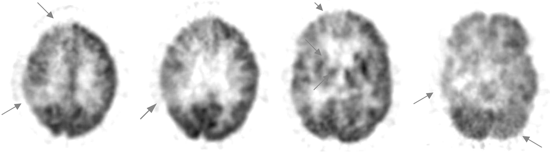
FIGURE 5.
18F-FDG PET images of vascular dementia. Hypometabolism affecting cortical, subcortical, and cerebellar areas is often seen in vascular dementia. This patient (65-y-old woman) was followed for 10 mo, and vascular dementia was diagnosed both clinically and by structural imaging. Arrows indicate hypometabolism of the right frontal cortex (far left, middle right), right parietal cortex (far left, middle left), right basal ganglia and thalamus (middle right), and right temporal cortex (far right). The hypometabolism of the left cerebellum (far right) is characteristic of cross-cerebellar diaschisis, caused by diminished afferent input from the contralateral cortex.
In this issue


Related Articles
Cited By...
- Early-Phase 18F-Florbetapir and 18F-Flutemetamol Images as Proxies of Brain Metabolism in a Memory Clinic Setting
- Usefulness of Dual-Point Amyloid PET Scans in Appropriate Use Criteria: A Multicenter Study
- Comparison of Early-Phase 11C-Deuterium-L-Deprenyl and 11C-Pittsburgh Compound B PET for Assessing Brain Perfusion in Alzheimer Disease
- MRI-Based Attenuation Correction for PET/MRI Using Multiphase Level-Set Method
- Everything you wanted to know about neuroimaging and psychiatry, but were afraid to ask
- Neuroimaging in dementia: a practical guide
- Early 11C-PIB Frames and 18F-FDG PET Measures Are Comparable: A Study Validated in a Cohort of AD and FTLD Patients
- Preclinical Properties of 18F-AV-45: A PET Agent for A{beta} Plaques in the Brain
- SPECT imaging in dementia
- Role of Neuroimaging in Alzheimer's Disease, with Emphasis on Brain Perfusion SPECT
- Visual Assessment Versus Quantitative Assessment of 11C-PIB PET and 18F-FDG PET for Detection of Alzheimer's Disease
- Hypometabolism Exceeds Atrophy in Presymptomatic Early-Onset Familial Alzheimer's Disease