

FIGURE 2.
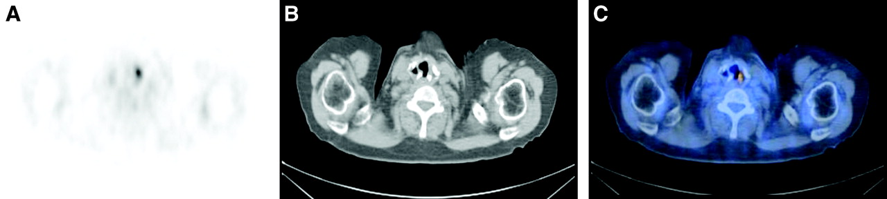
FIGURE 2.
Lung carcinoma (not shown) in right lung with focal increased 18F-FDG accumulation in left lower neck. (A) Transverse PET. (B) Transverse CT. (C) Integrated transverse PET/CT image of lower neck. Integrated PET/CT imaging ruled out N3 stage. PET/CT revealed that increased 18F-FDG accumulation in neck was localized in the intrinsic laryngeal muscles. Finding was result of compensatory laryngeal muscle activation caused by contralateral recurrent laryngeal nerve palsy from direct nerve invasion by lung cancer.
In this issue


Related Articles
Cited By...
- PET/MR Imaging: A Critical Appraisal
- MRI-Based Attenuation Correction for Hybrid PET/MRI Systems: A 4-Class Tissue Segmentation Technique Using a Combined Ultrashort-Echo-Time/Dixon MRI Sequence
- 18F-FDG PET Can Replace Conventional Work-up in Primary M Staging of Nonkeratinizing Nasopharyngeal Carcinoma
- Value of contrast-enhanced multiphase CT in combined PET/CT protocols for oncological imaging
- Introduction